Research Article
IN SILICO EVALUATION OF POTENT FOR PPAR
AGONIST OF LIGNAN DERIVATIVES FROM
Myristicafragrans HOUTT SEEDS
MUCHTARIDI MUCHRTARIDI
1*, KERI LESTARI
21Department of Pharmaceutical Analysis and Medicinal Chemistry, 2Department of Pharmacology and Clinic, Faculty of Pharmacy,
UniversitasPadjadjaran, Jl KM 21.5 Bandung-Sumedang, Jatinangor. *Email: [email protected]
Received: 26 Nov 2013, Revised and Accepted: 30 Dec 2013 ABSTRACT
Objective: Peroxisome proliferator-activated receptors gamma (PPAR) is a clinically established target for treatment of insulin resistance and has a
significant effect to improve the hyperglycemia and insulin resistance condition. In this investigation, lignan derivatives from nutmeg seeds (Myristicafragrans) was evaluated by in silico to know the potency of these compounds.
Methods: Molecular docking simulation was performed to screen out that the compounds had potent for PPAR agonist. Autodock 3.0.5 software
was employed to dock all ligand against PPAR and the all parameters of dcoking was validated by re-docking co-crystal ligand of
(2S)-2-(4-benzylphenoxy)-3-phenylpropanoic acid (PDB id : 3HOD) against to PPAR.
Results: Twenty compounds were favourably docked against PPAR agonist (PDB id: 3HOD). The tail of hydrophobic of lignan compounds also
favorable located in diphenyl pocket as well as TZD.
Conclusion: Macelignan and dihydro-di-isoegeunol (FEB -11.07 and -10.25 kkal/mol, respectively) could compete as agonist PPAR by connecting to
network hydrogen bond of His323, Tyr379, and Hist449, also formed hydrogen bond with Ser289 as mention thiazolidinediones (TZD) interacted
with PPAR, thus the both compounds might potent as agonist PPAR.
Keywords: Antidiabetes, Agonist, Myristicafragrans extract, PPAR
INTRODUCTION
WHO declares that Indonesia has become the fourth country that has the most diabetic patients in the world by following India, China and US [1]. WHO predicts this pandemic of the prevalence of diabetic in Indonesia that increasing from 8.4 million in 2000 to rise over 21.3 million in 2030.
More than 80 % of all diabetes is T2DM patients [2]. Based on epidemiology research, there are increasing the incidence and prevalence of T2DM due to ageing population structures in
developed countries and increasing obesity globally[3].
Furthermore, the WHO warns that T2DM diabetes is global pandemic [4].
T2DM is the multi factorial and multi genetic disease which occurs as combination metabolic disorder; insulin resistance and beta
pancreas cell insufficiency[5]. The both insulin resistance and
-pancreas cell insufficiency are caused by happening obesity and genetic factor [3, 6].
PPAR agonists have drawn great concern in the therapy
management of T2DM [7]. Sulfonylureas, metformin, acarbose, and thiazolidinones (TZDs) are current therapies for reducing plasma glucose [8, 9].
The antidiabetic effects of TZDs is due to the activation of the
peroxisome proliferator-activated receptors (PPAR) [9]. TZD is
high-affinity ligand for agonists of PPARγ and this phenomenon was
first reported by Lehman et al. [10]. However, this medicine gives side effect that may arise during treatment; TZDs also have side effects that increase the risk of heart attack and angina, fluid retention, weigh gain, and cardiac failure, thus TZDs use should be selective in diabetic patients who are not impaired liver and heart failure. For example, the treatment of TZD drugs such as rosiglitazone and pioglitazone, require monitoring to reduce the risk of adverse side effects, even troglitazone which is TZDs derivatives compounds have been withdrawn from the market because it showed an increased incidence of hepatitis induced by the drug [11, 12].
Based on the side effect story of TZD and derivatives, the
discovery of the other class drugs that selective into PPARγ and
PPAR agonist increases to reduce the risk of fatal side effect of
the drugs. The focus of this study is to development of new
PPAR agonists concentrate on structure-based design [13-15].
To date, there have been many crystal structures of PPAR
complexes available in Protein Data Bank (PDB) (www.rcsb.org).
Frachiolla et al. (2009) reported the design and synthesis of a novel
class of PPAR/ dual agonists, analogs of
-aryloxy-3-phenyl-propanoic acids compounds, and they published the crystal structure (PDB id : 3HOD) that declares the compounds are active in
nanomolar PPAR agonist [16].
Besides finding of novel synthetic compounds, the effort to explore alternative therapies using natural materials has been doing frequently in the community[17], because the materials relatively inexpensive and easily available as well as empirically shows efficacy for antidiabetic. However, the research that revealed the molecular mechanism of action of natural product antidiabetic has not much done, thus causing natural products potentially as antidiabetic cannot legally be used in treatment of diabetes.
We are encourage to develop nutmeg seeds as PPAR antagonist, it
contains chemical compounds derived of
2-aryloxy-3-phenyl-propanoic acids that proven as antagonist PPAR [18] such as lignan
and neolignan derivatives even macelignan is established and patented by Jae-Kwang et al [19, 20].
Here we screened nutmeg seeds lignan compounds-derived by using in silico by molecular docking simulation. Nutmeg seeds has been used traditionally as a spice and for medicinal purposes in Indonesia and other Asian countries [21-24]. In this study, evaluation of lignan
derivatives against PPARmay have not published yet, however one
of active compound of nutmeg seeds published by Jae-Kwang
796
the co-crystal ligand of
(2S)-2-(4-benzylphenoxy)-3-phenylpropanoic acid is complexed with PPARof homo sapiens that
expressed in E. Coli. The crystal structures are selected should have best resolution or lower resolution value, and also have R-free and R-value lower than 0.25 [25, 26]. The 3D structures of lignan derivatives compounds were constructed using Hyperchem 7, then were optimized using Austin Model 1 (AM1).
Molecular Docking Simulation
MGL tools program package 1.5.4. (Molecular Graphics Laboratory, The Scripps Research Institute) was used to prepare protein structures, ligand structures, grid parameter file and docking parameter file; furthermore, the AutoGrid v 3.05 program (The Scripps Research Institute) is used to prepare the grid, the Autodock 3.05 (http://autodock.scripps.edu) was employed to simulate the docking process under Linux program. As proposed by Brown and Ramaswamy (2007), qualified crystal structures should have the best resolution or lower resolution value, and also have R-free and R-value lower than 0.25 [25].
The chemical structures for the lignan derivatives of nutmeg seeds were obtained from literatures [27-30]. Twenty lignan compounds that contained in nutmeg seeds had been virtually screened via molecular docking (Autodock 3.0.5) [31].
The ligands and proteins were prepared by AutoDockTools (ADT).
Molecular docking was carried out on PPAR, PDB ID: 3HOD [18].
Ligand and protein available in the PDB structure were converted to PDBQ and PDBQS format by adding charges, hydrogens and assigning ligand fexibility. Kollman charges and solvation parameter were assigned using default value to the protein while Gasteiger charges were added to each ligand. A grid box of 60 x 60 x 60 points, with a spacing of 0.375 Å and a precise coordinate 29.305, 12.570, -20.539 along the x, y and z axes pertaining the centre of the active site was built around the binding region. Population size of 50 and 250 000 energy evaluations were used for 100 search runs via Lamarckian Genetic Algorithm (LGA) [26]. Docking result were analyzed based on the lowest free energy binding chosen from the most populated cluster and saved in dlg file for visualizationn. TZD
was used as control docking that imposed against PPAR(PDB
ID:3HOD)[18].
RESULTS AND DISCUSSION
Validation of Docking Method
Interaction TZDs as Control Ligand Against of PPAR
The protein target of PPARthat used in this study has diphenyl
pocket . This pocket is new L-shaped region of the PPARγ that
formed by forming several favourable hydrophobic interactions. This pocket increases the stabilization of the helix H3, inducing a conformation of the LBD less favourable to the recruitment of
co-activators required for full activation of PPARγ [ 8]. As shown in Fig. 1.a, the tail of aryloxy phenyl-propanoic acids (co-crystal ligand) and
TZD occupied the diphenyl pocket in PPARR assume a different
slope into the cavity in order to maintain the carboxylate H-bond network because of the longer protrusion of the Y314 side chain
( in PPARγ . PPARformsstrong hydrophobic contacts with several lipophilic residues such as Cys285, Leu330, Ile341, Met348 and Met364.
In the bottom of PPAR binding site (blue colored helix in Fig. 1), there are the loop 11/12 and is contoured sidewise by H3 and H11.
This loop stabilize diphenyl pocket that located between ( and
loop 11/12.
Interaction TZDs as Control Ligand Against of PPAR
Binding interaction ofTZD against PPAR explained well by some
literature [32-34]. In this study, TZD was used as control ligand, three-dimension of TZD structure was built by modelling, further
docked into PPAR(3HOD). TZD imposed against co-crystal
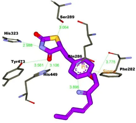
ligand (2S)-2-(4-benzylphenoxy)-3-phenylpropanoic acid (PDB id: 3HOD) as shown in Fig. 2. The hydrogen bond network of His323, His449, and Tyr473 interacted with the polar head of TZD as shown Fig. 2.
Fig. 1: Binding interaction pattern of TZD docked (purple) and imposed against co-crystal ligand (aryloxy phenyl-propanoic
acids-yellow) with H3 and H11 in complex with PPARγ. Blue light colored is Loop 11/12, and wire-grey colored is diphenyl
pocket .
Yu et al. (2003) analysed that the polar moiety of TZD (troglitazone) form five hydrogen bonds with Gln286, His449, Tyr473, His323 and Ser289, whereas the tails of the hydrophobic of TZD are located in
the hydrophobic pocket of PPAR[35]. The electrostatic interaction
appeared through pi-pi cation interaction (line orange colored in Fig.
2) between Phe282 of PPARand aromatic ring of TZD.
Fig. 2: Hydrogen bond network (His323, His444, and Tyr473) and hydrophobic interaction of TZD docked in complex with
PPARγ. Green colored carbon is hydrophobic residue.
Binding Interaction Prediction of Lignan derivatives of Nutmeg Seeds of Agonist PPAR
As in our previous study [36], we studied that nutmeg seeds extract might have potential as antidiabetic agent from natural product. The
nutmeg seeds extract gave increasing PPARγ-dependent luciferase activity, however this is not as good compared to TZD in enhancing
the activity of PPARγ-dependent luciferase.
Base on that results, we hypothesized that lignan derivatives compounds in nutmeg seeds containing might play role in the
797 lignan compounds in nutmeg as reported by Hattori et al [27], Orabi
et al. [29], Miyazawa et al. [37], Yang [38], and [39], prompted us to
explore of the potency of these compounds as PPARagonist.
Molecular docking simulation was employed to predict the potency these compounds. Autodock 3.0.5 was employed in this study [40]. Protein structure with PDB ID of 3HOD was selected as representing
PPAR ligand binding domain (LBD) and complexes with
aryloxypropanoic acid as ligand. TZD was became control ligand in this methods. Subsequently, TZD and co-crystal ligand of 2-aryloxy-3-phenyl-propanoic acids were docked to 1F8B thus produced RMSD less than 2.0 Å.
This resultsshowed that Lamarckian Genetic Algorithm using in AutoDock 3.0.5 was efficient and effective to predict true binding modes of TZD in grid dimension, which cover all important residues with a proper algorithm run amount, besidesthat, in all variations, RMSD values are equal or less than 2 Å, then Autodock was valid in docking simulation[41], even all the lowest crystallographic RMSD
values were 0.89 Å or less, indicating that low-energy structures found by the force field were very similar to the corresponding crystal structure [42].
All lignan derivatives from nutmeg seeds was favorably docked against
PPAR (3HOD). Interestingly, macelignan gave the smallest of binding
free energies (-11.07 kcal/mol), while neolignan had the highest free energy (FEB) (-8.00) kcal/mol (Table 1). This fact might be connected with the previous findings that macelignan has important role in the
activity to increase PPARγ-dependent luciferase [19].
However, the FEB of co-crystal ligand (PDB id: 3HOD) (-12.02 kcal/mol ) was less than macelignan, even TZD had lower than them (-12.65 kcal/mol). The all lignan derivatives had good binding
interaction against PPARas shown in Fig. 3 and Table 1. The lignan
compounds (grey colored carbon in Fig 3.) might imposed into TZD (purple colored carbon) in same position. The tail hydrophobic of lignan derivative interacted against hydrophobic pocket and
occupied diphenyl pocket as well as TZD.
Fig. 3: Imposition of 20 lignan derivatives compounds (grey colored carbons), ariloxypropanoic acid (yellow colored carbon) and TZD (purple colored carbon)of nutmeg seeds against PPAR.
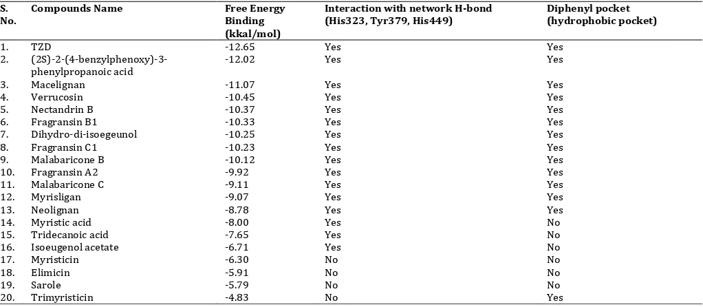
Table 1: Molecular Docking results of Lignan Derivatives Compounds of Nutmeg Seeds and Compared than TZD and co-crystal ligand.
S. No.
Compounds Name Free Energy Binding (kkal/mol)
Interaction with network H-bond (His323, Tyr379, His449)
Diphenyl pocket (hydrophobic pocket)
1. TZD -12.65 Yes Yes
2.
(2S)-2-(4-benzylphenoxy)-3-phenylpropanoic acid
-12.02 Yes Yes
3. Macelignan -11.07 Yes Yes
4. Verrucosin -10.45 Yes Yes
5. Nectandrin B -10.37 Yes Yes
6. Fragransin B1 -10.33 Yes Yes
7. Dihydro-di-isoegeunol -10.25 Yes Yes
8. Fragransin C1 -10.23 Yes Yes
9. Malabaricone B -10.12 Yes Yes
10. Fragransin A2 -9.92 Yes Yes
11. Malabaricone C -9.11 Yes Yes
12. Myrisligan -9.07 Yes Yes
13. Neolignan -8.78 Yes Yes
14. Myristic acid -8.00 Yes No
15. Tridecanoic acid -7.65 Yes No
16. Isoeugenol acetate -6.71 Yes No
17. Myristicin -6.30 No No
18. Elimicin -5.91 No No
19. Sarole -5.79 No No
798 Macelignan had the lowest FEB than others lignan compounds. As
mention TZD, macelignan also formed hydrogen bond with network hydrogen bond (His323, Tyr473, and His449), and also interact with Ser389. Aryloxy of diphenyl moiety of macelignan interacted with Ser464 through hydrogen bond interaction.
The diphenyl tail of macelignan also fitted into the bottom of the cavity of the Met463 side-chain of the loop 11/12 (light blue colored). This loop accommodated of the benzyl-phenoxy and
phenethyl-phenoxy groups faces of the terminal end. This interaction made strong interactions between the methyl and the p-cloud (aromatic ring). There were pi-pi interaction that indicated the electrostatic interactions occurred between aromatic ring and the ether group (-C-O-) of the Gln283 side chain. In the bottom cavity of
PPARγ, the Gln 86 side chain is also engaged in such interactions.
The residues of Met463 and Gln283 contacted well with the aromatic rings of hydrophobic tail of some lignan compounds. It was
evidence that macelignan was active as agonist PPARγ [19].
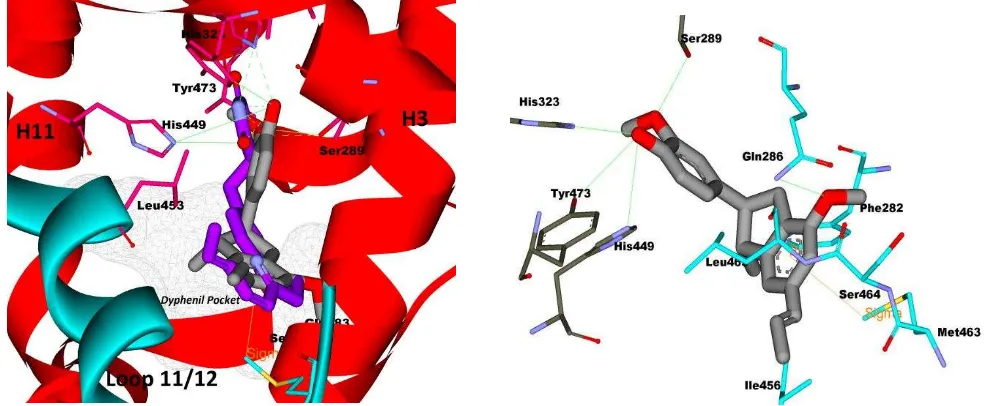
Fig. 4: Binding interaction of macelignaninti PPAR. (a) Macelignan imposed against TZD in same position, (b) hydrogen bond interaction (green colored line) of macelignan and hydrogen bond network of PPAR(His323, Tyr473, His449, and Sr289) and hydrophobic
interaction of macelignan tail and hydrophobic pocket.
The other interesting of lignan derivatives, the new potency of
dihydro-di-isoegeunol as agonist PPARwas shown in this study.
Molecular docking result of dihydro-di-isoegeunol almost coincided with TZD and co-crytal ligand s shon in Fig. 4. All the important
residue of PPAR interacted well with dihydro-di-isoegeunol. The
polar head of ortho-methoxyphenol of dihydro-di-isoegeunol imposed against azo group of TZD and connected with hydrogen bond network of His323, Tyr473, and His449.
The ortho-methoxyphenol group of dihydro-di-isoegeunol also formed hydrogen bond with Ser289 thus this group might important
role in bioactivity as agonist PPAR. There were electrostatic
interactions between Met346 of Loop 11/12 with aromatic ring of dihydro-di-isoegeunol through pi-pi interaction (orange colored line) as shown in Fig. 5. The previous study, the connecting between the aromatic ring and Met463 is evidenced by continuous electron density between the two interacting groups [16].
Fig. 5: (a) Dihydro-di-isoegeunol imposed against TZD in same position, (b) hydrogen bond interaction (green colored line) of macelignan and hydrogen bond network of PPAR(His323, Tyr473, His449, and Sr289) and hydrophobic interaction of dihydro-di-isoegeunol tail and
799 Based on the molecular docking simulation results, the binding
interaction of macelignan and dihydro-di-isoegeunol was similar compare than TZD thus the both macelignan and
dihydro-di-isoegeunol had potent as PPARagonist.
CONCLUSION
Lignan derivatives compounds of nutmeg seeds had favorably
docked against PPAR. The some lignan compounds interacted with
important residues of PPARLBD. Macelignan and
dihydro-di-isoegeunol formed hydrogen bond network of His323, Tyr379, His449, and Ser489. The hydrophobic tail of macelignanand
dihydro-di-isoegeunol fitted into diphenyl pocket , thus the both
compounds might potent as agonist PPAR.
REFERENCES
1. Sicree, R.; Shaw, J.; Zimmet, P., The Global Burden : Diabetes
and Impaired Glucose Tolerance. In Diabetes Atlas, International Diabetes Federation: 2010; Vol. 4, pp 1-105.
2. Moller, D. E.; Greene, D. A., Peroxisome proliferator-activated
receptor (PPAR) gamma agonists for diabetes. Adv Protein
Chem 2001, 56, 181-212.
3. Abate, N.; Chandalia, M., The impact of ethnicity on type 2
diabetes. J Diabetes Complications 2003, 17, (1), 39-58. 4. Hall, L. M. L.; Sattar, N.; Gill, J. M. R., Risk of metabolic and
vascular disease in South Asians: potential mechanisms for
increased insulin resistance. Future Lipidology 2008, 3, (4),
411-424.
5. Riddle, M. C., Glycemic management of type 2 diabetes: an
emerging strategy with oral agents, insulins, and combinations. EndocrinolMetabClin North Am 2005, 34, (1), 77-98.
6. Gloyn, A. L., The search for type 2 diabetes genes. Ageing Res Rev 2003, 2, (2), 111-27.
7. Pearson, E. R., Pharmacogenetics and future strategies in
treating hyperglycaemia in diabetes. Front Biosci 2009, 14,
4348-62.
8. Smith, U., Pioglitazone: mechanism of action. Int J
ClinPractSuppl 2001, (121), 13-8.
9. Hauner, H., The mode of action of thiazolidinediones. Diabetes
Metab Res Rev 2002, 18 Suppl 2, S10-5.
10. Lehmann, J. M.; Moore, L. B.; Smith-Oliver, T. A.; Wilkison, W. O.;
Willson, T. M.; Kliewer, S. A., An antidiabetic thiazolidinedione is a high affinity ligand for peroxisome proliferator-activated
receptor gamma (PPAR gamma). J BiolChem 1995, 270, (22),
12953-6.
11. Belfort, R.; Harrison, S. A.; Brown, K.; Darland, C.; Finch, J.; Hardies, J.; Balas, B.; Gastaldelli, A.; Tio, F.; Pulcini, J.; Berria, R.; Ma, J. Z.; Dwivedi, S.; Havranek, R.; Fincke, C.; DeFronzo, R.; Bannayan, G. A.; Schenker, S.; Cusi, K., A placebo-controlled trial
of pioglitazone in subjects with nonalcoholicsteatohepatitis. N
Engl J Med 2006, 355, (22), 2297-307.
12. Krentz, A. J.; Friedmann, P. S., Type 2 diabetes, psoriasis and thiazolidinediones. Int J ClinPract 2006, 60, (3), 362-3.
13. Sundriyal, S.; Bharatam, P. V., Important pharmacophoric
features of pan PPAR agonists: common chemical feature analysis and virtual screening. Eur J Med Chem 2009, 44, (9), 3488-95.
14. Rathi, L.; Kashaw, S. K.; Dixit, A.; Pandey, G.; Saxena, A. K., Pharmacophore identification and 3D-QSAR studies in
N-(2-benzoyl phenyl)-L-tyrosines as PPAR gamma agonists. Bioorg
Med Chem 2004, 12, (1), 63-9.
15. Lewis, S. N.; Bassaganya-Riera, J.; Bevan, D. R., Virtual Screening
as a Technique for PPAR Modulator Discovery. PPAR Res 2010, 2010, 861238.
16. Montanari, R.; Saccoccia, F.; Scotti, E.; Crestani, M.; Godio, C.; Gilardi, F.; Loiodice, F.; Fracchiolla, G.; Laghezza, A.; Tortorella, P.; Lavecchia, A.; Novellino, E.; Mazza, F.; Aschi, M.; Pochetti, G., Crystal structure of the peroxisome proliferator-activated receptor gamma (PPARgamma) ligand binding domain complexed with a novel partial agonist: a new region of the
hydrophobic pocket could be exploited for drug design. J Med
Chem2008, 51, (24), 7768-76.
17. Shivashankar, M.; Mani, D., A Brief Overview of Diabetes. Int J
Pharm PharmacSci 2011, 3, (4), 22-27.
18. Fracchiolla, G.; Laghezza, A.; Piemontese, L.; Tortorella, P.;
Mazza, F.; Montanari, R.; Pochetti, G.; Lavecchia, A.; Novellino, E.; Pierno, S.; Conte Camerino, D.; Loiodice, F., New 2-aryloxy-3-phenyl-propanoic acids as peroxisome proliferator-activated receptors alpha/gamma dual agonists with improved potency and reduced adverse effects on skeletal muscle function. J Med Chem 2009, 52, (20), 6382-93. Hwang, J. K., Therapeutic potential of peroxisome proliferators--activated receptor-alpha/gamma dual agonist with alleviation of endoplasmic reticulum stress for the treatment of diabetes. Diabetes 2008, 57, (3), 737-45.
21. Olajide, O. A.; Ajayi, F. F.; Ekhelar, A. I.; Awe, S. O.; Makinde, J. M.; Alada, A. R., Biological effects of Myristicafragrans (nutmeg) extract. Phytother Res 1999, 13, (4), 344-5.
22. Muchtaridi; Subarnas, A.; Apriyantono, A.; Mustarichie, R.,
Identification of Compounds in the Essential Oil of Nutmeg Seeds (MyristicafragransHoutt.) That Inhibit Locomotor Activity in Mice. Int J MolSci 11, (11), 4771-4781.
23. Ram, A.; Lauria, P.; Gupta, R.; Sharma, V. N., Hypolipidaemic
effect of Myristicafragrans fruit extract in rabbits. J
Ethnopharmacol 1996, 55, (1), 49-53.
24. Jain, N.; Goyal, S.; Ramawat, K. G., Evaluation Of Antioxidant
Properties And Total Phenolic Content Of Medicinal Plants Used In Diet Therapy During Postpartum Healthcare In Rajasthan. Int J Pharm PharmacSci 2011, 3, (3), 248-253.
25. Brown, E. N.; Ramaswamy, S., Quality of protein crystal structures.
ActaCrystallogr D BiolCrystallogr 2007, 63, (Pt 9), 941-50.
26. Musfiroh, I.; Nursamsiar, N.; Muhtadi, A.; Kartasasmita, R. E.;
Tjahyono, D. H.; Ibrahim, S., In Silico Study of Asiatic Acid Interaction With Inducible Nitric Oxide Synthase (INOS) and Cyclooxygenase-2 (COX-2). Int J Pharm PharmacSci 2013, 5, (1), 204-207.
27. Hattori, M.; Hada, S.; Kawata, Y.; Tezuka, Y.; Kikuchi, T.; Namba, T.,
New 2,5-Bis-aryl-3,4-dimethyltetrahydrofuran Lignans from the Aril of Myristicafragrans. Chem Pharm. Bull1987, 35, (8), 3322.
28. Chung, J. Y.; Choo, J. H.; Lee, M. H.; Hwang, J. K., Anticariogenic
activity of macelignan isolated from Myristicafragrans
(nutmeg) against Streptococcus mutans. Phytomedicine 2006,
13, (4), 261-6.
29. Orabi, K. Y.; Mossa, J. S.; el-Feraly, F. S., Isolation and
characterization of two antimicrobial agents from mace (Myristicafragrans). J Nat Prod 1991, 54, (3), 856-9.
30. Cho, J. Y.; Choi, G. J.; Son, S. W.; Jang, K. S.; Lim, H. K.; Lee, S. O.; Sung, N. D.; Cho, K. Y.; Kim, J. C., Isolation and antifungal activity of lignans from Myristicafragrans against various plant pathogenic fungi. Pest ManagSci 2007, 63, (9), 935-40.
31. M.M Garrett., e., al. ,Autodock : Automated Docking of Flexible
Ligands to Receptor version 3.05. Scipps Research Institute: U.S.A., 2000; p 51.
32. Liberato, M. V.; Nascimento, A. S.; Ayers, S. D.; Lin, J. Z.; Cvoro,
A.; Silveira, R. L.; Martinez, L.; Souza, P. C.; Saidemberg, D.; Deng, T.; Amato, A. A.; Togashi, M.; Hsueh, W. A.; Phillips, K.; Palma, M. S.; Neves, F. A.; Skaf, M. S.; Webb, P.; Polikarpov, I., Medium chain fatty acids are selective peroxisome proliferator activated receptor (PPAR) gamma activators and pan-PPAR partial agonists. PLoS One 2012, 7, (5), 362-397.
33. Yu, C.; Chen, L.; Luo, H.; Chen, J.; Cheng, F.; Gui, C.; Zhang, R.; Shen, J.; Chen, K.; Jiang, H.; Shen, X., Binding analyses between Human PPARgamma-LBD and ligands. Eur J Biochem2004, 271, (2), 386-397.
34. Nolte, R. T.; Wisely, G. B.; Westin, S.; Cobb, J. E.; Lambert, M. H.;
Kurokawa, R.; Rosenfeld, M. G.; Willson, T. M.; Glass, C. K.; Milburn, M. V., Ligand binding and co-activator assembly of the
peroxisome proliferator-activated receptor-[gamma]. Nature
1998, 395, (6698), 137-143.
800 36. Lestari, K.; Hwang, J.; Kariadi, S. H.; Wijaya, A.; Ahmad, T.;
Subarnas, A.; Supriyatna, S.; Muchtaridi, M., Screening for PPAR
γ agonist from MyristicafragransHoutt seeds for the treatment of Type 2 diabetes by in vitro and in vivo. Med. Health. Sci. Journal 2012, 12, (3), 7-15.
37. Miyazawa, M.; Kasahara, H.; Kameoka, H., A New Lignan
(+)-Myrisfragransin from Myristicafragrans. Nat Prod Lett 1996, 8, (1), 25-26.
38. Yang, X. W.; Huang, X.; Ahmat, M., [New neolignan from seed of
Myristicafragrans]. ZhongguoZhong Yao ZaZhi 2008, 33, (4),
397-402.
39. Purushothaman, K. K.; Sarada, A., Chemical examination of the
aril of Myristicafragrans. Indian J Chem 1990, 19 (B), 236-238.
40. Morris, G. M.; Goodsell, D. S.; Halliday, R. S.; Huey, R.; Hart, W.
E.; Belew, R. K.; Olson, A. J., Automated docking using a Lamarckian genetic algorithm and an empirical binding free energy function. J Com Chem 1998, 19, (14), 1639-1662.
41. Morris, G. M.; Lim-Wilby, M., Molecular docking. Methods
MolBiol 2008, 443, 365-82.
42. Morris, G. M.; Huey, R.; Olson, A. J., Using AutoDock for
ligand-receptor docking. CurrProtoc Bioinformatics 2008, Chapter 8,