Establishment of
naional laboratory-based surveillance
© World Health Organizaion 2011
All rights reserved. Requests for publicaions, or for permission to reproduce or translate WHO publicaions, whether for sale or for noncommercial distribuion, can be obtained from Publishing and Sales, World Health Organizaion, Regional Oice for South-East Asia, Indraprastha Estate, Mahatma Gandhi Marg, New Delhi-110 002, India (fax: +91-11-23370197; e-mail: publicaions@ searo.who.int).
The designaions employed and the presentaion of the material in this publicaion do not imply the expression of any opinion whatsoever on the part of the World Health Organizaion concerning the legal status of any country, territory, city or area or of its authoriies, or concerning the delimitaion of its froniers or boundaries. Doted lines on maps represent approximate border lines for which there may not yet be full agreement.
The menion of speciic companies or of certain manufacturers’ products does not imply that they are endorsed or recommended by the World Health Organizaion in preference to others of a similar nature that are not menioned. Errors and omissions excepted, the names of proprietary products are disinguished by iniial capital leters.
All reasonable precauions have been taken by the World Health Organizaion to verify the informaion contained in this publicaion. However, the published material is being distributed without warranty of any kind, either expressed or implied. The responsibility for the interpretaion and use of the material lies with the reader. In no event shall the World Health Organizaion be liable for damages arising from its use.
This publicaion contains the collecive views of an internaional group of experts and does not necessarily represent the decisions or the stated policy of the World Health Organizaion.
en
t o
f n
ai
on
al
lab
or
at
or
y-bas
ed
s
ur
ve
ill
an
ce
o
f an
im
ic
ro
bi
al
re
si
st
an
ce
Contents
Introducion
1. 1
Surveillance of animicrobial resistance
2. 3
Establishing a naional surveillance system
3. 5
Selecion of organisms for surveillance
4. 8
Selecion of animicrobial agents 1
5. 3
Protocols for animicrobial suscepibility tesing 1
6. 7
Quality control of animicrobial suscepibility tesing 2
7. 2
Troubleshooing for disc difusion tests 2
8. 6
Data collecion analyses and disseminaion 3
9. 1
Further reading 33
Annexes
Contributors 3
1. 5
WHO Model List (March 2010) Essenial medicines (excerpts) 3
During the past six decades animicrobial agents1 have played a criical
role in reducing the burden of communicable diseases all over the world. However, the emergence of resistance and its rapid spread is negaing the impact of these drugs, and hindering efecive applicaion of modern technologies in miigaing human misery. While the appearance of resistance is a continuous phenomenon in microorganisms, its ampliicaion and spread is through an array of pracices conducted by human beings. Improper uilizaion of animicrobial agents, especially in high disease-burden seings, results in strong selecion pressure that allows the resistant strain to grow and rapidly replace the suscepible isolates.
Diseases due to resistant organisms take longer to heal, and require expensive and at imes toxic drugs for longer periods, oten making the disease untreatable. The resistant organisms can also move across countries through travel and trade. In that sense, animicrobial resistance is a global challenge requiring concerted eforts at naional and internaional levels to preserve the available animicrobial agents. This is possible through treatment policies such as combinaion therapy, raional prescripion, paient adherence, a strong regulatory mechanism coupled with educaional aciviies, along with an eicient surveillance system that monitors the emergence and spread of resistance as well as the uilizaion of animicrobial agents.
To facilitate this acivity at the country level, WHO has developed a strategy that is simple, pracical and easy to scale up. The regional
1 In the strict sense, all animicrobials are not anibioics since some of these are chemically synthesized. However, for ease of reading these two terms will be used synonymously in this document.
2
Es
tab
lis
hm
en
t o
f n
ai
on
al
lab
or
at
or
y-bas
ed
s
ur
ve
ill
an
ce
o
f an
im
ic
ro
bi
al
re
si
st
an
ce
strategy aims to accord paricular atenion to intervenions involving the introduc ion of legislaion and policies governing the use of animicrobial agents, ensuring the raional use of these drugs at all levels of health-care seings and establishing laboratory-based net works for surveillance of resistance.
Scope of the document
This document aims to provide an overview of the steps that can be initiated to establish national laboratory-based surveillance of animicrobial resistance. The document also provides guidance on key elements of a good surveillance system. The document relies heavily on the experience gained in Thailand where the naional animicrobial resistance surveillance programme has been in operaion for several years.
Guideline development process
The WHO Regional Oice for South-East Asia commissioned the WHO Collaboraing Centre on Animicrobial Resistance, Naional Insitute of Health, Ministry of Public Health, Thailand to develop the irst drat of the guidelines. The objecives were to provide a tool to developing countries in establishing procedures and practices for a national system for a laboratory-based surveillance of animicrobial resistance which should generate evidence for forming policies and programmes for the raional use of anibioics. The guidelines were reviewed by several experts at the WHO Collaboraing Centre and subsequently by colleagues in the WHO Regional Oice for South-East Asia and WHO Country Oice for India.
Guidelines development team
Surveillance is defined as “the ongoing and systematic collection,
analysis and interpretaion of health data essenial to the planning, implementation, and evaluation of public health practice, closely integrated with the imely disseminaion of these data to those who
need to know”.
In simpler terms surveillance is data collecion for acion.
Surveillance of animicrobial resistance
Animicrobial resistance (AMR) surveillance data will help to formulate, monitor and idenify the prevailing and emerging problem, which can be contained by efecive strategy. Understandably, the majority of surveillance programmes are laboratory -based. Very few clinical data are collected and the data obtained by most surveillance programme are not useful to implement control and/or prevenion measures. One strategy to improve collecion, collaion and disseminaion for efecive use in the hospital/community is to integrate this funcion of animicrobial resistance surveillance aciviies into the exising disease surveillance aciviies.
Need for a naional animicrobial resistance
surveillance programme
The purpose of surveillance at the national level is to monitor suscepibility paterns of microorganisms to animicrobial agents which will reveal the animicrobial resistance status. This will provide reliable data that can be uilized by policy-makers or health administrators to
4
Es
tab
lis
hm
en
t o
f n
ai
on
al
lab
or
at
or
y-bas
ed
s
ur
ve
ill
an
ce
o
f an
im
ic
ro
bi
al
re
si
st
an
ce
review and revise the recommendaions for empirical treatment for community or hospital-acquired infecions. The regular disseminaion of animicrobial resistance informaion to physicians may improve the empirical selecion of animicrobial agents when treaing community or hospital-acquired infecions.
Informaion on animicrobial resistance derived from the surveillance can be used as an indicator of the quanitaive consumpion of anibioics in a paricular area covered by the network of hospitals. Data on the prevalence and resistance paterns of diferent pathogens derived from the surveillance will lead medical authoriies to produce prioriized steps and recommendaions or guidelines at the naional level on the control of community and hospital-acquired infecions and reduce the rate of resistance. In addiion, informaion on emerging and the increasing animicrobial resistance obtained from coninuous surveillance will alert both medical personnel and the people in the country, raise awareness and generate a common commitment to systemaically combat the animicrobial resistance crisis.
Advantages of animicrobial resistance surveillance
Animicrobial resistance surveillance is necessary to:
Understand when, where, how and why drug resistance is
emerging.
Reveal animicrobial eicacy.
Ensure the beter management of paients and infecion control
in hospital seings.
Improve management of community infecion control.
Inform policy-makers on evidence-based acion in developing
drug policy, essential medicines lists, standard treatment guidelines, procurement strategies, resource allocaion, health professional curricula and training.
Improve the empirical selecion of animicrobial agents when
treaing community or hospital acquired infecions.
en
t o
f n
ai
on
al
lab
or
at
or
y-bas
ed
s
ur
ve
ill
an
ce
o
f an
im
ic
ro
bi
al
re
si
st
an
ce
To establish naional animicrobial resistance surveillance, the following components and mechanisms are essenial:
Naional commitment
The Government or the highest public health authority should make a commitment and provide support to improve laboratory capacity for undertaking surveillance. This should be part of a naional agenda for the control and prevenion of animicrobial resistance.
Designate naional coordinator
The irst criical step to establish naional animicrobial resistance surveillance is to designate a naional coordinaing centre and give it the mandate to coordinate naionwide aciviies.
Forge a naional network
The naional coordinaing centre shall set up a naional network of selected representaive laboratories from hospitals and public health insitutes in diferent regions of the country. For beter representaion the members of the network may be from the public, private and not-for-proit organizaions as well as insitutes in diferent seings where animicrobial suscepibility paters are determined. These laboratories should ideally have quality microbiological faciliies for the determinaion of causative organisms, pathogen identification and susceptibility paterns.
Establishing a naional
surveillance system
6
Es
tab
lis
hm
en
t o
f n
ai
on
al
lab
or
at
or
y-bas
ed
s
ur
ve
ill
an
ce
o
f an
im
ic
ro
bi
al
re
si
st
an
ce
These regional laboratories shall provide uniform and validated data to the naional coordinaing centre to collate animicrobial resistance paterns, analyse these and disseminate imely surveillance informaion to potenial users on a regular basis.
Ensure uniformity of tesing and reporing
The members of the network need to:
Use standardized methods on collecion of clinical specimens,
laboratory test and reporing.
Assure quality of the microbiological tesing.
Be well versed with data input, analysis of data and contribuion
of informaion.
Reach an agreement on the mechanism to develop a naional/
internaional centralized database and ensure accurate and imely output as well as feedback.
Quality of surveillance data
The naional coordinaing centre should be responsible to provide support to paricipaing units in assuring quality of data through the following aciviies:
Organizaion of External Quality Assessment Scheme.
Supply of essenial materials, ie. animicrobial suscepibility
disks, transport media, reference control bacterial strains, tesing reagents.
Follow-up visits by experts/auditors.
Provision of referral services for the conirmaion of results.
Organizaion of training courses as per the requirements.
Disseminaion of technical informaion
en
t o
f n
ai
on
al
lab
or
at
or
y-bas
ed
s
ur
ve
ill
an
ce
o
f an
im
ic
ro
bi
al
re
si
st
an
ce
Types of surveillance
To set up naional resistance surveillance the country should decide the appropriate type of surveillance required to be set up depending on its needs, available infrastructure and resources and the feasibility of meeing the targets.
In a country with limited resources, surveillance may be performed by collecing primary laboratory data of common bacterial pathogens from the rouine tesing of representaive hospitals without seeking addiional reports from the primary data collectors (passive surveillance). If suicient resources are available, passive surveillance can be enhanced by the collection of additional data, which may help in initiating appropriate acions.
Laboratory-based surveillance
Prerequisites for establishing laboratory-based surveillance
Idenify organisms that should not be monitored.
Identify organisms that may be included in resistance
surveillance.
Finalize the animicrobial agents to be used for each isolate.
Develop a protocol for the determinaion of suscepibility.
Establish quality system.
Establish an informaion sharing mechanism.
Establishment of naional laboratory-based surveillance of animicrobial resistance
A large number of microorganisms infect human beings. Not all of these need to be included in the surveillance programme. Based upon local prevalence of diseases and the importance of these in speciic health care or community seings, selecion criteria should be applied. During the iniial phase of the surveillance network, it may be beter to have fewer organisms. The spectrum can be gradually expanded to include addiional organisms.
Given below is a brief descripion of some organisms with their characterisics to facilitate decision-making:
Organisms that should not be monitored
These include the followings:
Organisms for which no standardized disk difusion method is (a)
currently available for important animicrobial agents, and hence may not be included in rouine surveillance aciviies.
Some of these are:
Streptococcus viridans
group versus penicillin. Streptococcus pneumoniae versus
cephalosporins/carba-penems.
Staphylococcus
spp versus vancomycin.
Neisseria meningiidis
versus penicillin.
Burkholderia pseudomallei versus
all animicrobial agents.
Selecion of organisms for
surveillance
en
t o
f n
ai
on
al
lab
or
at
or
y-bas
ed
s
ur
ve
ill
an
ce
o
f an
im
ic
ro
bi
al
re
si
st
an
ce
Unusual organisms which are rarely involved in serious infecions. (b)
These include: Achromobacter.
Flavobacterium.
Bacillus.
Haemophilus
non-inluenzae/non-parainluenzae.
Organisms which do not pose serious treatment problems. These (c)
include:
Bordetella.
Corynebacterium.
Listeria.
Bacteria which are doubful enteric pathogens and rarely cause (d)
outbreaks. These include
Aeromonas.
Plesiomonas.
Edwardsiella.
Enteric pathogens for which no anibioic treatment is recommended. (e)
These include:
All categories of diarhhoeagenic
Escherichia coli.
Organisms that may be included in resistance
surveillance
The following groups of organisms may be included in resistance surveillance:
Organisms which are proven pathogens and not commensal
or contaminants.
Organisms that have high potenial for spread in the community
and the hospital seing.
Organisms that are known to acquire resistance against
currently used and recommended anibioics.
Organisms that have standard interpretaion of suscepibility
test.
Organisms that are widespread in the surveillance area and are
10
Es
tab
lis
hm
en
t o
f n
ai
on
al
lab
or
at
or
y-bas
ed
s
ur
ve
ill
an
ce
o
f an
im
ic
ro
bi
al
re
si
st
an
ce
Guidelines for the choice of organisms
Table 1 shows suggested organisms that are to be considered for inclusion in the surveillance system
Table 1: Organisms to be included in the surveillance system Respiratory pathogens and
agents of meningiis
Streptococcus pneumoniae
from respiratory isolates (i.e.
•
sputum, ear, sinus) and invasive isolate (i.e. blood, CSF, pleural luid). Haemophilus inluenzae
from respiratory isolates
•
(i.e.sputum, ear, sinus) and invasive isolates (ie. blood, CSF, pleural luid).
Sexually transmissible diseases
Neisseria gonorrhoeae
Gram-posiive cocci Staphylococcus aureus:
community acquired.
•
nosocomial acquired.
•
Staphylococcus saprophyicus: novobiocin resistance urinary
•
isolates.
Coagulase-negaive staphylococci: only from sterile site.
•
Enterococcus faecalis. Enterococcus faecium.
Enterococcus species.
Pathogens of diarrhoeal diseases
Shigella dysenteriae.
Shigella boydii. Shigella lexneri.
Shigella sonnei. Salmonella Typhi. Salmonella Paratyphi A. Salmonella (typhoid,
en
t o
f n
ai
on
al
lab
or
at
or
y-bas
ed
s
ur
ve
ill
an
ce
o
f an
im
ic
ro
bi
al
re
si
st
an
ce
Other members of family Enterobacteriaceae (This group is responsible for community-acquired urinary tract infecions and for all type of nosocomial infecions)
Escherichia coli.
Enterobacter:
Three categories are recommended,
•
E. cloacae, E. aerogenes and
Enterobacter spp. for those laboratories that make no speciic ideniicaion.
Klebseilla:
Two categories are suggested
•
K. pneumoniae, (only subspecies
pneumoniae should be monitored), and K. oxytoca (indole posiive). Serraia spp.
Proteus:
Proteus mirabilis
• (indole negaive).
P. penneri, P vulgaris
• (indole
posiive).
Morganella morganii.
Providencia retgeri. Providencia Stuarii.
Providencia spp.
Citrobacter freundii. Other Gram-negaive bacilli Pseudomonas aeruginosa
Acinetobacter spp. Acinetobacter
• spp. OF-glucose posiive, and non-hemolysis tentaively ideniied as A.
calcoaceicus-baumannii complex,whereas Acinetobacter spp. (OF-glucose negaive) presumpively ideniied as Acinetobacter sp. other than A. baumannii.
The following list can act as a guide for prioriizing the organisms that may be included in the naional surveillance system:
Acinetobacter calcoaciicus-baumannii complex (OF - glucose posiive, non hemolysis, growth at 44°C) Acinetobacter sp. (OF - glucose negaive) Citrobacter freundii
12
Es
tab
lis
hm
en
t o
f n
ai
on
al
lab
or
at
or
y-bas
ed
s
ur
ve
ill
an
ce
o
f an
im
ic
ro
bi
al
re
si
st
an
ce
Enterococcus faecalis Enterococcus faecium
Enterococcus spp.
Escherichia coli community-acquired (urine
isolates)
Escherichia coli hospital-acquired (non-urine
isolates)
Haemophilus inluenzae invasive infecions (encapsulated)
Haemophilus inluenzae respiratory isolates (sputum, oiis, sinusiis)
Klebsiella oxytoca
Klebsiella pneumoniae (subspecies pneumoniae) Neisseria gonorrhoeae
Proteus mirabilis (indole negaive)
Proteus vulgaris (indole posiive)
Providencia retgeri Providencia stuarii
Providencia spp.
Pseudomonas aeruginosa
Salmonella non-Typhi-Paratyphi A Salmonella Paratyphi A
Salmonella Typhi
Serraia spp. Shigella boydii
Shigella dysenteriae
Shigella lexneri
Shigella sonnei
Staphylococcus aureus community-acquired (out -
paients)
Staphylococcus aureus hospital-acquired
Staphylococcuscoagulase negaive signiicant repeated blood
isolates Staphylococcus saprophyicus (urine)
Streptococcus pneumoniae invasive isolates (blood,
cerebrospinal luid)
Streptococcus pneumoniae respiratory isolates (sputum, ear,
sinus) Vibrio cholerae O1
en
t o
f n
ai
on
al
lab
or
at
or
y-bas
ed
s
ur
ve
ill
an
ce
o
f an
im
ic
ro
bi
al
re
si
st
an
ce
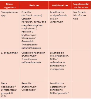
The organisms menioned in Chapter 4 and the animicrobial agents for which these need to be tested can also be categorized in a basic set, addiional set as well as supplemental set only for urinary pathogens (Table 2)
Table 2: Suggested animicrobial agents for animicrobial resistance surveillance
Micro-organisms Basic set Addiional set
Supplemental set for urine
Staphylococcus spp.
Oxacillin
(for Staph. aureus) Cefoxiin
(for Staph. aureus and coagulase negaive staphylococci) Penicillin G Erythromycin1 Clindamycin1 Gentamicin Trimethoprim-sulfamethoxazole
Levoloxacin or ciproloxacin MIC of vancomycin
Norloxacin Nitrofuran -toin
S. pneumoniae Oxacillin for penicillin Erythromycin1 Trimethoprim-sulfamethoxazole
Levoloxacin MIC of penicillin, MIC of
cefotaxime or cetriaxone or meropenem
Beta-haemolyic3, 2
Streptococcus group A, B, C, G,
Penicillin Erythromycin1 Clindamycin1
Levoloxacin Cefotaxime or cetriaxone MIC of penicillin4
Selecion of animicrobial agents
14 Es tab lis hm en t o f n ai on al lab or at or y-bas ed s ur ve ill an ce o f an im ic ro bi al re si st an ce
Micro-organisms Basic set Addiional set
Supplemental set for urine
Streptococcus spp.Viridans group3, 2
MIC of Penicillin Cefotaxime Cetriaxone
Enterococcus spp.
Penicillin or Ampicillin
Gentamicin (120 µg) β-lactamase test
Vancomycin or teicoplanin
Norloxacin or ciproloxacin or levoloxa -cin Nitrofuran -toin Enterobacteri -aceae Ampicillin Amoxicillin-clavulinic acid or ampicillin-sulbactam Cefazolin Cefoxiin Gentamicin Trimethoprim-sulfamethoxazole Ciproloxacin Cefuroxime Cefotaxime or cetriaxone Cefoperazone Cefepime or cefpirome Amikacin Imipenem Ertapenem Meropenem Cefoperazone - Salbactum Norloxacin or oloxacin Nitrofuran -toin
Shigella spp.5 Ampicillin Trimethoprim-sulfamethoxazole Ciproloxacin or Norloxacin Levoloxacin Salmonella spp5.
Ampicillin Trimethoprim-sulfamethoxazole Nalidixic acid6
Ciproloxacin or norloxacin6 Cefotaxime or cetriaxone5 Levoloxacin6 MIC of ciproloxacin6
en t o f n ai on al lab or at or y-bas ed s ur ve ill an ce o f an im ic ro bi al re si st an ce
Micro-organisms Basic set Addiional set
Supplemental set for urine
Pseudomonas aeruginosa Cetazidime Gentamicin Ciproloxacin Piperacillin-tazobactam Cefoperazone Cefepime or cefpirome Imipenem Meropenem Amikacin Cefoperazone - Salbactum Norloxacin or oloxacin Acinetobacter spp. Cefotaxime or cetriaxone Cetazidime Gentamicin Ciproloxacin Ampicillin-sulbactam Piperacillin- tazobactam Cefoperazon- sulbactam Cefepime or Cefpirome Imipenem Meropenem Amikacin Burkholderia cepacia Trimethoprim-sulfamethoxazole Cetazidime Meropenem Haemophilus inluenzae (CSF isolate) Ampicillin (β-lactamase test) Cefotaxime or cetriaxone Meropenem Haemophilus inluenzae, H. parainluenzae (respiratory isolate) Ampicillin (β-lactamase test) Amoxicillin-clavulanic acid or Ampicillin-sulbactam Trimethoprim-sulfamethoxazole Azithromycin or clarithromycin Ciproloxacin Neisseria meningiidis2 Cefotaxime Cetriaxone Meropenem MIC of penicillin
16
Es
tab
lis
hm
en
t o
f n
ai
on
al
lab
or
at
or
y-bas
ed
s
ur
ve
ill
an
ce
o
f an
im
ic
ro
bi
al
re
si
st
an
ce
Micro-organisms Basic set Addiional set
Supplemental set for urine
Burkholderia pseudomallei
MIC of trimethoprim-sulfamethoxazole MIC of amoxicillin-clavulinic acid MIC of cetazidime MIC of imipenem MIC of tetracycline or doxycycline
Moraxella catarrhalis
β-lactamase test
Foot notes
Not rouinely reported on organisms isolated from the urinary tract. 1.
Use Mueller Hinton agar added with 5% sheep blood for suscepibility test. 2.
The beta-haemolyic group includes the large colony-forming pyogenic strains of streptococci with 3.
Group A (Strept. pyogenes), C or G anigens and strains with group B (Strept. agalaciae) anigen. Small-colony-formimg beta-hemolyic strains with Group A, c, F, or G anigens (S. anginosus group) are considered part of the viridians group, and interpreive criteria for the viridians group should be used.
Determine MIC only on isolates from sterile sites. 4.
When fecal isolates of
5. Salmonella and Shigella spp. are tested, only ampicillin, a fuoroquinolone and trimethoprim-sulfamethoxazole should be reported rouinely. In addiion, chloramphenicol and a third-generaion cephalosporin should be tested and reported for extraintesinal isolates of Salmonella spp.
Fluoroquinolone-suscepible strains of
6. Salmonella that test resistant to nalidixic acid may be associated with clinical failure of delayed response in luoroquinolone-treated paients with extraintesinal salmonellosis. Extraintesinal isolates of Salmonella should also be tested for resistance to nalidixic acid. For isolates that test suscepible to luoroquinolones and resistant to nalidixic acid, the physician should be informed that the isolate may not be eradicated by luoroquinolone treatment, A consultaion with an infecious disease praciioner is recommended.
en
t o
f n
ai
on
al
lab
or
at
or
y-bas
ed
s
ur
ve
ill
an
ce
o
f an
im
ic
ro
bi
al
re
si
st
an
ce
Several laboratory techniques are available to determine suscepibility of microorganisms to animicrobial agents. Some of these are
Kirby-Baeur disc difusion method.
Stokes’ disc difusion method.
Minimum inhibitory concentraion (MIC) determinaion.
E-test.
Several variaions of these are now in use. The Clinical Laboratory Standards Insitute–USA (CLSI) has provided standards for disc difusion method for determinaion of animicrobial suscepibility of most of the pathogens. These standards are also updated regularly. The laboratories need to procure these standards along with interpretaion charts from CLSI for use in their faciliies.
The protocol should address the following issues for each of which the laboratory must have standard operaing procedures:
a. Preparaion of agar media
Some of the media used in laboratories are:
Mueller-Hinton agar (MHA)
MHA+ 5% sheep blood agar (MHB)
Gonococcus (GC) agar + 1% deined growth supplement
Haemophilus
test medium (HTM)
b. Preparaion of reagent
c. Preparaion or procurement of animicrobial discs
Animicrobial discs should be purchased from reliable companies which are shown by the given ceriicate of analysis inside the cartridges. These
Protocols for animicrobial
suscepibility tesing
18
Es
tab
lis
hm
en
t o
f n
ai
on
al
lab
or
at
or
y-bas
ed
s
ur
ve
ill
an
ce
o
f an
im
ic
ro
bi
al
re
si
st
an
ce
using, animicrobial discs should be placed at room temperature to have the same ambient temperature. This minimizes the condensaion of warm air to the cold discs. Once a cartridge is unsealed, the cartridge should then be kept at +4°C, in a box containing silica gel to ensure the anhydrous condiion. This refrigerated condiion is for a small working supply which should not be stored longer than one week. Some discs with labile drugs (e.g. imipenem, cefaclor and clavulanic acid combinaions) may retain beter stability if stored frozen unil it is used. Discard discs that reach the expiraion date stated on the label.
d. Standard turbidity using 0.5 McFarland Turbidity Standard
A 0.5 McFarland Standard is used to compare the standard turbidity. It is available commercially or can be prepared in the laboratory.
e. Measuring zone of inhibiion and interpreing results
The result is obtained by measuring the diameter of the zone of inhibiion, including the diameter of a disc in mm. A sliding caliper or ruler should be used and the zone on the back of the inverted agar plate must be measured with the following excepions:
The zone margin is the area showing no obvious growth. If
the faint growth or iny single colonies are seen, measure the colony-free inner zone.
Ignore the swarming growth of
Proteus spp. and measure the
obvious zone of inhibiion.
On MHA containing blood, measure the growth inhibiion zone
not the zone of inhibiion of haemolysis.
Interpretaion is done by comparing the measured diameter
to the breakpoint diameter shown in CLSI Tables.
Report the bacteria as suscepible, intermediate or resistant to the animicrobial agents and record the zone size of the test and control strains in the computer using WHONET programme.
Protocol for rapidly growing aerobic pathogens
Rapidly growing aerobic pathogens include Staphylococcus spp., Enterococcus spp., Salmonella spp., other Enterobacteriaceae,
en
t o
f n
ai
on
al
lab
or
at
or
y-bas
ed
s
ur
ve
ill
an
ce
o
f an
im
ic
ro
bi
al
re
si
st
an
ce
The direct colony suspension method is recommended for these organisms for the CLSI approved protocol.
Protocol for tesing fasidious organisms
Streptococcus pneumoniae
and other
Streptococcus spp.
MHA containing 5% sheep blood is recommended for suscepibility tesing of S. pneumoniae and other streptococci. The test method is
Kirby Bauer and direct colony suspension method, with the following excepions:
Prepare inoculum by suspending pure culture grown on sheep
blood agar for 18-20 hours in saline or MHB to have the turbidity of 0.5 McFarland standard.
Take the animicrobial disc (according to the list in Table 2) from
the cartridge by using sterile forceps. Place not more than ive animicrobial discs onto the surface of the agar plate (diameter of 100 mm) or no more than 12 discs on a 150 mm plate. Incubate at 35
°C in 5% CO2 atmosphere or in the candle jar for 20-24 hours.
Measure diameter of the zone of inhibition and interpret by comparing with the breakpoints shown in CLSI tables. Record the zone size of the test and control strains in the computer using WHONET programme.
Table 3. Zone diameter interpretaive standards for
Streptococcus pneumoniae (in mm)
Animicrobial agent Resistant Intermediate Suscepible
Oxacillin 1 µg* - - ≥ 20
Erythromycin 15 µg ≤ 15 16-20 ≥ 21 Co-trimoxazole 25 µg ≤ 15 16-18 ≥ 19
Levoloxacin 5 µg ≤ 13 14-16 ≥ 17
20
Es
tab
lis
hm
en
t o
f n
ai
on
al
lab
or
at
or
y-bas
ed
s
ur
ve
ill
an
ce
o
f an
im
ic
ro
bi
al
re
si
st
an
ce
Susceptibility test of other streptococci to oxacillin is not recommended. For beta-haemolytic streptococci only penicillin or ampicillin disc is used to do the test. In contrast, both penicillin and ampicillin discs are not reliable for viridans group streptococci. A penicillin MIC should be performed on the Viridans group streptococci isolated from sterile sites (e.g., CSF, blood, bone).
Haemophilus inluenzae and H. parainluenzae
HTM is recommended for suscepibility tesing of H. inluenzae and H. parainluenzae. Mueller-Hinton chocolate agar is not appropriate for animicrobial suscepibility tesing of Haemophilus spp. The test
method is Kirby Bauer and direct colony suspension method with the following excepions:
Inoculum is prepared by suspending pure culture grown on
chocolate agar for 20-24 hours in saline or MHB to have the turbidity of 0.5 McFarland standard.
Take the animicrobial disc (according to the list in Table 1) from
the cartridge by using sterile forceps. Place not more than ive animicrobial discs onto the surface of the agar plate (diameter of 100 mm) or no more than 12 disks on a 150 mm plate. Incubate at 35
°C in 5% CO2 atmosphere or in the candle jar for 16-18 hours.
Measure diameter of the zone of inhibiion and interpret by
comparing with the breakpoints shown in CLSI Tables.
Record the zone size of the test and control strains in computer using WHONET programme.
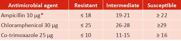
Table 4: Zone diameter interpretaive standards for Haemophilus inluenzae (in mm)
Animicrobial agent Resistant Intermediate Suscepible
Ampicillin 10 µg* ≤ 18 19-21 ≥ 22 Chloramphenicol 30 µg ≤ 25 26-28 ≥29 Co-trimoxazole 25 µg ≤ 10 11-15 ≥ 16
* Most of the ampicillin resistance among H. inluenzae is caused by the presence of a β-lactamase. In some seings, less than 1% of the clinical isolates of H. inluenzae are ampicillin-resistant due to another mechanism (altered penicillin binding proteins). For this reason, a negaive β-lactamase test can be reported as suscepible to ampicillin (without performing the disc test). Since many other animicrobial agents have a predictable acivity against H. inluenzae (3rd generaion cephalosporins, quinolones, amoxicillin-clavulanic
en
t o
f n
ai
on
al
lab
or
at
or
y-bas
ed
s
ur
ve
ill
an
ce
o
f an
im
ic
ro
bi
al
re
si
st
an
ce
Neisseria gonorrhoeae
GC agar containing 1% deined growth supplement is recommended for susceptibility testing of N. gonorrhoeae. Cysteine–free growth
supplement is not required for disc difusion test and chocolate agar enriched with other supplements is not appropriate for suscepibility tesing of N. gonorrhoeae.
The test method is Kirby Bauer and direct colony suspension method with the following excepions:
Inoculum is prepared by suspending pure culture grown on
chocolate agar for 20-24 hours in 5% CO2 atmosphere, in saline or MHB to have the turbidity of 0.5 McFarland standard. Take the animicrobial disc (according to the list in Table 1)
from the cartridge by using sterile forceps. Place not more than ive animicrobial discs onto the surface of the agar plate (diameter of 100 mm) or no more than 12 discs on a 150 mm plate. For animicrobial disks that produce vary large zone e.g., luoroquinolones or cephalosporins, only two to three discs may be tested per 100 mm diameter plate.
Incubate at 35
°C (do not exceed 37°C) in 5% CO2 atmosphere or in the candle jar for 20-24 hours.
Measure diameter of the zone of inhibition and interpret by comparing with the breakpoints shown in CLSI Tables. Record the zone size of the test and control strains in computer using WHONET programme.
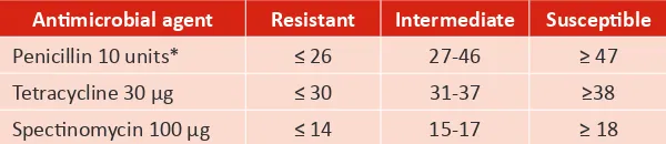
Table 5. Zone diameter interpretaive standards for Neisseria gonorrhoeae (in mm)
Animicrobial agent Resistant Intermediate Suscepible
Penicillin 10 units* ≤ 26 27-46 ≥ 47 Tetracycline 30 µg ≤ 30 31-37 ≥38 Specinomycin 100 µg ≤ 14 15-17 ≥ 18
Establishment of naional laboratory-based surveillance of animicrobial resistance
Quality control (QC) in the laboratories should cover all diagnosic tests from the collection and evaluation of specimen through the interpretaion and accuracy of the test results. In this way, guideline for laboratory pracices should be established and revised consistently and concomitant with the establishment of internal quality control of all reagents, media, and equipment used.
The main objecives of quality control of animicrobial suscepibility by the disc difusion method are to control the following:
The precision and accuracy of the test method. (1)
The quality of reagents, medium, and antimicrobial discs (2)
used.
Laboratory performance, reading, and interpretaion of test (3)
results.
With the use of reference bacterial strains recommended by CLSI, this process will ensure the highest achievement in the control of precision and accuracy of the test method. The standard bacterial strains should be obtained from reliable sources (e.g., American Type Culture Collecion or Culture Collecion Center, Naional Insitute of Health, Department of Medical Sciences, Nonthaburi, Thailand).
Storing and testing quality control strains should follow the procedure described by CLSI.
Quality control of animicrobial
suscepibility tesing
en
t o
f n
ai
on
al
lab
or
at
or
y-bas
ed
s
ur
ve
ill
an
ce
o
f an
im
ic
ro
bi
al
re
si
st
an
ce
Internal quality control for the rapid-growing aerobic
pathogens
Quality control of the test method is performed by using the following standard bacterial strains.
: Staphylococcus aureus ATCC 25923 (DMST 8840)
: Escherichia coli ATCC 25922 (DMST 4212)
: Pseudomonas aeruginosa ATCC 27853 (DMST 4739)
: Enterococcus faecalis ATCC 29212 (DMST 4737)
The Enterococcus standard strain is used to test the minimum level
of thymidine and thymine in Mueller-Hinton medium for suscepibility tesing of bacteria to co-trimoxazole. The high level of thymidine or thymine gives rise to the false negaive result. Thus, all batches of this medium should be tested by performing the suscepibility of E. faecalis ATCC 29212 or 33186 to co-trimoxazole. Suicient low level of thymidine and thymine is indicated by an inhibiion zone of ≥ 20 mm. This QC should be done once a week or whenever the new batch of medium is prepared or whenever the new disc cartridge is opened.
The acceptable minimum and maximum inhibiion zones for a single QC test are shown in corresponding CLSI Tables. If the QC result is out of range only one out of 20 tests, it is acceptable. If the QC result is out-of-control more than one test, correcive acion is required immediately.
Internal quality control for fasidious organisms
Streptococcus pneumoniae
Standard bacterial strain used for QC is S. pneumoniae ATCC 49619.
Perform the same test procedure as described for clinical isolates of
S. pneumoniae (Mueller–Hinton sheep blood agar, incubated at 35°C in 5%
CO2 atmosphere or in the candle jar for 20-24 hours).The minimum and maximum inhibiion zones for a single QC test are shown in CLSI Table.
Neisseria gonorrhoeae
Standard bacterial strain used for QC is N. gonorrhoeae ATCC 49226.
Perform the same test procedure as described for clinical isolates of
N. gonorrhoeae (GC agar base plus 1% deined growth supplement,
24
Es
tab
lis
hm
en
t o
f n
ai
on
al
lab
or
at
or
y-bas
ed
s
ur
ve
ill
an
ce
o
f an
im
ic
ro
bi
al
re
si
st
an
ce
the candle jar for 20-24 hours).The minimum and maximum inhibiion zones for a single QC test are shown in CLSI Table.
Haemophilus inluenzae
Standard bacterial strain used for QC is H. inluenzae ATCC 49766. Perform the same test procedure as described for clinical isolates of H. inluenzae (HTM consising of 15 µg/ml β–NAD;15 µg/ml bovine hemain; and 5 g/L yeast extract; adjust pH to 7.2 to7.4 incubated at 35°C in 5% CO2 atmosphere or in the candle jar for 16-18 hrs).The minimum and maximum inhibiion zones for a single QC test are shown in CLSI Table.
Table 6 shows the list of standard bacterial strains collected at the Culture Collecion Center, NIH, Department of Medical Sciences, Nonthaburi, Thailand (DMST-CC). The detail of strains used for QC is also described.
Table 6. List of standard bacterial strains for QC of animicrobial suscepibility tesing
Species Strain No. Primary use/characterisics
1 Enterococcus
faecalis
DMST 4736 =ATCC 29212
Sensiive to vancomycin, high level gentamicin (16-23 mm), and high-level streptomycin (14-20 mm)
Use to check media that is acceptable for tesing sulfonamides, trimethoprim, and their combinaion, use to test vancomycin screen plate (will not grow) and high-level aminoglycoside disk and MIC tests for enterococci.
2 Enterococcus
faecalis
DMST 4737 =ATCC 51299
Resistant to high level gentamicin, high -level streptomycin, and vancomycin. Use to test vancomycin screen plate (will grow) and high-level aminoglycoside disc and MIC tests for enterococci.
3 Enterococcus
faecium
DMST 4743 =UCLA 192
en t o f n ai on al lab or at or y-bas ed s ur ve ill an ce o f an im ic ro bi al re si st an ce
4 Escherichia coli DMST 4212
=ATCC 25922
Sensiive to all gram negaive organisms
5 Escherichia coli DMST 7948
=ATCC 35218
Ampicillin-resistant but sensiive to β-lactamase/β-lactamase inhibitor combinaions. Use to test Amp/Clav (17-22 mm), Ticar/Clav, and other combinaion drugs.
6 Haemophilus
inluenzae
DMST 7943 =ATCC 49247
Recommended reference strain for animicrobial suscepibility tests 7 Haemophilus inluenzae DMST 7944 =ATCC 49766 Ampicillin suscepible,
recommended reference strain for animicrobial suscepibility tests
8 Pseudomonas
aeruginosa
DMST 4739 =ATCC 27853
Use to monitor caion content and pH of media. Aminoglycosides are especially sensiive to caions.
9 Staphylococcus aureus
DMST 8840 =ATCC 25923
Oxacillin sensiive (18-24 mm) and penicillin sensiive -β-lactamase negaive.
Use to test gram-posiive drugs with disc difusion test and as negaive control for the oxacillin agar screen plate (will not grow).
10 Streptococcus pneumoniae
DMST7945 =ATCC 49619
Use to test disc difusion and E-test, MIC test for
Streptococcus spp. including S. pneumoniae.
DMST = Department of Medical Sciences , Thailand ATCC = American Type Culture Collecion UCLA = University of California, Los Angeles
External quality assessment scheme
Establishment of naional laboratory-based surveillance of animicrobial resistance
Various factors that inluence the results of the disc difusion tests are summarized below:
Element Check possible reason
Standard bacterial strain
Use of wrong strain
-
Improper maintenance, e.g. storage and
-
frequent subculture
Contaminaion or changes in the geneic
-
characterisics Tesing supplies - Improper storage
Contaminaion or use of the expired materials
-
Use of damaged or defecive agar plates
-
Tesing procedure
Use of wrong incubaion temperature or length
-
of ime
Use of incorrectly prepared inocula (too light or
-
too heavy)
Use of deteriorated or expired turbidity
-
standard
Inoculums prepared from the uncontrolled pH
-
Mueller–Hinton medium or from selecive or diferenial agar medium
Use of the wrong disk or placement of disk
-
provides inadequate contact with agar Reading errors - Inhibiion zone is too small or a faint growth
appeared (paricular with sulfonamide, trimethoprim and their combinaion). This may due to the inhibitory substances in the medium. Equipment - No calibraion or calibraion funcioning
improperly
Troubleshooing for disc
difusion tests
en
t o
f n
ai
on
al
lab
or
at
or
y-bas
ed
s
ur
ve
ill
an
ce
o
f an
im
ic
ro
bi
al
re
si
st
an
ce
Quality control by result veriicaion
A clinical bacteriologist should review all test results criically in addiion to the regular quality control of test procedures. This is done to verify whether the results are suspect or illogical, which usually results from:
Reading errors or use of the wrong discs
Incorrectly ideniied clinical organisms
Improper test performance
Some bacteria provide the typical or consistent suscepibility
proile, e.g.
Proteus
species. are known to be nitrofurantoin and tetracycline-resistant bacteria.
Streptococcus pyogenes
usually shows no resistance to penicillin.
Serraia
, Citrobacter, Enterobacter, and Klebsiella pneumoniae are usually resistant to ampicillin.
Most
staphylococci and streptococci are susceptible to vancomycin. (If resistant strains are found, recheck the ideniicaion of the organisms. Lactobacillus, Leuconostoc, and Pediococcus which have similar cell morphology to staphylococci
and streptococci are resistant to vancomycin)
Shigella
spp. is usually norloxacin suscepible.
The clinical isolates that give the suspected or illogical results and are important for epidemiology e.g., norloxacin-resistant Shigella,
vancomycin-resistant Staphylococcus, should be sent for conirmaion at
the reference laboratory. A clinical bacteriologist should be informed of the informaion on typical, atypical, impossible or unusual test results regularly and revise as necessary.
Examples of the suspect test results
Clinical isolates show resistance to all antimicrobial discs
(possible contaminaion with yeast).
Clinical isolates express resistance to amikacin but are suscepible
28
Es
tab
lis
hm
en
t o
f n
ai
on
al
lab
or
at
or
y-bas
ed
s
ur
ve
ill
an
ce
o
f an
im
ic
ro
bi
al
re
si
st
an
ce
to gentamicin and tobramycin or gram-negaive bacilli show resistance to the third generation cephalosporin but are suscepible to the irst or second generaion cephalosporin.
Staphylococcus aureus
that is oxacillin-resistant but suscepible to penicillin.
The important thing in animicrobial suscepibility tesing using disc difusion test is the precision of test result reporing. Clinical isolates that are suscepible but are reported as drug resistant will result in the administraion of higher drug classes to the paients. A very major error is reporing suscepibility instead of resistance which leads to administraion of anibioics to paients which will have no efect on disease causing organisms.
Unusual test results
Table7:Example of phenotypes that have not been reported
Gram-negaive Organisms
Animicrobial agent
Gram-posiive organisms
Animicrobial agent
Citrobacter freundii Enterobacter spp.
Serraia
marcescens
Ampicillin, cefazolin, or cephalothin-S
E. faecalis Ampicillin or penicillin-R Daptomycin-NS Linezolid-R Quinuprisin-dalfoprisin-S Klebsiella
spp. Proteus vulgaris Providencia
spp.
Ampicillin-S E. faecium Daptomycin-NS Linezolid-R
Stenotroph-omonas malto-philia
Carbapenem-S S. aureus Daptomycin-NS
Linezolid-NS Quinuprisin-dalfoprisin-I or R
en t o f n ai on al lab or at or y-bas ed s ur ve ill an ce o f an im ic ro bi al re si st an ce Gram-negaive Organisms Animicrobial agent Gram-posiive organisms Animicrobial agent Haemophilua inluenzae Aztreonam-NS Carbapenem-NS
3rd generaion
cephalosporin-NS Fluoroquinolo -ne-NS Staphylococcus, coagulase-negaive Streptococcus, viridans group Daptomycin-NS Linezolid-NS Vancomycin-I or R Neisseria gonorrhoeae
3rd generaion
cephalosporin-NS
S. pneumoniae Fluoroquinolo
-ne-R Linezolid-NS Vancomycin-NS Neisseria menigiidis Aztreonam-NS 3rd generaion
cephalosporin-NS
Meropenem-NS Minocycline-NS Chlorampheni
-col-I or R Fluoroquinolo
-ne-I or R Rifampin-I or R
Streptococcus,
beta group
Ampicillin or penicillinc -NS
3rd generaion
cephalosporin -NS
Daptomycin-NS Linezolid-NS Vancomycin-NS
Any organism Resistant to all agents rouinely tested
Any organism Resistant to all agents rouinely tested
Note: R: Resistant; I: Intermediate suscepible; S: Suscepible; NS: nonsuscepible are used for animicrobial agents for which only suscepible interpreive criteria are provided.
When these phenotypes are observed, they should be veriied as follows:
30 Es tab lis hm en t o f n ai on al lab or at or y-bas ed s ur ve ill an ce o f an im ic ro bi al re si st an ce
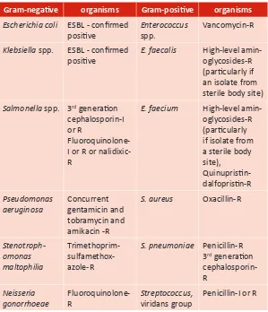
Table 8: Example of uncommon phenotypes possibly resuling from technical errors
Gram-negaive organisms Gram-posiive organisms
Escherichia coli ESBL - conirmed
posiive
Enterococcus
spp.
Vancomycin-R
Klebsiella spp. ESBL - conirmed posiive
E. faecalis High-level amin
-oglycosides-R (paricularly if an isolate from sterile body site)
Salmonella spp. 3rd generaion
cephalosporin-I or R
Fluoroquinolone-I or R or nalidixic-R
E. faecium High-level amin
-oglycosides-R (paricularly if isolate from a sterile body site), Quinuprisin-dalfoprisin-R Pseudomonas aeruginosa Concurrent gentamicin and tobramycin and amikacin -R
S. aureus Oxacillin-R
Stenotroph-omonas maltophilia Trimethoprim-sulfamethox -azole-R
S. pneumoniae Penicillin-R
3rd generaion
cephalosporin- R Neisseria gonorrhoeae Fluoroquinolone-R Streptococcus, viridans group
Penicillin-I or R
Note: R: Resistant; I: Intermediate suscepible
When these phenotypes are observed on individual paient isolates, the veriicaion steps as outlined ater Table 7 should be considered in a given situaion. Reports of Salmonella spp. that are intermediate
or resistant to 3rd generaion cephalosporins and/or intermediate or
en
t o
f n
ai
on
al
lab
or
at
or
y-bas
ed
s
ur
ve
ill
an
ce
o
f an
im
ic
ro
bi
al
re
si
st
an
ce
WHONET software which can be freely downloaded from (http:// www.who.int/drugresistance/whonetsotware/en/) is recommended to input the data of suscepibility test results. WHONET is a Windows-based database sotware developed for the management and analysis of microbiology laboratory data with a special focus on the analysis of animicrobial suscepibility test results.
Essenial data set
The recommended minimal data to be inputed by hospital laboratory is as follows:
Paient idenity (hospital number)
Sex
Age
Specimen idenity (laboratory number)
Locaion (ward where paient admited)
Locaion type (whether the paient is inpaient, outpaient,
intensive care, or from community ) Specimen type (site of infecion)
Name of isolate (genus species, genus only or group of
species)
Quanitaive suscepibility data (diameter of inhibiion zone or
minimum inhibiion concentraion)
Data collecion analyses
and disseminaion
32
Es
tab
lis
hm
en
t o
f n
ai
on
al
lab
or
at
or
y-bas
ed
s
ur
ve
ill
an
ce
o
f an
im
ic
ro
bi
al
re
si
st
an
ce
AMR Surveillance: Questionnaire for Assessment of National (1)
Networks 2003. WHO/CDS/CSR/RMD/2003.1 Centers for Disease Control and Prevention.
(2) Manual for the
Laboratory ideniicaion and animicrobial suscepibility tesing of bacterial pathogens of public health concern in the developing
world. World Health Organizaion.WHO/CDS/CSR/ EPH.2002.15
Clinical and Laboratory Standards Insitute.
(3) Performance Standards
for Animicrobial Disk Suscepibility Tests, 10th ed., Approved Standard M02-A10. Volume 29 Number 1, Clinical and Laboratory Standards Insitute, Villanova, PA. 2009.
Clinical and Laboratory Standards Insitute.
(4) Performance Standards
for Animicrobial Disk Suscepibility Tesing; Twenieth Informal Supplement M100-S21. Volume 31 Number 1, Clinical and Laboratory Standards Insitute, Villanova, PA. 2011.
Guidelines for the Collecion of Clinical Specimens during Field
(5)
Invesigaion of Outbreaks. World Health Organizaion, 2000. WHO/CDS/CSR/EDC/ 2000.4
Nugent R., Back E., Beith A.
(6) The Race against Drug Resistance: A Report of the Center for Global Development’s Drug Resistance
Working Group. The Center for Global Development, 2010.
Protocol for the Assessment of Naional Communicable Disease
(7)
Surveillance and Response Systems: guidelines for assessment
teams. World Health Organization, 2001. WHO/CDS/CSR/
ISR/2001.2
Surveillance Standards for Animicrobial Resistance
(8) WHO/CDS/
CSR/DRS/2001.5 Vandepite J et al.
34
Es
tab
lis
hm
en
t o
f n
ai
on
al
lab
or
at
or
y-bas
ed
s
ur
ve
ill
an
ce
o
f an
im
ic
ro
bi
al
re
si
st
an
ce
World Health Organizaion, 1991. ISBN 92 4 154425 2
Vandepite, J., El-Nageh, M., Tikhomirov, E., Stelling, J.M., Estrela, (10)
A. Guidelines for Animicrobial Resistance Surveillance. Geneva, World Health Organizaion, 1996.
WHO Recommended Surveillance Standards.
(11) World Health
Organizaion,1999. WHO/CDS/CSR/ISR/99.2
World Health Organizaion and Centers for Disease Control and (12)
Prevenion. Laboratory Methods for the Diagnosis of Epidemic Dysentery and Cholera. Centers for Disease Control and Prevenion, 1999. WHO/CDS/CSR/EDC/99.8
WHO Regional Strategy for Prevention and Containment of (13)
en
t o
f n
ai
on
al
lab
or
at
or
y-bas
ed
s
ur
ve
ill
an
ce
o
f an
im
ic
ro
bi
al
re
si
st
an
ce
Annex 1
Contributors
Ms Surang Dejsalert Director
WHO COllaboraing Centre on Animicrobial Resistance and Training Chief of Miscellaneous Bacteriology Secion Naional Insitute of Health Department of Medical Sciences Ministry of Public Health, Nonthaburi, Thailand
Ms Wantana Paveentkiiporn Chief of Legionella
Laboratory Miscellaneous Bacteriaology Secion Naional Insituteof Health Department of Medical Sciences Ministry of Public Helath
Nonthaburi, Thailand
and several scienists working in WHO CC on AMR and Training
Dr Sangeeta Joshi Head of Microbiology Manipal Hospital Bangalore
Dr Vikas Manchanda Head of Microbiology Chacha Nehru Bal Chikitsalya Shahdara
Delhi
Dr Anuj Sharma NPO (Laboratories) WHO Country Oice India New Delhi
Dr Rajesh Bhaia
36
Es
tab
lis
hm
en
t o
f n
ai
on
al
lab
or
at
or
y-bas
ed
s
ur
ve
ill
an
ce
o
f an
im
ic
ro
bi
al
re
si
st
an
ce
Annex 2
WHO Model List (March 2010)
Essenial Medicines
(excerpts)
16th ediion (updated)
The core list presents a list of minimum medicine needed for a basic
health-care system, lising the most eicacious, safe and cost-efecive medicines for priority condiions. Priority condiions are selected on the basis of current and esimated future public health relevance, and potenial for safe and cost-efecive treatment. Some examples of anibioics in core list are amoxicillin, gentamicin, cefalexin etc.
The complementary list presents essenial medicines for priority