Durian Consumption Effect on Plasma Malondialdehyde Level as
Biomarker of Stress Oxidative in Rats
Anugrah Aulia Ulil Amri1, Ani Melani Maskoen2, Syarief Hidayat3
1Faculty of Medicine Universitas Padjadjaran, 2Department of Biochemistry and Molecular
Biology, Faculty of Medicine Universitas Padjadjaran, 3Department of Cardiology and Vascular
Medicine Faculty of Medicine Universitas Padjadjaran/Dr. Hasan Sadikin General Hospital Bandung
Abstract
Background: Excessive consumption of durian (Durio zibethinus Murray) in Indonesia is often connected with its effect on health. This study aims to understand the effect of durian consumption to malondialdehyde (MDA) in plasma as oxidative stress biomarker.
Methods: The study used an experimental research design on animal models, in the Biochemistry and Molecular Biology Department, Faculty of Medicine, Universitas Indonesia, July–August 2012. Thirty two Sprague-Dawley rats were used, divided into four groups: control, treatment week 1, 2, and 3. Each treatment group was given 20 gram durian fruit diluted with water until 20 ml volume per oral, divided into two doses (10 ml each) with 4 hours interlude between doses for 1 week, 2 weeks, and 3 weeks. All groups got normal diet and water ad libitum. Plasma MDA level was measured by TBARS method, then analyzed using Kurskal-Wallis and Mann-Whitney tests.
Results: Seventeen samples were successfully decapitated (5 for control; 6 for week 1; 3 for week 2; 3 for week 3). Average plasma MDA level for control treatment week 1, 2 and 3 groups were 0.707 nmol/ml,
0.432 nmol/ml, 0.312 nmol/ml, and 0.746 nmol/ml respectively. Data was significant (p<0.05) with p=0.02. Compared with control group, a significant increase occurred in week 1 and 2 groups with p=0.028 and p=0.025 respectively.
Conclusions: Results of durian consumption show MDA level significantly decreases in week 1 and 2.
However, MDA level dramatically increases exceeding control group level in week 3. [AMJ.2016;3(1):22–8]
Keywords: Durian, malondialdehyde, oxidative stress
Correspondence: Anugrah Aulia Ulil Amri, Faculty of Medicine, Universitas Padjadjaran, Jalan Raya Bandung-Sumedang
Km.21, Jatinangor, Sumedang, Indonesia, Phone: +6282111838844 Email: [email protected]
Introduction
Durian is a fruit unique in Indonesia and other countries in Southeast Asia. With its unique taste and strong penetrating odor, durian is widely known and consumed in the society. However, there are rumours saying that the durian has an effect on health. People believe that the consumption of an inappropriate amount of durian may cause miscarriage in pregnant women, increase the cholesterol level and increase blood pressure or hypertension.
In contrast, currently the durian fruit is popular in daily utilization because of its health promoting compounds. The importance of durian is mostly connected with its composition
of antioxidant properties, flavanoid, flavanol,
ascorbic acid and tannin.1-6 Antioxidant is an
important compound found in both human and nature which act as scavengers of free radicals
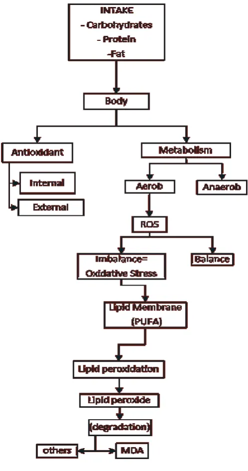
and reduce the oxygen toxicity. The imbalance between antioxidant and the reactive oxygen species (ROS) may result in oxidative stress. The ROS are free radicals and peroxides that are derived from the oxygen metabolism and present in all aerobic organisms. These
include superoxide radical (O2•-), peroxyl radical (HOO•), hydroxyl radical (OH•) and
hydrogen peroxyde (H2O2). The ROS play a
significant role in many biological processes.
The oxidative stress may affect molecules thus causing cell injuries which may lead to pathological processes in human.7
reactions form plenty of radicals including alkyl radical, peroxyl and alkoxyil. As the more stable product, lipid peroxidation produces many aldehydes including saturated aldehydes (propanal, butanal, hexanal, octanal, decanal), 2,3-trans-unsaturated-aldehydes (hexenal, octenal, nonenal, decenal and undecenal), and a series of 4-hydroxylated,2,3-trans-unsaturated aldehydes (4-hydroxyundecenal, and 4-hydroxinonenal (HNE)). Among the
metabolites produced, malonyldialdehyde (MDA) was considered for a long time as the most important lipid peroxidation metabolite.8 The products of lipid peroxidation can be used as biomarkers of stress oxidative (Figure 1).9
The purpose of this experiment was to measure the oxidative stress caused by the durian compounds antioxidant properties by studying rats which were fed with excessive amount of durian and observed the effect of
the treatment on plasma MDA level.
Methods
An experimental research design was conducted on animal models. The experiment waserformed in rats to investigate the effect of durian consumption on blood ROS level detected as plasma MDA level. This study
was conducted r five weeks starting from July
2012 to August 2012. The data were collected based on the results of the experiment. The study took place in the Faculty of Medicine Universitas Indonesia, Jakarta, Indonesia. The rats were divided into four different groups i.e. the control group for rats that did not consume durian; the group of week 1 for rats that consumed durian for 1 week, the group of week 2 for rats that consumed durian for 2 weeks, and the group of week 3 for rats that consumed durian for 3 weeks.
In order to obtain maximum validity, the experiment should be repeated several times. While to estimate the number of observations for each variable the Federer’s formula was used and resulted in 24 samples (Figure 2). Furthermore, the rats used in the experiment weighted between 100 and 200 gram. The rats were male young adult aged 7 week-old before adaptation with the type of Sprague Dawley rats. The rats were bought in Bogor, Jawa Barat, Indonesia, while the durian fruit used in the experiment was purchased from a local market in Pramuka Street, Jakarta.
All rats received a normal diet and ad libitum daily. Group of week 1, week 2, and week 3 were also given 20 mg of durian that has been diluted to 20 ml of volume with distilled water. Then, it given twice daily with each of 10 ml dilution and manually using a gastric tube. All methods were already approved by the Health Research Ethics Comittee.
At the end of each observation period,
every survived rat was sacrificed under deep
ether anesthesia. Next, the blood of each rat was collected directly by heart puncture and put in a heparinized tube. The plasma
Figure 2 Federer’s Formula
Note: n= Minimum number of repetition needed for each treatment, minimum repetition for this study is 6 x 4(treatments)= 24 samples, In the samples, it was added 10% for drop out criteria 24 + (10% x 24) = 27 rats
was obtained after separating the red blood cells by centrifugation at 3000 rpm for 15 minutes. All plasma were placed in -20oC until the MDA measurement. The plasma MDA was assayed using the Thiobarbituric Acid Reactive Substances (TBARS) assay.10 The
assay measures 2-TBARS which were naturally present in tissues and reported in MDA equivalents. The TBARS assay were based on the reaction of a chromogenic reagent, 2-thiobarbituric acid, with MDA at 25°C and pH 2-3.This reaction showed a pink-chromogen
color which has a λ(max) of 532 nm that was
able to be counted by the spectrophotometry. The plasma MDA level was analyzed using a computerized analysis of the Kurskall-Wallis non-parametrical test and Post-Hoc analysis of Mann-Whitney test.
Results
The study was initially conducted using 32 rats, and out of them, 15 rats were omitted and only 17 rats were successfully decapitated (Table 1 and 2).
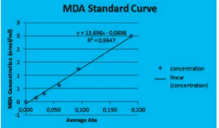
In order to establish the MDA concentration
Table 1 Number of Samples and the Percentage
Sample Group Initial Number Final Number Percentage
Control 5 5 100%
Week 1 9 6 67%
Week 2 9 3 33%
Week 3 9 3 33%
Figure 3 MDA Standard Curve Table 2 Body Weight of Rats Group
Sample Group Initial Body Weight (gram) Final Body Weight (gram)
Control 163.3 188
Week 1 141 131.7
Week 2 150 143.3
Week 3 195 215
Average 162.3 169.5
based on the absorption rate of sample plasma, the function of MDA standard curve is needed (Figure 3).
The plasma MDA level of rats established from the standard curve formula which was compared afterwards (Table 3 and 4).
Generally, the average of MDA concentration
data is significant according to the
Kurskal-Wallis non-parametrical test. The p value
was less than 0.05 (p=0.02). Compared to
the control group, the MDA concentration is decrease in the 1 week treatment group.
This data was significant according to the
Post-Hoc test using the Mann-Whitney test
with p=0.028. Similarly, the 2 week treatment group was also showed significantly decrease compared to the control group with p=0.025.
Moreover, the week 3 treatment group was rather increase compared to the control group
(p=0.655).
Discussion
Malondialdhyde (CH2(CHO)2) is a routinely
identified product of lipid peroxide chain
polyunsaturated fatty acid by itself therefore, starting a chain reaction which will damage the membrane thus, causing more extensive damage to the adjacent cells. Along the process a plenty of ROS were formed. In normal circumstances, the ROS need to be quenched by antioxidant in order to prevent extensive damage.
To date, the durian is majorly linked with
its antioxidant properties including flavonoid, flavonol, ascorbic acid and tannin.1–6 The durian consumption on rats was significantly
correlated with the plasma MDA level. In the rat experiment the plasma MDA levels of rats decreased in the week 1 and week 2 treatment groups compared to the control group. The decrease of the plasma MDA level on the week 1 and week 2 treatment groups is probably related to the antioxidant compounds in durians as stated by many studies including the comparative study held by Haruenkit R et al. The study asserted that durian nevertheless is a fruit that shows in vitro antioxidant activities and is the highest compared with the Mangosteen and Snake fruit.5 The antioxidant
in durian might be able to decrease the level of damage from ROS to the lipid, thus decreasing plasma MDA level.
On the contrary, after 3 weeks of durian consumption on the experimental rats, the
plasma MDA level was dramatically increased compared to the group of week 1 and week 2, and slightly exceeded the control group.
Although the data obtained was insignificant to control but clearly significant to week 2, the
increase of plasma MDA level on the week 3 treatment group was important.
Basically, the antioxidant is a compound that gives electron (electron donors). Biologically, the antioxidant is widely known as the scavenger of oxidants and free radicals. The action of antioxidant does not only depend on the dose and the duration of administration but also on the type of the antioxidant itself as well as on the environment. For instance, vitamin E can only act as an antioxidant when the pO2 is low. Furthermore, the antioxidant also has a capacity of becoming pro-oxidant or radicals as occurred in vitamin E, vitamin C,
and flavonoids.9
Flavonoids which can be found in the durian
including flavones, isoflavones, and flavanones
acted as antioxidants against peroxyl and hydroxyl radicals and served as pro-oxidants in the presence of Cu2+. Both the antioxidant and the copper-initiated pro-oxidant activities
of a flavonoid depend upon the number
of hydroxyl substitutions in its backbone structure. The single hydroxyl substitution at position 5 provides no activity, whereas the Table 3 Plasma MDA Level of Rats
Sample Group n MDA Level (nmol/ml) P Value*
Average MDA Level +SD
Note: * Kruskal-Wallis non-parametrical test
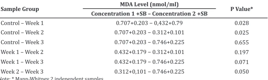
Table 4 Comparison
Sample Group MDA Level (nmol/ml) P Value*
Concentration 1 +SB – Concentration 2 +SB
Control – Week 1 0.707+0.203 − 0,432+0.79 0.028 Control – Week 2 0.707+0.203 − 0.312+0.101 0.025 Control – Week 3 0.707+0.203 − 0.746+0.225 0.655 Week 1 – Week 2 0.432+0.179 − 0.312+0.101 0.197 Week 1 – Week 3 0.432+0.179 − 0.746+0.225 0.071 Week 2 – Week 3 0.312+0,101 − 0.746+0.225 0.050
di-OH substitution at 3 and 4 is particularly
important to the peroxyl radical absorbing
activity of a flavonoid. The conjugation between
rings A and B is an important pro-oxidant
action of a flavonoid. The O-methylation of
the hydroxyl substitutions inactivates the antioxidant and the pro-oxidant activities of
the flavonoids.11,12
Moreover, the antioxidant is also produced endogenously in rats’ body, such as the glutathione (GSH). In the same rats, the
plasma GSH increased in the first and second
week and fell dramatically in the third week compared to the control. The plasma GSH level increased in response to the durian consumption considering the amino acid compound of durian (including glycine, cysteine and glutamic acid) is essential to GSH synthesis. The decrease of the plasma GSH level was strongly related the use of GSH to reduce damage caused by the ROS, meaning that pro-oxidant has been produced in the rats’ body.
This experiment successfully decapitated 17 rats as samples. At the beginning of the study, the Sprague-Dawley rats bought were 7 weeks old (before adaptation). Experimental
rats usually have a lifespan as long as 2−3.5
years (average 3 years).13 In the experiment,
rats died mostly during the period when the second dose has been administered to the
first dose on the next day. Other factors that
should be considered in this error included the effects of durian supplementation to the rats such as the mechanical trauma, the age of the experimental rats and the operator’s skill.
Before the interventions began the rats
were 8-week old, which was classified as young
adult rats, and the average weight was 162.3 gram. According to the weight measurement, it could be predicted that the rats were young adults. Considering this period, the
Sprague-Dawley rats have an average weight of 100−200
gram.13 Therefore, doubts regarding the age of
the rats could be at least cleared away.
The operator’s skill is certainly affecting the experimental rats. The durian consumption was given with a conventional gastric tube in which the tube was directly inserted from the mouth of the rat through to the stomach. A mechanical trauma could arise following a repetitive injury of the stomach, including of the throat, liver and other related organs. Besides, the durian aspiration could also cause a fatal damage. However, in this experiment the
definite cause of the death was not observed.
In conclusion, generally there are differences on the MDA serum level of rats
with durian consumption. The MDA level on durian consumption for 1 week and 2 weeks
have shown a significant decrease. However,
after 3 weeks of durian consumption, the MDA
level has increased insignificantly. The data
can be used as a prediction for the effect of durian consumption on health. The durian consumption is safe because it does not induce oxidative stress. However, continuous eating in excessive amount for a long period is still not recommended.
This study has some limitations including the effect of durian consumption on different doses which remains unclear and the number of rats that died during the intervention period were too many and with unknown cause of death.
In the future, to investigate the effect of durian consumption on experimental animals, some factors should be considered including observing the effect of durian consumption according to the dose, the initial training for the feeding operator before the intervention begin, and the use of a permanent feeding tube.
References
1. Avila AP, Toledo F, Park YS, Jung ST, Kang SG, Heo BG, et al. Antioxidant properties
of durian fruit as influenced by ripening. LWT-Food Sci Tech. 2006;41(10):2118–
125.
2. Leontowicz H, Leontowicz M, Haruenkit R, Poovarodom S, Jastrzebski Z, Drzewiecki J, et al. Durian (Durio zibethinus Murr.) cultivars as nutritional supplementation to rat’s diets. Food Chem Toxicol.
2008;46(2):581–89.
3. Toledo F, Avila AP, Park YS, Jung ST, Kang SG, Heo BG, et al. Screening of the antioxidant and nutritional properties,
phenolic contents and proteins of five
durian cultivars. Int J Food Sci Nut.
2008;59(5):415–27.
4. Haruenkit R, Poovardom S, Vearasilp S, Namiesnik J, Sliwka-Kaszynska M, Park Y, et al. Comparison of bioactive compounds, antioxidant and antiproliferative activities of Mon Thong durian ripening. Food Chem.
2010;118(3):540–7.
5. Haruenkit R, Poovarodom S, Leontowicz H, Leontowicz M, Sajewicz M, Kowalska T, et al. Comparative study of health properties and nutritional value of durian, mangosteen, and snake fruit: Experiments in vitro and in vivo. J Agric Food Chem.
2007;55(14):5842−9.
Buddhasukh D. GC–MS analysis of fatty acids in Thai durian aril. Chiang Mai J Sci. 2005;32(2):169–72.
7. Kumar V, Abbas AK, Fausto N, editors. Robbins and Cotran Pathologic Basis of
Disease, 8thed. Philadelphia: Elsevier Inc;
2005.
8. Repetto M, Semprine J, Boveris A. Lipid peroxidation: Chemical mechanism, biological implications and analytical determination. In: Catala A, editor. Lipid peroxidation. Rijeka: InTech; 2012. p.
1−21.
9. Purnomo Suryohudoyo. Oksidan, antioksidan dan radikal bebas. In: Ilmu kedokteran molekuler. Kapita Selekta.
Jakarta: Sagung Seto; 2000. p.31−46.
10. Linsley MD, Ekinci FJ, Ortiz D, Rogers E,
Shea TB. Monitoring thiobarbituric acid-reactive substances (TBARs) as an assay for oxidative damage in neuronal cultures and central nervous system. J Neurosci
Methods 2005;141(2):219−22
11. Cao G, Sofic S, Prior RL. Antioxidant
and prooxidant behavior of flavonoids:
Structure-Activity relationships. J Free Rad
Biol Med 2014;22(5):749−60.
12. Amid BT, Mirhosseini H, Kostadinović S. Chemical composition and molecular structure of polysaccharide-protein biopolymer from Durio zibethinus seed:
Extraction and purification process. Chem
Cent J. 2012;6(1):117.