Molecular Identification of Avian Influenza A Virus in House Flies (
Musca
domestica
Linnaeus) Collected from Different Poultry Farms in Indonesia
Identifikasi Molekuler Virus
Avian Influenza
A pada Lalat Rumah (
Musca domestica
Linnaeus) yang Diperoleh dari Beberapa Peternakan Ayam yang Berbeda
di Indonesia
1 2
Hastari Wuryastuty , R. Wasito
1
Department of Internal Veterinary Medicine, Faculty of Veterinary Medicine, Gadjah Mada University, Yogyakarta
2
Department of Pathology, Faculty of Veterinary Medicine, Gadjah Mada University, Yogyakarta
Email: [email protected]
Abstrak
Sampai saat ini, di Indonesia, wabah virus influenza A masih mengakibatkan kematian pada unggas. Virus avian influenza juga diduga telah menginfeksi dan mengakibatkan kematian pada manusia sehingga situasi avian influenza di Indonesia dianggap lebih serius dibandingkan negara Asia lainnya. Sumber utama infeksi AIV H5N1 adalah unggas. Karena lalat rumah merupakan lalat utama yang berada di peternakan ayam dan di rumah-rumah, serta juga merupakan kendaraan bagi berbagai macam mikroorganisme penyebab penyakit, kemungkinan adanya AIV H5N1 di dalam tubuh lalat rumah (Musca domestica L) perlu diteliti dengan tujuan utama menentukan kemungkinannya sebagai vektor mekanik dan/atau biologik dari AIV H5N1.
Reverse-transcriptase polymerase chain reaction (RT-PCR) tehnik digunakan untuk identifikasi AIV H5N1 dari bagian
abdomen lalat rumah. Adanya AIV H5N1 di dalam tubuh lalat rumah dibuktikan dengan RT-PCR menggunakan primer spesifik untuk gen NP, H5 and N1. Pada penelitian ini, semua sampel lalat rumah yang dianalisa secara molekuler memiliki ekspresi gen virus influenza tipe A subtipe H5N1. Isolat virus influenza A H5N1 dari sampel lalat rumah yang dikoleksi dari daerah asal yang berbeda (Sidrap, Sulawesi Selatan; Blitar dan Malang, Jawa Timut dan Karanganyar, Jawa Tengah) terdeteksi menggunakan RT-PCR. Semua provinsi tempat pengambilan sampel memiliki sejarah kejadian wabah AIV H5N1 yang berbeda. Sebaliknya, hasil uji RT-PCR menggunakan sampel lalat rumah yang dikoleksi dari daerah yang tidak pernah terjadi wabah AIV di Tuban, Jawa Timur menunjukkan hasil yang negatif terhadap ekspresi gen AIV H5N1. Hasil penelitian tersebut menunjukkan bahwa, di Indonesia, lalat rumah memiliki peranan penting dalam menyebarkan virus AI H5N1. Meskipun demikian, perlu dilakukan penelitian lanjutan untuk menentukan apakah lalat rumah merupakan vektor mekanik dan/atau biologik di dalam penyebaran virus avian influenza H5N1.
Abstract
To date, the outbreaks of avian influenza viruses (AIV) in Indonesia are still highly lethal to poultry. The AIV in Indonesia has also infected to humans and make the AIV situation is more serious than in other Asian countries. It is believed that the most likely source of AIV H5N1 infection is the chicken. Since the fly is a well-known cosmopolitan pest of the poultry farm and home as well as a valuable alternative vehicle of disease-causing microorganisms, the possibility of the presence of AIV H5N1 in the house flies (Musca domestica L) was investigated with the overall aim of determining the possible mechanical and/or biological vector of avian influenza virus. The RT-PCR was performed on abdominal parts of the house flies. The present of influenza A H5N1 was confirmed by RT-PCR with primers specific for NP, H5 and N1 genes. In the present study, all flies molecularly analyzed have gene expression of avian influenza virus of subtype H5N1. The RT-PCR detects AIV H5N1 isolates of the houseflies samples of the different geographic origin (Sidrap, South Sulawesi, Blitar and Malang, East Java and Karanganyar, Central Java). All those provinces had the different history of the AIV H5N1 outbreaks. On the other hand, results of the RT-PCR assay on the flies collected from Tuban, East Java in the area where never had any AIV outbreaks considered to be negative AIV H5N1 gene expression. Our findings suggest that, in Indonesia, houseflies may be important in the transmission of AIV H5N1. Further studies, however, still need to be done to decide whether houseflies is a mechanical and/or biological vector of avian influenza virus H5N1.
Key words: House flies (Musca domestica L), RT-PCR, AIV H5N1, mechanical vector, biological vector
Introduction
In Indonesia, outbreak of highly pathogenic avian influenza has occurred among poultry in almost all provinces since late 2003. The introduction of avian influenza into commercial poultry still continues to be a serious problem, but it appears infrequently and is unpredictable. Confirmed human cases of H5N1 were first reported in mid July 2005. By comparison, AIV H5N1 affected many more people in Indonesia, where the humans death due to AIV H5N1 infection is estimated to be 154 out of 196 cases (Anonymous, 2012).
Avian influenza (AI) is a contagious viral disease affecting the respiratory, digestive and/or nervous system of many species of birds. The virus causing AI is an influenza virus type A of the family
Orthomyxoviridae. Several virus subtypes exist,
haemagglutinin (H) and neuraminidase (N). At present, 16 H subtypes (H1-H16) and 9 N subtypes (N1-N9) have been recognized. The disease has become major concern throughout the world because of their medically and economically impacts (Fouchier et al., 2005, Murphy and Webster, 1996, Rohm et al., 1996).
bedding or carcasses (21.2%) and 5. Other indirect contacts through exchange of farm staff, caretakers etc. (9.4%) (Marangon and Capua, 2005). It was suspected that the movement of livestock and/or livestock products from affected areas to clear zones were causes for serious AIV outbreak in Indonesia.
The role of live vectors, such as infected wild birds, rodents or flies which may act as 'AIV mechanical and/or biological vectors' is largely undetermined and is still controversial. It was believed that flies do not constitute a major factor in transmitting AIV (Harder and Werner, 2006). Even though, the AI outbreak in Pennsylvania, USA in 1987 was suspected spread by garbage flies (Beard, 1998). In view of the fact that, anything that is crawling can be a vector for avian influenza has lead to a new hypothesis that house flies is an important mechanical and/or biological vector for AI viruses. In order to prove that hypothesis it is important to do a sequentially relevant research as follow: 1. Whole
fly-RT PCR AIV NP, H5 and N1 and 2. Cutting up flies-RT PCR AIV NP, H5 and N1.
Materials and Methods
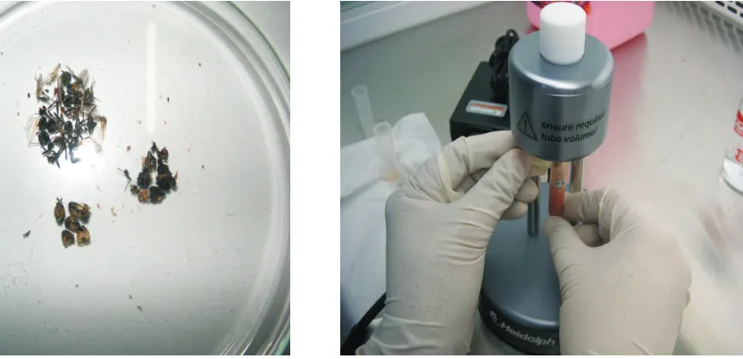
House flies (Musca domestica Linnaeus) were collected from poultry farm in 3 different provinces, such as East Java (Malang, Blitar, and Tuban), South Sulawesi (Sidrap) and Central Java (Karanganyar) in Indonesia. Flies were submitted at the Department of Internal Medicine, Faculty of Veterinary Medicine, Gadjah Mada University, Yogyakarta, Indonesia between October 2003 to August 2008. The flies were collected from infected flock during the AIV H5N1 outbreak (Karanganyar-Central Java) and also 3 months (Maros, South Sulawesi) – 1 year (Blitar, and Malang-East Java and Sidrap, South Sulawesi) after the AIV H5N1 outbreak. The house flies samples were al so collected from the poultry farms in Tuban - East Java that those farms never had any confirmated AIV H5N1, outbreaks.
Oligonucleotide primers were synthesized by a commercial source (Integrated DNA Technologies Inc., Coralville, Iowa USA). The NP, H5 and N1-specific primers were used for diagnostics biosurveillance for the occurrence of AIV H5N1 in flies in Indonesia.
Reverse-transcriptase polymerase chain reaction (RT-PCR) amplification to detect NP, H5 and N1 genes were respectively done as follows: a. The 45.0 µl of PCR mixtures containing 25 µl 2x reaction mix, 2.5 µl of 5 mM MgCl , 1 µl of forward 2 primer (5'AGC AAA AGC AGG GTA GAT AA3'), 1 µl of reverse primer (5'TCC TTG CAT CAG AGA GCA CA3'), 1 µl of SuperScript III RT/Platinum Taq
mix and 14.5 µl of DEPC treated water were pipetted into a sterile 200 µl thin wall flat cap PCR tube into which 5 µl of template was then added. The first cycle of the amplification program of RT-PCR
0
consisted of a 45-minute period at 48 C and a 15-0
minutes period at 95 C, followed by 36 cycles with
0 0
the following conditions: 94 C for 30 seconds, 52 C 0 SuperScript III RT/Platinum Taq mix and 14.5 µl of DEPC treated water were pipetted into a new 200 µl thin wall flat cap PCR tube into which 5 µl of template was then added. The first cycle of the amplification program of RT-PCR consisted of a
30-0
minutes period at 50 C and 15-minutes period at
0
95 C, followed by 36 cycles with the following
0 0
conditions: 94 C for 30 seconds, 51 C for 1 minutes;
0 0
72 C for 1 minutes, and 7 minutes extension at 72 C and c. The 45.0 µl of PCR mixtures containing 25 µl 2x reaction mix, 2.5 µl of 5 mM MgCl , 1 µl of 2 forward primer (5'TTG CTT GGT CAG CAA GTC CT3'), 1 µl of reverse primer (5'TCT GTC CAT CCA TTA GGA TCC3'), 1 µl of SuperScript III RT/Platinum Taq mix and 14.5 µl of DEPC treated water were pipetted into a new 200 µl thin wall flat cap PCR tube into which 5 µl of template was then added. The first cycle of the amplification program
0 of RT-PCR consisted of a 30-minutes period at 50 C
0
and 15-minutes period at 95 C, followed by 40 0
cycles with the following conditions: 94 C for 30
0 0
seconds, 55 C for 30 seconds; 72 C for 1 minute, 0
and 7 minutes extension at 72 C (Wuryastuti and Wasito, 2012).
Five microliters of each PCR products were loaded onto 1.5% low melting point agarose and stained after electrophoresis with ethidium bromide.
Results and Discussions
There are three types of influenza viruses (A, B, and C). The virus envelope glycoproteins have hemagglutinin (HA) and neuraminidase (NA) activity. These characteristics are used to subtype the A, B, and C viruses. Influenza A viruses are the cause of avian influenza. There are 16 different HA antigens (H1 to H16) and nine different NA antigens (N1 to N9) for influenza A. All known subtypes of influenza A can be found in birds, but only subtypes H5 and H7 have caused severe outbreaks of disease in birds populations known as highly pathogenic avian influenza (HPAI) (Bosch et al., 1981; Harder and Werner, 2006). Influenza A also occurs in pigs, horses and certain marine mammals (whales and seals), tigers and cats (Anonymous, 2005; Keawcharoen et al., 2004; Kuikeen, 2004). Human disease historically has been caused by three subtypes of HA (H1, H2, and H3) and two subtypes of NA (N1 and N2). All past influenza pandemics in humans have been caused by influenza A virus (Anonymous, 2005 ).
In Indonesia, since the introduction of avian influenza into commercial poultry in October 2003, the AIV infection in poultry continues to be a problem and appears infrequently and is unpredictable. It is suspected that high population exposures to live chickens are factors that may contribute to the spread in Indonesia. The infection and deaths of 159 of 191 humans infected with an H5N1 avian influenza virus in Indonesia (Anonymous, 2012) has even resulted in a serious consideration of the important role that the vectors, besides avian species have on the epidemiology of human influenza.
It has been known that Musca domestica spp. are the most impportant fly species at poultry farms (Axtell, 1999). More than 100 pathogens associated with the house fly may cause disease in humans and animals, including typhoid, cholera, bacillary dysentery, tuberculosis, anthrax ophthalmia and infantile diarrhea, as well as parasitic worms. An accumulated chicken manure in the poultry farm is a Fig. 2. One tube RT-PCR run on 1.5% agarose gel electrophoresis for AIV NP of 552 bp gene (A) for AIV H5 of 290 bp gene (B) and for AIV N1 of 616 bp gene (C) in the whole bodies the flies. Lane 1: Negative control, Lane 2: Positive control AIV from chicken sample, Lane 3: DNA marker 100 bp, Lane 4: Flies AIV positive isolated from Sidrap-South Sulawesi, Lane 5-6: Flies AIV positive isolated from Blitar and Malang-East Java, Lane 7: Flies AIV positive isolated from Karanganyar-Central Java and Lane 8: Flies AIV negative isolated from Tuban-East Java.
A B C
1 2 3 4 5 6 7 8 1 2 3 4 5 6 7 8 1 2 3 4 5 6 7 8
populations. This finding may suggest that flies have potency in spreading avian influenza in both poultry and humans. The health and economic impacts of this AIV H5N1 in the house flies in Indonesia, in particular, on eggs production and meat performance in chicken, and humans' health as well, remains to be defined in detail. .
In an area, where AIV is endemic, such as during an outbreak sound presumptive program can be made by house flies eradication. It is more likely, a routine and appropriate program of national bio-surveillance and bio-security allowing efficient identification of AIV H5N1 in flies and their elimination or eradication, respectively is essential to constraint further spread of AIV H5N1 in Indonesia. Our next research are focusing on study the fate of the AIV H5N1 throughout the flies life cycle and the fly ability to transmit the H5N1 to chicken.
Acknowledgments
We thank Dr. Roger K Maes, Professor of Virology, Michigan State University, East Lansing, MI, USA, for thoughtful discussion and reviewing our manuscript and providing the molecular reagents. The authors wish also to thank Dr. Ronny Mudigdo, previous Director for Animal Diseases Investigation Center Maros, South Sulawesi, Dr. Agus Sulistyo, Director for the PT Lisan Mulia and his staff for providing flies, (Late) Dr. Julianti Darjono for determining flies species. This work was supported in part by the Ministry of Agriculture for Indonesia.
good breeding place for the house flies. Therefore, the flies have good oppurtunities of picking up the AI viruses from the feces of infected chickens and then transferred on their mouthparts and other body parts, through their vomitus, feces and contaminated external body parts to human and animal food (Sanchez-Arroyo, 2003). The effectiveness of the transmission through regurgitation and defecation of the flies may depend upon the activity and the amount of the pathogen in the fly body.
By molecular diagnostic approach of the reverse transcriptase polymerase chain reaction (RT-PCR) we now report the isolation of avian influenza type A subtype H5N1 from the house flies (Musca domestica) in Indonesia. It is noteworthy, in fact that expression of AIV H5N1 in the whole tissues and the abdominal parts of the flies by applying the RT-PCR in the present study complement each other and the RT-PCR assay provides useful tool for detecting AIV H5N1 in flies. The RT-PCR is highly sensitive and reliable technique in quantifying AIV H5N1 gene expression in the flies.
References
Anonymous. (2005) Avian influenza (Bird Flu): Implications for human disease. Center for Infectious Disease Research & Policy (CIDRAP). Academic Health Center, University of Minnesota, USA.
Anonymous. (2012) Avian Influenza Update. Western Pacific Regional Office of the World Health Organization. October 2012.
Axtell, R.C. (1999) Poultry integrated pest management status and future. Integrated Pest
Management Reviews 4: 53-73.
Beard, C.W. (1998) Avian influenza (fowl plaque). In: US Animal Health Association, Committee on Foreign Animal Diseases. Ed 6. Richmond, VA: US Animal Health Assoc., USA.
Bosch, F.X., Garten, W., Klenk, H.D. and Rott, R. (1981) Prototype cleavage of influenza virus hemagglutinin: Primary structure of the connecting peptide between HA1 and HA2 determines proteolytic cleavability and pathogenicity of avian influenza virus. Virol. 113: 725-735.
Fouchier, R.A.M., Munster, V. and Wallensten A. (2005) Characterization of a novel influenza A virus hemagglutinin subtype (H16) obtained from black-headed gulls. J. Virol. 79: 2814-2822.
Harder, T.C. and Werner, O. (2006) Avian Influenza. In: Influenza Report. Kamps BS, Hoffmann, C and Preiser W (Eds.). p:1-47.
Kawaoka, Y., Nestorowicz, D.J., Alexander, D.J. and Webster, R.G. (1987) Molecular analyses of the hemagglutinin genes of H5 influenza viruses origin of a virulent turkey strain. Virol. 158: 218-227.
Keawcharoen. J., Oraveerakul, K., Kuiken, T., Fouchie,r R., Amonsin, A. and Payungporn, S. (2004) Avian influenza H5N1 in tigers and leopards. Emerg. Infect. Dis. 10: 2189-2191. Kuikeen, T., Rimmelzwaan, G., van Riel D., van
Amerongen G., Baars, M. and Fouchier, R.A. (2004) Avian H5N1 influenza in cats. Science 363: 587-593.
Marangon, S. and Capua, I. (2005) Control of AI in Italy: from “Stamping-Out”-strategy to emergency and prophylatic vaccination. In: Proc. Internat. Conf. on Avian Influenza, Paris; OIE p. 29.
Murphy, B.R. and Webster, R.G. (1996) Orthomyxoviruses, p. 1397-1445. In BN Fields, DM Knipe and Howley PM (ed.) Fields
rd
Virology 3 ed. Lippincott-Raven, Philadelphia, Pa.
Rohm, C., Zhou, N.A., Suss, J.C. and Webster, R.G. (1996) Different hemagglutinin cleavage site variants of H7N7 in an influenza outbreak in chickens in Leipzig, Germany. Virol. 217: 508-516.
Sanchez-Arroyo, H. (2003) House fly, Musca
domestica Linnaeus. IFAS Extension,
University of Florida, USA.