NSTEMI because of the absence of signifi cant ST elevation on the 12 standard ECG leads. ST elevation is the condition ‘sine qua non’ for diagnosing acute total coronary occlusion causing transmural infarction. However, ST elevation when there is circumfl ex artery occlusion is seen on the 12 standard ECG leads in fewer than 50% of patients. We reported a 77 years old women who diagnosed with NSTEMI. Twelve lead ECG showed ST depressed in V2-V5. On angiography we found a totaly ocluded of left circumfl ex as culprit lession.
Keywords: NSTEMI; culprit lession; total occlusion; Left Circumfl ex
Introduction
Prompt restoration of blood flow in the infarct-related artery is essential to myocardial salvage and mortality reduction. As the benefi ts of reperfusion decline rapidly with time, prompt and accurate diagnosis of acute myocardial infarction (AMI) is very important in determining the initiation of reperfusion therapy4. The 12 standard ECG leads has been an initial diagnostic tool in patients with suspected AMI in the emergency department (ED) and ideally should be performed and interpreted within 10 min of arrival to the ED. However, this conventional ECG has a very low sensitivity for the detection of AMI, especially if the culprit lesion is in the left circumfl ex artery (LCx)5.
ST elevation was not seen on the 12 standard ECG leads in up to 60% patients in LCx-related AMI and this could possibly lead to an unwarranted delay of therapeutic decisions. Because of lack of ECG presentation, these patients with LCx occlusion might be underdiagnosed by the physician in the emergency department.. It seems that the patients with an occluded LCx presented with less ST elevation, and their primary PCI was delayed or performed less5,8.
Case Illustration
We reported a 77 old woman who came to ED Sardjito General Hospital complaining of
short of breath (dyspnea) since 6 hours before. One day before, patient has chest pain along with diaphoresis, no dyspnea, nausea or vomitus. She went to ER in a private hospital. Standard 12-lead ECG was taken, unfortunately, we didn’t have the record. The laboratory test showed normal cardiac enzyme, so she was discharged. One day after, she came to ER complaining of dyspnea but no chest pain. The 12 lead ECG showed ST depressed in lead V2-V5 (fi gure 1). The laboratory test showed increased cardiac enzyme (CK: 3119 IU/L, CKMB: 587 IU/L and Troponin I: 20.49 IU/L). Patient was unconciousness, blood pressure was increased (190/110 mmHg) and rales in both of lung on physical examination.
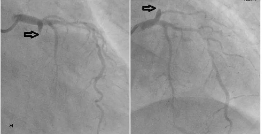
Patient was diagnoses as NSTEMI, hypertensive emergency and acute pulmonary e d e m a . T h e n , p a t i e n t w a s t r a n f e r e d t o catheterization laboratory. Coronary angiography demonstrated totaly occludded of LCx as culprit lession (fi gure 2). DES was inserted and patient was transferred to intensive cardiac care unit (ICCU).
Discussion
a
FIGURE 1. ECG recording on admission show ST depression in V2-V5
FIGURE 2. Coronary angiography show LCx total occlussion (arrow) a.. RAO 200 Caudal 200, b. RAO 100 Cranial 300
treatment patient with ACS. Autopsy studies have shown that plaque rupture causes approximately 75% of fatal MIs, whereas superfi cial en-dothelial erosion accounts for the remaining 25%. After
followed by platelet activation and aggregation and the subsequent formation of a thrombus. Two types of thrombi can form: a platelet-rich clot (referred to as a white clot) that forms in areas of high shear stress and only partially occludes the artery, or a fi brin-rich clot (referred to as a red clot) that is the result of an activated coagulation cascade and decreased fl ow in the artery. Red clots are frequently superimposed on white clots, and this characteristic causes total occlusion1.
T h e d i f f e r e n c e s i n t h e u n d e r l y i n g pathophysiology of UA/ NSTEMI and STEMI call for different therapeutic goals and approaches. In UA/ NSTEMI, the goal of antithrombotic therapy is to prevent further thrombosis and to allow endogenous fi brinolysis to dissolve the thrombus and reduce the degree of coronary stenosis. Revascularization is frequently used to increase blood fl ow and prevent reocclusion or recurrent ischemia. In contrast, in STEMI, the infarct-related artery is usually totally occluded, and immediate pharmacological or catheter-based reperfusion is the initial approach, with the goal of obtaining normal coronary blood fl ow1,6,9
Total oclussion of coronary artery was assosiated with STEMI, but total occlussion can be found in UAP or NSTEMI. Apps et al. in March
2013 published a study which show of 308 patient underwent immediate angiography and primary PCI, total aclussion was found in 75% patient ACS presenting with ST elevation, 73% in patient with ST depressed or T inverted and 63% total oclussion found in patient ACS without any ST-T changes1.
In this case, coronary angiography showed total oclussion of LCx. Our fi nding accordingly with study above. But, these case may be a STEMI inferobasal (posterior) which didn not diagnose because absence of ECG posterior lead as additional lead to diagnose this case. Approximatelly 48% total oclussion LCx related AMI has ST elevation on ECG recording, and 30% has not signifi cant ST-T changes2. According to algorithm management of ACS, this is potentially patients to be treated inappropriately as having a NSTEACS without having primary percutaneous coronary intervention or receiving early administration of fi brinolytic therapy8
Data from randomized clinical trials of STEMI have repeatedly shown that LCx is the least frequent culprit artery. But, failure to detect LCx related AMI have consequences because LCx supply signifi cant area of left ventricel7. LCx supplies the inferobasal area of the myocardium. FIGURE 3. Coronary angiography post PCI show fl ow
The term posterior, to refl ect the basal part of the left ventricle wall that lies on the diaphragm and it can easily assessed by leads V7–V9. Because the anterior leads are relatively opposite in direction to the inferobasal leads, anterior ST depression is often the mirror image of inferobasal ST elevation. None of the 12 standard ECG leads face the inferobasal wall so an isolated inferobasal STEMI often masquerades as a non-STEMI10
In this case, patient was diagnose as NSTEMI based on ECG recoding which showed ST depression in V2-V5. It is possible the ECG records in previous hospital show normal ECG, so the physician didn’t diagnose as ACS and patient didn’t receive ACS’s management.
The lack of ECG presentation in patients with LCx-related AMI is multi-factorial. One possible reason is that absence of ST segment changes was explained by smaller infarct size. A previous study showed total mass of myocardium lost in LCx-related AMI is smaller than in other anatomic distributions (notably anterior MI) . Infarct size could be assessed by the amount of serum cardiac marker (creatine kinase-MB, troponin) and ejection fraction5, but this case have increased cardiac marker and presence of acute pulmonary edema which reflected low ejection fraction. Second, LCx usually supplies the lateral and posterior walls of the left ventricle, which are areas not well detected by the 12 standard ECG leads. Third, there were some trials suggesting that patients without ST segment deviation were likely because of incomplete coronary occlusion due to thrombus or vasospasm5, but this study confi rmed complete LCx occlusion during coronary angiography. Forth, the coronary artery dominance, right coronary
dominance may act as protective factor in acute occlussion LCx by giving colateral or dual fl ow, minimize area of infarc which lead no changes in ECG recording5. This
fi nding suggested that this case may have large infarc size due to a STEMI inferobasal (posterior).
Recording the additional chest lead does not include in fi rst routine chart to diagnose ACS. The ECS guideline recommended to record additional ECG leads (V3R, V4R, V7–V9) when routine leads are inconclusive. Wong, 2011, recommend to record V7-V9 to diagnose STEMI inferobasal (posterior). Unfortunatelly, in this case, no posteror ECG was recorded. The management of patient with NSTEMI based on risk stratifi cation. Invasif management was recomennden when patient have high risk (table 1 and 2). In this case, invasif management was performed due to acute pulmonary edema and haemodinamic instability.
Conclussion
We reported a 77 years old women with diagnose as NSTEMI due to the 12 standard ECG leads record, but in the coronary angiography we found total occlussion LCx as culprit lession. The 12 standard ECG leads does not enough to diagnose LCx related AMI, so the physician must notice this condition. The clinician must role LCx when there is no ECG changes in patient suspected ACS.
Dichotomy patient to having ST elevation or not does not reflected having total or non total occlussion culprit lession because total occlussion can also be found in patient with UAP and NSTEMI.
Acknowlegment
The authors would like to thanks Head of Intensive Coronary Care Unit and Catheterization Laboraratorium Staff for assistance to conduct this case report.
References
Apps A, Malhotra A, Tarkin J, Smith R, Kabir 1.
patients without protocol positive ST segment elevation referred to an open access primary angioplasty programme. Postgrad Med J doi:10.1136/ postgradmedj-2012-130818 (Abstract)
From AM, Best PJM, Lennon RJ, Rihal 2.
CS, Prasad A. Acute Myocardial Infarction Due to Left Circumflex Artery Occlusion and Signifi cance of ST-Segment Elevation. American Journal of Cardiology 2010;106( 8):1081-1085
Hamm CW, Bassand JP, Agewall S, Bax 3.
J, Boersma E, Bueno H, Caso P, Dudek D, Gielen S, Huber K, Ohman M, Petrie MC, Sonntag F, Uva MS, Storey RF,Wijns W, Zahger D. ESC Guidelines for the manage-ment of acute coronary syndromes in patients presenting without persistent ST-segment elevation: the task force for the management of acute coronary syndromes (ACS) in patients presenting without persistent ST-segment elevation of the European Society of Cardiology (ESC). Eur Heart J 2011;32:2999-3054.
Hiasa Y, Morimoto, Wada T, Hamal K, 4.
Nakata Y, et al. Differentiation Between Left Circumfl ex and Right Coronary Artery Occlusions: Studies on ST-Segment Deviation
During Percutaneous Transluminal Coronary Angioplasty. Clin. Cardiol. 1990;13: 783-788. Kim SS, Choi HS, Jeong MH, Cho JG, Ahn YK, 5.
et al. Clinical outcomes of acute myocardial infarction with occluded left circumfl ex artery. Japanese College of Cardiology. 2011;1:14. Kumar A, Canon CP. Acute Coronary 6.
Syndromes: Diagnosis and Management, Part I. Mayo Clin Proc. 2009;84(10):917-938. O’Keefe JH Jr, Sayed-Taha K, Gibson W, 7.
Christian TF, Bateman TM, Gibbons RJ. Do patients with left circumfl ex coronary artery-related acute myocardial infarction without ST-segment elevation benefi t from reperfusion therapy? Am J Cardiol 1995; 75:718–720. Stribling WK, Kontos MC, Abbate A, Cooke R, 8.
Vetrovec JW, et al. Left Circumfl ex Occlusion in Acute Myocardial Infarction (from the National Cardiovascular Data Registry). The American Journal of Cardiology. 2011;108:959–963. Tanaka A, Shimada K, Sano T, et al. Multiple 9.
plaque rupture and C-reactive protein in acute myocardial infarction. J Am Coll Cardiol. 2005 May 17;45(10):1594-1599. Epub 2005 Apr 25. Wong CK, White HD. Patients with circumfl ex 10.