STRUCTURE ELUCIDATION OF ALKALOIDS FROM LEAVES OF
Voacanga foetida
(Bl.)
Rolfe OF LOMBOK ISLAND
Surya Hadi
1.*, Dina Asnawati
1, and Novi Febrianti
21
Study Program of Chemistry, Faculty of Mathematics and Natural Sciences, University of Mataram, Jl. Majapahit 62 Mataram-NTB 83125
2
Study Program of Biology, Faculty of Mathematics and Natural Sciences, University of Mataram, Jl. Majapahit 62 Mataram-NTB 83125
Received January 13, 2010; Accepted April 5, 2010
ABSTRACT
The leaves of Voacanga foetida (Bl.) Rolfe, have been used ethnomedically for the treatment of wounds, itches, and swellings particularly in Lombok island. A phytochemical study has been done to investigate chemical compounds responsible against the cause of the diseases. By separating alkaloidal fraction from the leaves was found voacristine 1 as the major alkaloidal compound, and voacangine 2 and coronaridine 3 as the minor components. The structure elucidation of the compounds was carried out on the basis of spectroscopy data. A structure revision of voacristine1was also reported.
Keywords:alkaloids, Voacanga foetida, Lombok
INTRODUCTION
The plant Voacanga foetida (Bl.) Rolfe (Apocynaceae), locally in Lombok Island known as “kumbi”, is distributed throughout Indonesia. It grows in areas about 400 m above sea level and reaches 10-15 m in height. In Lombok, an aqueous extract of the leaves or bark is used commonly to treat a wide range of skin conditions such as wounds, itches, and swellings. The leaves of V. foetida(Bl.) Rolfe, are also warmed over a fire and then placed on chronic leg sores; this is a common practice in many parts of Indonesia In Sumatra, the plant’s latex has been used externally to treat skin disorders [1].
An initial alkaloid screening showed that all parts of the plant contained high concentrations of alkaloids [2], although a previous report [3] indicated that only small amounts of alkaloids occurred in the bark, fruit rind, and seeds. Moreover, a thorough survey of the relevant literature indicated that no further information concerning structural properties of the alkaloids contained in this plant had been published. Other Voacanga species had been shown to yield a variety of indole alkaloids [4].
EXPERIMENTAL SECTION
Materials
The leaves of Voacanga foetida (Bl.) Rolfe were collected from Narmada, West Lombok with permission of the local government and in collaboration with the University of Mataram, Lombok, Indonesia. The
collection and botanical identification were carried out by botanists from the University of Mataram and the Research and Development Centre for Biology, Bogor, Indonesia. A voucher specimen was deposited at the Laboratory of Biology, the University of Mataram.
Instrumentation
NMR spectra of 1H, gCOSY, gHSQC, and gHMBC were recorded on a Varian Inova-500 MHz NMR spectrometer, unless otherwise stated. 13C-NMR and DEPT spectra were collected on a Varian Unity 300 spectrometer running at 75.42 MHz. Cl (reactant gas: isobutene) and El (at 70 eV) mass spectra were obtained on a Shimadzu QP-5000 by the direct insertion technique. HRCIMS were run on a Fisons/VG Autospec-oa-TOF Mass Spectrometer; relative intensities of peaks are given in brackets after the m/z values. The UV absorption spectra (solvent corrected)
were recorded on a Shimadzu UV-265
Procedure
Isolation and Purification
Finely-powdered, air-dried leaves (2.0 kg) of V. foetida (Bl.) Rolfe extracted with cold MeOH (3 x 4 L) with occasional swirling produced a dark green extract (290.5 g). Further steps used to isolate alkaloids from this extract followed the procedure acid-base extraction and produced a dark-green, crude alkaloid extract (692.8 mg). Two known alkaloids, voacristine (18.3 mg, major) and voacangine (2.1 mg, minor), and coronaridine (4.2 mg,minor) were isolated from the crude extract by
repeated PTLC on silica gel
(DCM:MeOH:conc.NH4OH(aq) / 90:9:1).
Voacristine 1: brown solid; m.p 168-169 °C (m.p. 167-169 °C; [5]); UV λmax(nm, CHCl3): 278 (logmax= NH);gHSQC (CDCl3, 500 MHz), 3.02 & 2.82/50.8(C-3), 3.43 & 3.15/52.1 (C-5), 3.12 & 3.06/21.7 (C-6), 6.78/109.5 (C-9), 7.32/119.3 (C-10), 6.78/109.5 (C-11), 2.03/28.8 (C-14), 1.84 & 1.70/24.1 (C-15), 2.55 & 1.96/ 36.8 (C-17), 1.28/22.39 (C-18), 3.91/70.8 (C-19), 1.40/24.2 (C-20), 4.08/54.5 (C-21), 3.73/56.0 (OMe), 3.83/52.4 (COOMe), LRCIMS, m/z 385 (MH+);LREIMS, m/z (relative intensity, %) 384 (46), 369 (20), 367 (20), 366 (60), 339 (10), 323 (7), 297 (9), 297 (10), 279 (10), 265 (9), 245 (10), 244 (34), 225 (16), 224 (20), 212 (20), 198 (10), 184 (46), 160 (46), 152 (57), 140 (41);
HRCIMS, C22H29N2O4 (measured 385.2105, calc. 385.2127, for MH+).
Voacangine 2: yellow solid; m.p 135-136 °C (m.p. 136-137 °C; [6]); UV λmax(nm, CHCl3): 272 (logmax= 3.789), 286 (logmax = 3.835) 293 (logmax = 3.767);
LRCIMS, 369 (MH+),LREIMS, m/z (relative intensity, %) 368 (48), 253 (11), 338 (20), 323 (8), 309 (7), 283 (9), 245 (9), 244 (15), 225 (7), 208 (20), 195 (12), 184 (33), 167 (20), 160 (31), 154 (41), 136 (100); HRCIMS, C22H29N2O3 (measured 369.2162, calc. 369.2178, for MH+).
Coronaridine 3: yellow amorphous solid; m.p. 236-238 °C (m.p. 237-239 °C; [7]);UVλmax(nm, CHCl3): 283 C21H27N2O2 (measured 339.2069, calc. 339.2073, for MH+).
RESULT AND DISCUSSION
The following section discusses the structural elucidation of the above isolated compounds.
Voacristine
A brown solid was also isolated from the alkaloid mixture, which was found to be the alkaloid, voacristine
1. This compound absorbed UV light with maxima at 278 and 298 nm, characteristic of the presence of a ring A-substituted indole nucleus. The LRCIMS for voacristine showed a major peak at 385 (MH+) while EIMS produced peaks at m/z 384, 366, 339, 244, and 184. Initial identification of this compound was based on its ion fragmentation pattern, which was characteristic of iboga type alkaloids. HRCIMS indicated the formula C22H28N2O4 (found 385.2104, calc. 385.2127, for MH+), supportive of voacristine 1. The structural assignment of 1 was then established from1H-NMR, gCOSY, HSQC, HMBC experiments and by comparison of the spectra of 1 with the spectra of (19R)-voacristine [8]. To this author’s knowledge, however, this is the first report to elucidate the structure of voacristine on the basis of 2D-NMR experiments.
As a starting point for the structural confirmation of voacristine, focus was placed on the aromatic region. The signal corresponding to the NH proton, existed as a broad singlet at 7.73 ppm. Also in this region of the spectrum, other signals were evident which were typical of a substituted indole moiety. Closer inspection of the aromatic signals permitted the assignment of H-10 as a multiplet at 7.32 ppm integrating for one proton, and another multiplet centralised at 6.78 ppm integrating for two protons, and ascribed as H-9 and H-11 respectively. The gCOSY confirmed the connectivity between the two signals, while the gHSQC spectrum provided a straightforward identification of the attached carbons resonating at 7.32 ppm (H-10)/ 119.3 ppm (C-10);
N H
N
H
COOMe OH
OMe
5 6 9
18 20 21 3
14 15 17
10
11
12
16 7
2 13
19
1: Voacristine
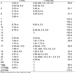
Table 1.Assignment of1H and13C-NMR data of voacristine1by gCOSY, gHSQC and gHMBC
Position H (ppm), multiplicity,
J(Hz)
Reference (H (ppm), multiplicity,
J(Hz)) [8]
C (ppm)
3 3.02 m 3.02 ddd, 2.4, 3.6, 9.2 50.8
2.82 bd, 8.5 2.82 bd, 9.2
5 3.43 m 3.40 m 52.1
3.15 m 3.25-3.0 m
6 3.12 m 3.25-3.0 m 21.7
3.06 m
7 123.0
8 - 136.2
9 6.78 m 6.92 d, 2.5 109.5
10 7.32 m - 119.3
11 6.78 m 6.48 dd, 2.5, 8.8 109.5
12 *) 156.9
13 - 140.6
14 2.03 bs 2.03 bs 24.1
15 1.84 m 1.81-1.73 m 28.8
1.70 m 1.81-1.73 m
17 2.55 bd, 13.5 2.58 bd, 13.5 36.8
1.96 m 1.97, ddd, 13.5, 4.0, 2.4
18 1.28 d, 6.0 1.28 d, 6.6 22.4
19 3.91 m 3.89 dq 2.4, 6.6 70.8
20 1.40 m 1.42 dddd, 10.2, 8.1, 2.4, 0.5 24.2
21 4.08 bs 4.09 bs 54.5
NH 7.73 bs *)
OMe 3.73 s *) 56.0
COOMe 3.83 s *) 52.4
COOMe *) 175.0
*) Not mentioned in the reference [8]
literature [9-8]). It is possible that the alkaloid isolated in our work is an isomer of voacristine, or alternatively the original structure is incorrect. The original data is from a 60 MHz NMR spectrometer and it was difficult to make a detailed comparison of the 1H-NMR spectra in the aromatic region. The position of the methoxy group was confirmed by a gHMBC experiment, as evidenced by a weak cross peak between7.37 ppm (H-10) and carbon signal at 140.6 ppm (C-13). The position of the quaternary carbon, C-13, was determined by comparison with the spectra of analogous compounds [10]. The carbon signal of the methoxy substituent was observed at 56.0 ppm according to the gHSQC spectrum. Other indole alkaloids, e.g. fuchsiaefoline, are known with a methoxy group in the same position as proposed structure1[11].
Fig 1.Selected fragment ions of voacangine2
methylene protons at1.84 and 1.70 ppm (H-15, H-15’), which had a correlation to24.1 ppm (C-15).
The proton signals at 3.02 ppm, which shared a cross peak with a signal at2.82 ppm, were assigned as H-3, 3’ and were connected to a carbon resonating at
50.8 ppm (C-3), while a peak at 3.02 ppm (H-3) showed a correlation with a proton signal at 2.03 ppm (H-14) and also to a carbon signal at24.1 ppm (C-14). The C-14 proton gave a cross peak to1.96 ppm (H-17) coupled to a proton signal at2.55 ppm (H-17), while the gHSQC spectrum showed both signals had cross peaks indicating that they were coupled to a carbon signal at
36.8 ppm (C-17).
The protons attached to the C-5 carbon, adjacent to Nb, appeared as two multiplets centred around 3.43
(H-5) and 3.15 (H-5’) and were coupled to the 13C signal at 52.1 ppm. The two protons (H-5) were observed to have a connection to 3.12 ppm (H-6), which showed a further a cross peak indicating a coupling to the proton signal at 3.06 ppm (H-6). The gHSQC spectrum showed H-6 was connected to a carbon signal at21.7 ppm.
The positions of the quaternary carbons were determined by a gHMBC long range carbon coupling experiment. In the aromatic region, the gHMBC spectrum showed the proton signal at7.32 ppm (H-10) coupled to carbon signals at156.90 ppm (C-9),140.6 ppm (weak, C-13), 136.2 ppm (C-8), and 109.5 ppm (C-9a and C-11). The presence of a quaternary signal at
156.9 ppm suggested methoxy substitution at this carbon in the aromatic ring [10]. The proton signal at
6.78 ppm correlated to 156.9, 136.4, 123.0, and
109.5 ppm. From here it can be suggested that quaternary carbons at C-7 and C-8 gave rise to signals at 123.0 and 136.4 ppm, respectively. The signal attributable to the protons associated with the methyl ester moiety was observed as a singlet at 3.83 ppm,
which correlated to the peak at175.0 ppm (C=O). The NMR spectroscopic data for the compound are summarized in Table 1.
Voacristine obtained from V. africana, was first reported by Renner and Thomae in 1957 [5]. Tremorigenic activity has been observed in several iboga alkaloids. It was studied that the change in activity against change in functional groups for various intracerebrally injected tremorigenic indole alkaloids including voacristine and found that the tremorigenic potency was increased by the presence of a methoxy group and decreased by a hydroxyl or carbomethoxy group [12].
Voacangine
Voacangine 2, a minor component in the aerial parts (bark and leaves) of the plant V. foetida, was isolated as a yellow solid. It absorbed UV light with maxima at 272, 286, and 293 nm, characteristic of a substituted indole moiety [6]. The identification of this compound was based on ion fragmentations by LREIMS, which showed most of the simple ion fragments observed for voacangine [6]. Several important fragment ions of voacangine 2 are depicted in structures a-e of Fig 1. From the1H- NMR spectrum, the pattern of peaks in the aromatic region was identical to that reported for voacangine suggesting the presence of a 2,3,5-substituted indole nucleus. The other peaks are not presented due to the weakness of the1H-NMR spectrum observed as a result of the small amount of material available as well as the presence of a significant amount of impurities. The molecular formula of3was than confirmed by HRCIMS.
N H
N
C O2M e
R
7 9
12
5 6
8 10
11 2
3
1 6 21
1 8 19
20
1 7 1 4 1 5
3: Coronaridine (R = H)
4: 18-Hydroxycoronaridine (R = OH)
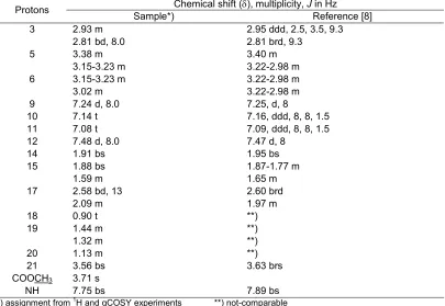
Table 2.The comparison of1H-NMR spectrum of coronaridine sample and that of reference4 Chemical shift (), multiplicity,Jin Hz
Protons
Sample*) Reference [8]
3 2.93 m 2.95 ddd, 2.5, 3.5, 9.3
2.81 bd, 8.0 2.81 brd, 9.3
5 3.38 m 3.40 m
3.15-3.23 m 3.22-2.98 m
6 3.15-3.23 m 3.22-2.98 m
3.02 m 3.22-2.98 m
9 7.24 d, 8.0 7.25, d, 8
10 7.14 t 7.16, ddd, 8, 8, 1.5
11 7.08 t 7.09, ddd, 8, 8, 1.5
12 7.48 d, 8.0 7.47 d, 8
14 1.91 bs 1.95 bs
15 1.88 bs 1.87-1.77 m
1.59 m 1.65 m
17 2.58 bd, 13 2.60 brd
2.09 m 1.97 m
18 0.90 t **)
19 1.44 m **)
1.32 m **)
20 1.13 m **)
21 3.56 bs 3.63 brs
COOCH3 3.71 s
NH 7.75 bs 7.89 bs
*) assignment from1H and gCOSY experiments **) not-comparable
et al. [15], demonstrated the CNS-stimulating activity of some iboga type alkaloids and suggested that the presence of methoxy substituents increased the activity, while it was lowered by the presence of methoxycarbonyl groups.
Coronaridine
Coronaridine 3 was isolated as a yellow amorphous solid absorbing UV atmax 283 and 310 nm consistent with an indole chromophore. Initial identification of the sample was achieved using the ion fragmentation patterns observed in the LREIMS spectrum, which showed the sample’s ion fragmentation pattern was identical to that of coronaridine. The molecular formula of coronaridine was obtained by HRCIMS and found to be C21H26N2O2 (measured
339.2069, calc. 339.2073, for MH+). Further structure elucidation was carried out by utilizing 1H-NMR and gCOSY experiments and by comparing the compound’s spectrum with that of an authenticated sample of a known [8] analogous compound, namely 18-hydroxycoronaridine4, as shown in Table 2.
From the 1H NMR spectrum, the compound clearly had an indole moiety, with a characteristic signal for an N-H and a 1,2,3,4-aromatic proton pattern. The signal for the N-H appeared as a broad singlet at
7.75 ppm while the aromatic signals appeared as a doublet at 7.48 ppm (H-12) which correlated to a triplet at 7.08 ppm (H-11); the triplet ascribed to H-11 also coupled to another triplet at7.14 ppm (H-10) as shown by the gCOSY spectrum. Another doublet at
deduced from the appearance of a triplet at 0.90 ppm (H-18), with an integral appropriate for three protons, coupled to two multiplets resonating at1.44 and1.32 ppm respectively (H-19, H-19’). Furthermore, a cross peak confirmed coupling between the C-19 protons and a multiplet at1.13 ppm (H-20), correlating in turn with a multiplet at3.56 ppm (H-21) and two multiplets at1.88 and 1.59 ppm assigned as C-15 protons. The C-3 protons appeared as a multiplet at 2.93 ppm and a broadened doublet at 2.81 ppm (J = 8.0 Hz), from which both signals correlated to a broad singlet at1.91 ppm (H-14). The proton at C-14 gave a correlation to a broadened singlet at 1.88 ppm (H-15). A proton signal ascribed to a proton at C-5 appeared to overlap with a C-6 proton giving a multiplet signal at 3.15-3.23 ppm. Other proton signals of C-5H and C-6H appeared at
3.38 and3.02 ppm, respectively.
Coronaridine 3, an iboga alkaloid isolated from
Ervatamia species, was first reported by Gorman et al. [7]. Iboga alkaloids arise biogenetically from tryptophan or its equivalent and two head-to-tail mevalonate residues [16]. Coronaridine was reported to have potent antileishmanial activity, inhibiting promastigote and amastigote growth [17]. Some iboga alkaloids including coronaridine have been found showing anti-addictive properties [18]. It has also been reported that coronaridine, like voacangine, produced analgesic and hypothermic effects in mice [18]. However, coronaridine was also found to display cytotoxic activity.
ACKNOWLEDGEMENT
I would like to give my sincere thanks to my supervisor, Prof. John B. Bremner, University of Wollongong for supporting spectroscopic data. Thanks are extended to University of Mataram and AusAid for funding this study.
REFERENCES
1. Perry, L.M., and Metzger, J., 1980, Medicinal Plants of East and Southeast Asia: Attributed Properties and Uses, The MIT Press, Cambridge, 620
2. Hadi, S., and Bremner J.B., 2001, Molecules, 6, 117-129.
3. Bisset, N.G., 1958,Ann, Bogor,3, 105-236.
4. Southon, I.W., and Buckingham, J., 1989, Editors
Dictionary of Alkaloids, Chapman and Hall 1230 p, Ltd, London, 1161.
5. Renner, U., 1957,Experientia, 13, 468-469.
6. Tantivatana, P., Ponglux, D., Wongseripipatana, S., and Phillipson, J.D., 1980, Planta Med., 40, 299-301.
7. Gorman, M., Neuss, N., Cone, N.J., and Deyrup, J.A.,1960,J. Am. Chem. Soc.,82, 1142-1145. 8. Van Beek, T.A., and Verpoorte, R., 1985,
Fitoterapia, 56, 304-307.
9. Govindachari, T.R., Joshi, B.S., Saksena, A.K., Sathe, S.S., and Viswanathan, N., 1965,
Tetrahedron Lett., 3873-3878.
10. Gunasekera, S.P., Cordell, G.A., and Farnsworth, N.R., 1980,Phytochem.,19, 1213-1218.
11. Braga, R.M., and Reis, F. de A.M., 1987,
Phytochem.,26, 833-836.
12. Singbarti, G., Zetler, G., and Schlosser, L., 1973,
Neuropharmacology,12, 239-244.
13. Rastogi, N., Abaul, J., Goh, K.S., Devallois, A., and Philogene, E., 1998, FEMS Immunol. Med. Microbiol.,20, 267-273.
14. Okuyama, E., Gao, L.H., and Yamazaki, M., 1992,
Chem. Pharm. Bull., 40, 2075-2079.
15. Bert, M., Marcy, R., Quermonne, M.A., Cotelle, M., and Koch, M., 1988,Planta Med.,54, 191-192. 16. Battersby, A.R., 1969, Biochem. J.,113, 26P-27P 17. Delorenzi, J.C., Attias, M., Gattass, C.R., Andrade,
M., and Rezende, C., 2001. Antimicrob. Agents Chemother.,45,1349-1354.