MAGNETIC RESONANCE IMAGING AND MAGNETIC
RESONANCE ANGIOGRAPHY IN THE MANAGEMENT OF
PATIENTS WITH ISCHEMIC STROKE IN VERTEBROBASILAR
CIRCULATION
Martina pero, Miljenko Kalousek, Josip Hat, Darko Bedek and Miljenko Marotti
Section of Neuroradiology, Department of Interventional and Diagnostic Radiology, Sestre milosrdnice University Hospital, Zagreb, Croatia
SUMMARY Vertebrobasilar occlusion is a life-threatening event that requires prompt diagnostic evaluation and subsequent therapy. Advanced magnetic resonance imaging (MRI) methods, including diffusion-weighted imaging and magnetic resonance angiography (MRA), are highly sensitive for the detection of ischemic tissue injury, and for the detection and localization of intracranial arterial occlusion and stenosis. In the era of thrombolytic therapy, MRI and MRA provide useful information for therapeutic decision making in the early stage of stroke evaluation. This retrospective review included patients with posterior circulation symptomatology examined at our Department between July 2002 and January 2005, 8 female and 11 male, mean age 54.9 years. The aim was to present the possibilities of MRI and MRA in the management of patients with ischemic stroke in posterior circulation. In 19 patients with an ischemia in the vertebrobasilar circulation detected by MRI of the brain, MRA identified 8 cases of basilar artery occlusion, 4 cases of basilar artery stenosis, 3 cases of multiple atherosclerotic stenoses of the vertebral arteries with 2 cases of concurrent vertebral artery occlusion, 2 cases of vasculitis in the posterior circulation, 1 case of proximal posterior cerebral artery occlusion, and 1 case of posterior cerebral artery stenosis. In 8 patients with basilar artery occlusion, the site of occlusion was proximal in 3 cases, proximal and middle in 2 cases, middle and distal in 2 cases, and distal in 1 case. MRI is a powerful tool to detect ischemic changes in stroke immediately upon stroke onset, while MRA is highly sensitive for the detection of occlusive disease in large intracranial arteries as well as in posterior circulation. In the acute stroke setting, MRI and MRA are useful for: 1) early and reliable identification of ischemic stroke; 2) improved choice of treatment modality by helping exclude from thrombolysis patients at high risk of hemorrhage and by identifying those patients most likely to benefit from it; 3) pinpoint the vascular origin of ischemic stroke; 4) determination of neurologic consequences of stroke, including final infarct size, clinical outcome and hemorrhagic risk.
Key words: Cerebrovascular accident diagnosis; Cerebrovascular circulation diagnosis; Cerebral arteries pathology; Ischemic attack diagnosis; Magnetic resonance imaging methods; Magnetic resonance angiography
Correspondence to: Martina pero, MD, Section of Neuroradiology, Department of Interventional and Diagnostic Radiology, Sestre milosrdnice University Hospital, Vinogradska c. 29, HR-10000 Zagreb, Croatia
E-mail: martinasp@yahoo.com
Received April 27, 2005, accepted July 22, 2005 Introduction
Ischemia in the vertebrobasilar region may cause involvement of the pons, midbrain, cerebellum,
thala-mus and occipital lobes with sudden or stuttering onset of symptoms from the large group of heterogeneous con-ditions usually lumped together under the term verte-brobasilar ischemia or insufficiency.
Basilar artery occlusion (BAO) is an infrequent cause of stroke: it is an important neurologic emergency re-quiring rapid diagnostic evaluation and subsequent ther-apy. When recognized late and/or untreated, it may lead to progressive neurologic deficits or death. The devel-opment of new, safe, noninvasive diagnostic tools such
as spiral computed tomography with computed tomog-raphy angiogtomog-raphy (CTA), and magnetic resonance im-aging (MRI) with diffusion-weighted imim-aging (DWI) and perfusion-weighted imaging (PWI) and magnetic resonance angiography (MRA) as well as the advent of new treatments such as systemic, intravenous and local intra-arterial thrombolysis (LIT) and percutaneous transluminal angioplasty (PTA), have facilitated diag-nosis and treatment of BAO and basilar artery stediag-nosis (BAS).
The aim of the study was to present the possibili-ties of MRI and MRA in the management of patients with ischemic stroke in posterior circulation through evaluation of our own results.
Patients and Methods
Medical records and reports of head CT, MRI and MRA findings of 19 patients examined at our Depart-ment between July 2002 and January 2005, and diag-nosed as having occlusive disease of the vertebrobasilar artery system were retrospectively reviewed. Eight (42.1%) female and 11 (57.9%) male patients, mean age 54.9 years, were admitted to our hospital with clinical signs suggestive of ischemic stroke in the posterior cir-culation: 18/19 had sudden onset of symptoms (one pa-tient had been hospitalized at another institution for a month and was transferred to our hospital for neuroradi-ologic diagnostic procedures), and 1/19 had transient symptoms.
Eleven of 19 patients underwent emergency CT scan of the head within few hours of arrival (in eight patients emergency head CT was unavailable for technical rea-son). Emergency brain CT studies were performed us-ing a conventional Shimadzu Intellect 4800 CT scan-ner. CT scans were unenhanced, with a slice thickness of 5 mm throughout the skull base and posterior fossa. Head CT findings were classified as positive with, and negative without signs of acute ischemic stroke in the posterior circulation.
All patients were submitted to MRI and MRA stud-ies of the brain within 2 to 10 days of admission to the hospital. MRI and MRA studies were performed with a 1.0-T MR imaging system (25 mT/m, Magnetom Har-mony, Siemens, Erlangen, Germany), using a standard head coil. The standard MRI study included diffusion weighted echo-planar sequence (DWI) in transverse plane, T1-weighted (T1W) spin-echo (SE) sequence in sagittal plane, T2-weighted (T2W) fast SE sequence in transverse plane, fluid attenuated inversion recovery (FLAIR) fast SE sequence in transverse plane, and T2*-weighted gradient-echo sequence in transverse plane. MRAs of the intracranial arteries were performed with a standard three-dimensional time-of-flight technique (3D TOF MRA): 3D TOF angiograms were reconstruct-ed using maximum-intensity projection (MIP) images. Ischemic lesions on MRI were categorized as tha-lamic, midbrain, pons, posterior cerebral artery (PICA) territory, and cerebellar [subdivided into superior, ante-rior infeante-rior, and posteante-rior infeante-rior cerebellar artery
Fig. 1. A 74-year-old female patient with right frontotemporal headache, vertigo, nausea and vomiting, ataxia: (a) transverse T2W image: acute ischemia in the right posterior inferior cere-bellar artery territory involving dorsolateral medulla oblonga-ta; (b) 3D TOF MRA: right vertebral artery occlusion.
(PICA) territories]. The sites of basilar artery (BA) oc-clusion were classified according to Archer and Horen-stein1 following the three anatomic segments of the BA:
proximal, from the confluence of the vertebral arteries to the origin of the anterior inferior cerebellar artery (AICA); middle, from the origin of the AICA to the ori-gin of the superior cerebellar artery (SCA); and distal, distal to the SCA. The length of occlusion was classi-fied as short if only one segment of the BA was cluded, and long if two or more segments were oc-cluded.
Results
Eleven patients were evaluated by head CT on ad-mission. CT findings were negative in 6 and positive in 5 cases with acute brain infarction involving vertebro-basilar territory. MRI and MRA studies were performed in 19 patients with suspected or previously confirmed acute stroke in the posterior circulation. On MRI stud-ies, abnormal parenchymal signals related to ischemic stroke were localized as follows: 8 in the cerebellum, 5 in the pons, 3 in the PICA territory, 1 in the PICA terri-Table 1. Magnetic resonance imaging (MRI) and magnetic resonance angiography (MRA) findings
MRI findings MRA findings
Cerebellum 8/19 Basilar artery occlusion 8/19
PICA territory 5/8 proximal, short 3/8
AICA territory 2/8 distal, short 1/8
SCA territory 1/8 proximal and middle, long 2/8
middle and distal, long 2/8
Pons 5/19 Basilar artery stenosis 4/19
Midbrain 1/19 Multiple atherosclerotic stenoses of vertebral arteries 1/19 with vertebral artery occlusion 2/19 PICA territory 3/19 PICA territory, proximal occlusion 1/19 PICA territory and thalamus 1/19 PICA territory, proximal stenosis 1/19
Thalamus 1/19 Vasculitis in posterior circulation 2/19
PCA = posterior cerebral artery; AICA = anterior inferior cerebellar artery; PICA = posterior inferior cerebral artery; SCA = superior cerebellar artery
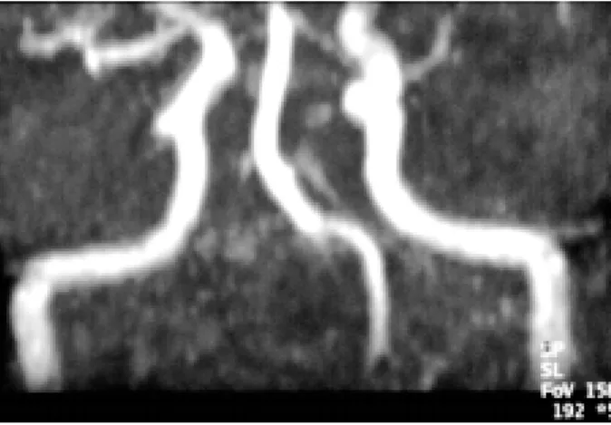
Fig. 2. A 53-year-old male patient with vertigo, right hand and leg paresthesias, horizontal nystagmus, transient dysarthria: (a) transverse T2W image: acute ischemia of the left paramedial pons and crura cerebri; (b) 3D TOF MRA: occlusion of the proximal and middle basilar artery.
tory and thalamus, 1 in the thalamus, and 1 in the mid-brain. Five cerebellar infarctions were localized in the territory of PICA, 2 in the territory of AICA, and 1 in the territory of SCA. Results are summarized in Table 1.
In 19 patients with ischemia in the vertebrobasilar circulation, MR angiograms identified 8 cases of BA oc-clusion, 4 cases of BA stenosis, 3 cases of multiple athero-sclerotic stenoses of the vertebral arteries with 2 cases of concurrent vertebral artery occlusion, 2 cases of vasculi-tis in the posterior circulation, 1 case of proximal PICA occlusion, and 1 case of proximal PICA stenosis. In 8 pa-tients with BA occlusion, the site of occlusion was proxi-mal in 3, distal in 1, proxiproxi-mal and middle in 2, and
mid-dle and distal in 2 cases. Long and short BAO was found in 4 cases each. Results are summarized in Table 1.
In 18 of 19 study patients, the onset of symptoms was sudden (one patient had been hospitalized at an-other institution for a month and was transferred to our hospital for neuroradiologic diagnostic procedures), whereas one patient had intermittent symptoms. The stroke pattern observed in 19 patients with stroke in the vertebrobasilar circulation included vertigo (n=12), headache (n=8), nausea and vomiting (n=6), diplopia (n=4) and nystagmus (n=3), ataxia (n=3), astasia-aba-sia (n=2), dysarthria (n=2) and dysphaastasia-aba-sia (n=1), psy-choorganic changes (n=2), coma (n=2), respiratory in-sufficiency (n=1), and singultus (n=1). Results are summarized in Table 2. One patient presented with the locked-in syndrome and multiple transient ischemic attacks (TIAs) preceding infarction each.
All patients were monitored and treated at the in-tensive care unit according to the Recommendations for Stroke Management issued by the Croatian Society for Neurovascular Disorders of the Croatian Medical Asso-ciation and the Croatian Stroke Society2. One patient
died, one patient was discharged from the hospital with severe disability, while 17 patients regained complete or partial recovery with moderate or minor disability.
Discussion
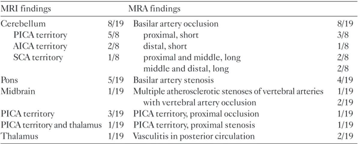
BAO is a life-threatening event, therefore it is cru-cial to expedite investigations, confirm the diagnosis, Fig. 3. A 71-year-old female patient with sudden onset of obtundation, respiratory insufficiency and left-sided hemiparesis that progressed to quadriparesis during several hours: (a) and (b) 3D TOF MRA: occlusion of the middle and distal basilar artery.
Table 2. Clinical symptoms of vertebrobasilar ischemia
Symptom n/N
Vertigo 12/19
Headache 8/19
Nausea and vomiting 6/19
Diplopia 4/19 Nystagmus 3/19 Ataxia 3/19 Astasia-abasia 2/19 Dysarthria 2/19 Dysphasia 1/19 Psychoorganic changes 2/19 Coma 2/19 Respiratory insufficiency 1/19 Singultus 1/19
and commence life-saving treatment through the early involvement of a number of disciplines including neu-rology, radiology and intensive care. This condition usu-ally has poor outcome and is associated with high mor-tality rates of 75% to 86%3-8 without thrombolysis, and a
survival rate of approximately 50%9-11 with thrombolytic
therapy. The most common causes of BAO are thrombo-sis on atherosclerotic lesion, cranial embolism, and trau-matic dissection or arteriosclerosis at the origin and in-tracranial segment of the vertebral arteries; proximal and middle BAO tend to be atherothrombotic, whereas dis-tal BAO tends to be embolic.
The initial clinical condition, etiology, time of on-set, age, location and length of occlusion on angiogra-phy, presence or absence of recanalization, and degree of collateral circulation have been reported as factors related to favorable outcome. According to Devuyst et al.11, four clinical features present on patient admission,
i.e. consciousness disorders, dysarthria, pupillary disor-ders and bulbar symptoms, are highly significantly as-sociated with poor outcome. Poor prognosis has been reported in patients aged 75 years and older, regardless of whether or not the occlusion was recanalized, mostly because of the reduction of cerebral recoverability be-cause of aging4,12,13.
Intra-arterial digital subtraction angiography (DSA) is an invasive radiologic method, whereas transcranial color-coded duplex sonography (TCCD) and transcra-nial Doppler ultrasound (TCD)14, CT and CTA, and
MRI and MRA are noninvasive radiologic methods that can be used in the assessment of acute ischemia in the posterior circulation, with their advantages and limita-tions. While the validity of DSA for the diagnosis and exclusion of BAO is beyond question, in many centers it has been replaced by noninvasive techniques; the main reasons are its invasiveness, hazards, limited availabili-ty, high costs, time consumption, and request for good cooperation of the patient or general anesthesia. Dop-pler sonography (DS) has become a standard vascular assessment tool. Unfortunately, certain technical prob-lems, e.g., technically insufficient visualization of the distal parts of the BA or adipose necks, limit the validi-ty of DS for the diagnosis and exclusion of BAO. How-ever, the ability of DS to provide information on flow dynamics, its usefulness as a bedside tool applicable in unstable patients and for repeat flow monitoring are advantages that may be used for therapy and follow-up. In vertebrobasilar territory, primarily a disturbance of the brainstem function threatens life, thus relatively smaller volumes of ischemic brainstem tissue can cause Fig. 4. A 24-year-old female patient, previously healthy, with sudden weakness and visual field defects on the left eye, bilateral temporal headache, nausea, vomiting; on the day before the symptom onset excessively consumed alcohol, tobacco and canabis: (a) transverse diffusion-weighted image, and (b) sagittal TWI image: right occipital subacute ischemia;
death; in contrast to the middle cerebral artery territo-ry, CT is not suited to show early ischemic edema in the brainstem because of its technical limitations.
Com-pared to CT, MRI has a greater sensitivity for the de- tection of ischemic lesions in the vertebrobasilar circu-lation, but in most institutions CT is available around the clock, whereas MRI is not, therefore CT with CTA still plays a major role in the emergency work-up.
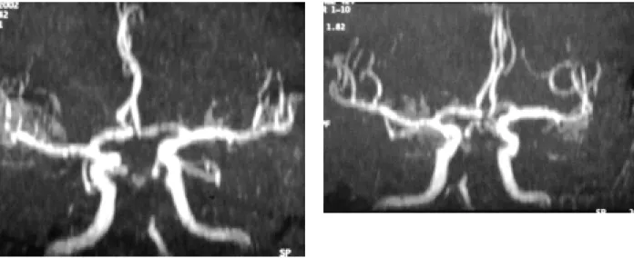
DWI, PWI and MRA, as new MRI techniques, can reliably identify the clinically relevant lesion in the acute stroke setting, increase diagnostic confidence, lead to a more focused evaluation of the underlying cause of Fig. 4. (c) 3D TOF MRA: multiple, short segmental stenosis of
the right posterior cerebral artery local vasculitis.
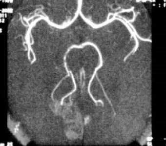
Fig. 5 (c) 3D TOF MRA: occlusion of the proximal basilar ar-tery.
Fig. 5. A 51-year-old male patient with sudden weakness, verti-go, nausea, nystagmus, astasia-abasia: (a) transverse diffusion-weighted image,
Fig. 5. (b) sagittal T2W image: acute ischemia of the medulla oblongata;
stroke, and may alter patient management. Acute ischemic tissue injury is currently best identified with DWI that delineates the extent of irreversible tissue damage fairly accurately. Identification of potentially salvageable tissue at risk surrounding the irreversibly damaged ischemic core requires PWI in addition to DWI. DWI, PWI and MRA lead to improvements in patient selection for intravenous or local intra-arterial thrombol-ysis15. Stenosis or occlusion in the vertebrobasilar
terri-tory can easily be demonstrated by MRA: the reported sensitivity and specificity of MRA in outlining high grade stenoses range between 86% and 100%16. MRA is used
to visualize vessel dissections, stenoses based on vascu-litis16 as well as fetal remnants of anastomoses between
the carotid arteries and the vertebral system, e.g., per-sistent trigeminal artery, and collateral circulation in the posterior circulation17,18. Bhadelia et al.19 have shown that
MRA has a good correlation with DSA in the detection and characterization of occlusive disease in the verte-brobasilar system.
MRI has several practical limitations: (1) modern scanners operating at least at 1.0 T capable of ultrafast imaging methods are required; (2) for acute stroke man-agement, emergency access to these scanners is need-ed and currently is only possible in specializneed-ed centers; (3) exclusion of contraindications for MRI, e.g., cardiac pacemakers, and metal implants, is occasionally diffi-cult, especially in unaccompanied aphasic or unconscious patients; (4) staff performing the scanning need to be specially trained, and furthermore, some of the tech-niques including PWI require substantial postprocess-ing, which can be time-consuming; (5) the cost of MRI currently still exceeds the cost of CT. In our hospital, there is only an emergency access to CT scanner for acute stroke management.
Angiographically, the site and length of occlusion and collateral status3,4,5,8,10,11,17,20,21 have been shown to have
an important impact on prognosis. In several studies, the tops of basilar occlusions were associated with the most favorable outcome because of preservation of flow into the cerebellar arteries and the arteries penetrating the brainstem, whereas short occlusions and good col-lateral flow may restrict the hypoperfused tissue vol-ume in the brainstem and enable survival. To limit he-morrhagic transformation of infarction, it is a standard practice in the anterior circulation to restrict intrave-nous thrombolysis to the first 3 hours and intra-arterial thrombolysis to the first 6 hours of the symptom onset, however, in case of BAO the time window for therapeu-tic intra-arterial thrombolysis is prolonged and exceeds
6 hours. It is explained by the preserved collateral flow and the possibility that the brainstem is tolerant to long-er plong-eriods of ischemia than the clong-erebral cortex5,13.
In five of 11 patients examined by emergency head CT at admission, CT finding was positive, while all 19 patients had signs of ischemia in the posterior circula-tion on MRI. Using MRA, we identified 11 cases of oc-cluded and 5 cases of stenosed artery in the posterior circulation, with 2 cases of vasculitis and 1 case of athero-sclerotic changes as well. In case of BAO, the proximal and middle segment of BA were pathologically changed, with generally favorable outcome.
Patients with BAO have a poor prognosis when reca-nalization does not occur. It can be achieved using intra-venous or local intra-arterial thrombolysis. Intraintra-venous thrombolysis has been criticized as being a shotgun ap-proach because it ignores specificity, whereas LIT was found to enhance the recanalization rate giving the pa-tient with recanalization a fair chance of favorable out-come and significantly reducing mortality6,22-24.
Howev-er, there are no randomized trials comparing intravenous thrombolysis with LIT. According to indirect comparison of intravenous and intra-arterial thrombolysis6, a cautious
statement would be that LIT is at least as effective and safe as intravenous thrombolysis with tPA and can be ap-plied with a longer time window. The risk of symptomat-ic intracranial hemorrhage does not seem to increase or at least not as much as in systemic thrombolysis6.
Current-ly, there are no established guidelines for selecting pa-tients with suspected basilar occlusion for intra-arterial thrombolysis based on clinical or MRI criteria. Different studies have shown that patients who benefit from LIT in case of BAO are: 1) young patients (<75 years) with-out any infarction in brainstem before the start of treat-ment13; 2) patients with low baseline NIHSS on
admis-sion and recanalization of BAO after early initiated LIT6,22,23; and 3) patients with good collateral flow4,10 or
distal clot location4,5,8. De Rochemont et al.24 report that
patients with only relatively small or no DWI lesions have a potentially favorable outcome if reperfusion is achieved rapidly with LIT, and that small DWI lesions, even if lo-cated in the brainstem, do not exclude a favorable out-come. In Croatia, intravenous thrombolysis with tPA has been approved since September 2004. None of these 19 patients received either intravenous or intra-arterial thrombolysis.
In summary, advanced MRI methods are a preferred investigative mode in the management of patients with ischemic lesion in the vertebrobasilar territory for
re-vealing or excluding artery occlusion or stenosis nonin-vasively and showing the extent of severe ischemic tis-sue injury in critical brain structures. Intravenous throm-bolytic therapy is the method of choice in the early treat-ment of ischemic stroke in the posterior circulation, but at large institutions with an interventional neuroradio-logical service, LIT should be considered as a method of choice as well.
References
1. ARCHER CR, HORENSTEIN S. Basilar artery occlusion. Stroke 1977;8:383-90.
2. DEMARIN V, LOVRENÈIÆ-HUZJAN A, ERIÆ V, VARGEK-SOLTER V, TRKANJEC Z, VUKOVIÆ V, LUPRET V, KALOUSEK M, DESYO D, KADOJIÆ D, LUIÆ I, DIKANOVIÆ M, VITAS M. Recommendations for stroke management. Acta Clin Croat 2001;40:127-54.
3. BECKER KJ, MONSEIN LH, ULATOWSKI J, MIRSKI M, WILLIAMS M, HANLEY DF. Intraarterial thrombolysis in vertebrobasilar occlusion. Am J Neuroradiol 1996;17.255-62. 4. BRANDT T, von KUMMER R, MULLER-KUPPERS M, HACKE
W. Thrombolytic therapy of acute basilar artery occlusion. Variables affecting recanalization and outcome. Stroke 1996;27:875-81. 5. CROSS DT, MORAN CJ, AKINS PT, ANGTUACO EE,
DIRIN-GER MN. Relationship between clot location and outcome after basilar artery thrombolysis. Am J Neuroradiol 1997;18:1221-8. 6. GÖNNER F, REMONDA L, MATTLE H, et al. Local intra-arterial
thrombolysis in acute ischemic stroke. Stroke 1998;1894-900. 7. NIGHOGHOSSIAN N, DEREX L, TURJMAN F, et al.
Hyperacute diffusion-weighted MRI in basilar artery occlusion treated with intra-arterial t-PA. Cerebrovasc Dis 1999;9:351-4. 8. SLIWKA U, MULL M, STELZER A, DIEHL R, NOTH J.
Long-term follow-up of patients after intraarterial thrombolytic therapy of acute vertebrobasilar artery occlusion. Cerebrovasc Dis 2001;12:214-9.
9. ZEUMER H, FREITAG HJ, GRZYSKA U, NEUNZIG HP. Local intra-arterial fibrinolysis in acute vertebrobasilar occlusion. Neuroradiology 1989;31:336-40.
10. CROSS DT, MORAN CJ, AKINS PT, ANGTUACO EE, DERDEYN CP, DIRINGER MN. Collateral circulation and outcome after basilar artery thrombolysis. Am J Neuroradiol 1998;19:1557-63.
11. DEVUYST G, BOGOUSSLAVSKY J, MEULI R, MONCAYA J, de FREITAS G, van MELLE G. Stroke or transient ischemic attacks with basilar artery stenosis or occlusion: clinical patterns and outcome. Arch Neurol 2002;59:567-73.
12. UEDA T, SAKAKI S, KUMON Y, OHTA S. Multivariable analysis of predictive factors related to outcome at 6 months after intra-arterial thrombolysis for acute ischemic stroke. Stroke 1999;30:2360-5.
13. EZAKI Y, TSUTSUMI K, ONIZUKA M, KAWAKUBO J, NOBUHIRO Y, SHIBAYAMA A, TOBA T, KOGA H, MIYAZAKI H. Retrospective analysis of neurological outcome after intra-arterial thrombolysis in basilar artery occlusion. Surg Neurol 2003;60:423-30.
14. ALEXANDROV AV, DEMARIN V. Insonating techniques and diagnostic criteria for transcranial doppler sonography. Acta Clin Croat 1999;38:97-108.
15. NEUMANN-HAEFELIN T, MOSELEY ME, ALBERS GW. New magnetic resonance imaging methods for cerebrovascular disease: emerging clinical applications. Ann Neurol 2000;47:559-70. 16. KRUG B, TERSTEGGE K, NEVELING M, ZÄHRINGER M,
KUGEL H, LACKNER K. MRA in patients with cerebrovascular disease. Contraindications of clinical effectiveness. Acta Radiol 2000;41:1-7.
17. BAUMGARTNER RW, MATTLE HP, AASLID R. Transcranial color-coded duplex sonography, magnetic resonance angiography, and computed tomography angiography: methods, applications, advantages and limitations. J Clin Ultrasound 1995;23:89-111. 18. WILMS G, BOSMANS H, DEMAEREL Ph, MARCHAL G.
Magnetic resonance angiography of the intracranial vessels. Eur J Radiol 2001;38:10-8.
19. BHADELIA RA, BENGOA F, GESNER L, PATEL SK, UZUN G, WOLPERT SM. Efficacy of MR angiography in the detection and characterization of occlusive disease in the vertebrobasilar system. J Comput Assist Tomogr 2001;25:458-65.
20. BRANDT T, KNAUTH M, WILDERMUTH S, et al. CT angiography and Doppler sonography for emergency assessment in acute basilar artery ischemia. Stroke 1999;30:606-12. 21. WELSH LW, WELSH JJ, LEWIN B. Basilar artery and vertigo. Ann
Otol Rhinol Laryngol 2000;109:615-22.
22. ECKERT B, KUCINSKI T, PFEIFFER G, GRODEN C, ZEUMER H. Endovascular therapy of acute vertebrobasilar occlusion: early treatment onset as the most important factor. Cerebrovasc Dis 2002;14:42-50.
23. ARNOLD M, NEDELTCHEV K, SCHROTH G, BAUMGART-NER RW, REMONDA L, LOHER TJ, STEPPER F, STURZEN-EGGER M, SCHUKNECHT B, MATTEL HP. Clinical and radiological predictors of recanalisation and outcome of 40 patients with acute basilar artery occlusion treated with intra-arterial thrombolysis. J Neurol Neurosurg Psychiatry 2004;75:857-62. 24. de ROCHEMONT RM, NEUMANN-HAEFELIN T,
BERKE-FELD J, SITZER M, LANFERMANN H. Magnetic resonance imaging in basilar artery occlusion. Arch Neurol 2002;59:398-402.
Saetak
MAGNETSKA REZONANCA MOZGA I MAGNETSKA ANGIOGRAFIJA U ZBRINJAVANJU BOLESNIKA S ISHEMIJSKIM MODANIM UDAROM U VERTEBROBAZILARNOJ CIRKULACIJI
M. pero, M. Kalousek, J. Hat, D. Bedek i M. Marotti
Vertebrobazilarna okluzija je za ivot opasno stanje koje zahtijeva brzu dijagnostièku obradu i terapiju. Suvremene metode magnetske rezonance (MR) mozga, ukljuèujuæi difuzijski mjerenu sliku i magnetsku angiografiju (MRA), imaju visoku osjetljivost u otkrivanju ishemijske lezije modanog parenhima, te u otkrivanju i lokalizaciji okluzije i stenoze intrakranijskih arterija. U doba trombolitiène terapije MR mozga i MRA daju korisne podatke bitne za donoenje odluke o izboru terapije u procjeni ranog stadija ishemijskog modanog udara. Proveden je retrospektivni pregled bolesnika sa simptomatologijom stranje cirkulacije koji su na naem Zavodu pregledani u razdoblju od srpnja 2002. do sijeènja 2005. godine, 8 ena i 11 mukaraca srednje ivotne dobi od 54,9 godina. Cilj je bio pokazati moguænosti MR mozga i MRA u zbrinjavanju bolesnika s ishemijskim modanim udarom stranje cirkulacije. U 19 bolesnika s ishemijskim modanim udarom vertebrobazilarnog sliva, koji je dokazan pomoæu MR mozga, MRA je otkrila 8 okluzija bazilarne arterije, 4 stenoze bazilarne arterije, 3 sluèaja viestrukih aterosklerotskih stenoza vertebralnih arterija s 2 sluèaja istodobne okluzije vertebralne arterije, 2 vaskulitisa u stranjoj cirkulaciji, 1 okluziju proksimalnog dijela i 1 stenozu stranje modane arterije. Meðu 8 bolesnika s okluzijom bazilarne arterije mjesto okluzije bilo je proksimalni dio arterije u 3, proksimalni i srednji dio u 2, srednji i distalni dio u 2 sluèaja i distalni dio bazilarne arterije u 1 sluèaju. MR mozga je moæno sredstvo u otkrivanju ishemijskih promjena neposredno nakon nastupa modanog udara, dok MRA ima visoku osjetljivost za otkrivanje okluzivne bolesti velikih intrakranijskih arterija. Kod zbrinjavanja akutnog modanog udara MR mozga i MRA su korisne zbog: 1) brzog i sigurnog otkrivanja ishemije; 2) sigurnijeg izbora oblika terapije pomauæi da se tromboliza ne primijeni kod bolesnika s visokim rizikom za razvoj krvarenja te da se otkriju bolesnici koji æe imati najvie koristi od iste; 3) moguænosti toènog odreðivanja vaskularnog podrijetla ishemijskog modanog udara; 4) odreðivanja neurolokih posljedica modanog udara ukljuèujuæi konaènu velièinu ishemijske lezije, klinièki ishod i rizik od krvarenja.
Kljuène rijeèi: Modani udar dijagnostika; Cerebrovaskularna cirkulacija dijagnostika; Modane arterije patologija; Ishemijski udar dijagnostika; Prikazivanje magnetskom rezonancom metode; Magnetska angiografija