Characterisation of metal threads in Renaissance tapestries
A.-M. Hacke
a, C.M. Carr
a, A. Brown
ba
Department of Textiles and Paper, University of Manchester Institute of Science and Technology, PO Box 88, Manchester, M60 1QD, United Kingdom
b
CSMA Ltd, Queens Road, Penkhull, Stoke-on-Trent, Staffordshire ST4 7LQ, United Kingdom
Abstract
The MODHT (Monitoring of Damage in Historic Tapestries) project investigated the history, composition and deterioration of metal threads with a textile core.
Metal thread manufacture has been investigated by Scanning Electron Microscopy (SEM) and characteristic deformations established on historic and “model” metal threads. Rare examples of triple wrapped silver gilt threads were observed in some 16th century tapestries of the Royal Spanish collection.
SEM, Energy Dispersive X-ray analysis (EDX), X-ray Photoelectron Spectroscopy (XPS) and depth profiling Secondary Ion Mass Spectrometry (SIMS) were utilized for chemical compositional analysis of the metal layers and alloys and the corrosion products of original metal thread samples.
Accelerated tarnishing tests on silver and copper were performed to determine the influence of the degradation of the dyed wool and silk fibres on the formation and composition of corrosion products. Metal threads and aged metal foils were analysed by SEM in order to establish the morphology of corrosion growth due to natural and accelerated ageing.
Keywords: metal thread, gold layer, tapestry, corrosion, XPS, SIMS
Corresponding author: Tel.: 0044 161 2003893 email: [email protected]
1. Introduction
1.1 History of metal threads
Decorative metals have been incorporated into textiles for thousands of years, with an early reference found in the Old Testament, Exodus 39:2-3; approximately 12th to 13th centuries BC.
The earliest metal threads were thin strips of gold, which were cut from a beaten metal foil and directly woven or embroidered into textiles. Later these strips were wound around a fibrous core; this would introduce more flexibility to the thread and make its uses more versatile. The core fibres were usually silk, dyed according to the colour of the metal wrapping, e.g. yellow or red for gold threads, undyed white silk for silver threads; though metal threads with a linen, cotton, animal hair or sinew core have also been reported (Braun-Ronsdorf 1961; Budney and Tweedle 1985; Indictor and Blair 1990; Skals 1991). Early examples of wound metal threads are a 3rd century textile fragment found in a Roman sarcophagus in Hungary (Geijer and Thomas 1964-65) and the 4th century remains of gold filament wrappings from the grave of a woman at London’s Spitalfield market (King 2003).
Published data on surviving threads from the 12th century and before suggest that either pure gold or alloys with a very high gold content were used (Braun-Ronsdorf 1961; Jaro 1990; King 2003). It is not clear when silver or silver-copper gilt metal threads were first introduced but most surviving threads of the late Medieval and Renaissance period are of that type (Hoke and Petrascheck-Heim 1977; Darrah 1987; Garside 2002; Hacke et al. 2003).
century (Oddy 1977). Towards the end of the 14th century production centres in Italy and Spain took up the manufacture of cast, drawn and rolled metal threads (Braun-Ronsdorf 1961) and from the late 15th until the 17th century this technique gradually replaced the beaten and cut method in Europe (Jaro et al. 1990; Montegut et al. 1992; Jaro et al. 2000; Garside 2002; Tronner et al. 2002).
There were several possible methods of gilding the blocks or rods of silver; the earliest is believed to involve hammering the gold onto the silver surface, while later methods included soldering, welding or fire-gilding with mercury amalgam (Hoke and Petrascheck-Heim 1977; Schreier and Bresee 1979; Jaro et al. 1990; Jaro and Toth 1994).
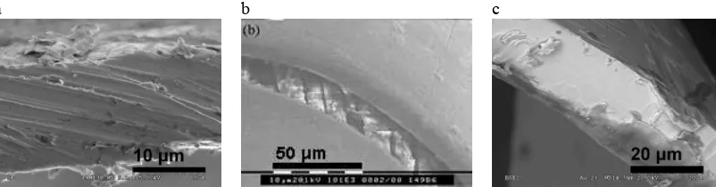
In the 11th or 12th century the so called “Cyprus gold” began to spread to Europe from Byzantium, western Asia or northern Africa through the ports of Cyprus (Braun-Ronsdorf 1961). Cyprus gold was a membrane thread, based on a gilt leather or animal gut filament wound around a fibrous core. Membrane threads of Chinese or Japanese origin usually had a paper substrate (Darrah 1987). Membrane threads are more flexible and lighter than solid metal threads but regarded of inferior quality due to the fragility of the gold layer to abrasion and the lower luster compared to solid metal threads. By the 13th or 14th century production of membrane threads was also developed in Europe but their importance declined by the 16th century. The most common gilding technique was the application of gold leaf (Jaro et al. 1990), possibly with animal glue or fat as an adhesive (de Reyer et al. 2002). Determination of the manufacturing technique is often made by means of SEM analysis of the edges of metal threads. Sharp angles with tool marks left from cutting indicate “beaten and cut” threads, while smooth and round edges and longitudinal striations are characteristic for “cast, drawn and rolled” threads. However, SEM analysis of model metal threads, produced, in this study, from flattened (rolled) wires, showed that this characterisation can be misleading. Figure 1 shows the edges on the model metal threads were also often rough with sharp angles. Therefore, an unambiguous determination of the manufacturing technique is limited to the analysis of metal filaments with single- or double- sided gilding.
a b c
Figure 1. SEM micrographs of edges of metal threads. a: “beaten and cut”, b:”cast, drawn and rolled”, c: model metal filament
1.2 Description of metal threads in this study
The metal threads investigated in this study were taken from five European Renaissance tapestries; their details of origin and manufacture are listed below:
• ‘Abraham and Melchizedek’ (HCP), Hampton Court Palace, woven by Willem de Kempeneer in Brussels, ca. 1535, 112 metal thread samples taken.
• ‘Christ before Pilate’ (BXL2), National Museum, Brussels, woven in Brussels, ca. 1520, 21 metal thread samples taken.
• ‘Dédalo e ĺcaro’ (PNM1), Patrimonio Nacional Madrid, Wilhelm van Pannemaker, Brussles, ca. 1545, 27 metal thread samples taken.
• ‘Júpiter y Ganimedes’ (PNM2) Patrimonio Nacional Madrid, Wilhelm van Pannemaker, Brussles, ca. 1545, 17 metal thread samples taken.
The thread diameter, width of metal foil and number of coils per 5 mm thread were measured manually through digital imaging analysis. All µm measurements were rounded to the nearest 50 due to the relative imprecision of the measurement technique.
All metal thread samples in this study consist of solid metal strips wound around a silk core, which is white or light yellow for silver threads and dyed in darker yellow shades for gilt threads. All but one sample exhibit S twist direction of the metal coils and are categorized according to thread diameter and colour of metal strip in Table 1. The metal strip width and number of coils per unit length were found to vary with thread diameter. Large diameter threads (≥ 700 µm) generally have wider metal strips and fewer coils than medium (< 650 > 350 µm) or small diameter threads (≤ 350 µm), which show the finest metal strip width, the most coils per unit length and are also the least corroded, Figure 2. The relationship between diameter, width and coil number was also observed in the silver threads and those that were heavily corroded.
Table 1. Categorization of metal threads from five MODHT Renaissance tapestries.
Group Number of samples in tapestry
HCP PNM1 PNM2 PNM5 BXL2 Total
gilt large diameter (≥ 700 µm) 14 - - - - 14
Figure 3. Triple wrapped metal threads.
Metal threads in tapestries are in close proximity to wool and silk and it has been suggested that the degradation of textile fibres influences the tarnishing of metals (Hoke and Petrascheck-Heim 1977; Howell et al. 1999). This study investigates the corrosion products on metal coupons tarnished by accelerated ageing, as well as historical metal threads. The combination of X-ray Electron Spectroscopy (XPS), Secondary Ion Mass Spectrometry (SIMS) and Secondary Electron Microscopy coupled with Energy Dispersive X-ray analysis (SEM/EDX) have been applied to investigate the bulk and surface metal alloy contents and corrosion products. These techniques have been previously used to study metal threads and provide a powerful complementary approach to characterise surface and sub-surface composition and morphology (Howell et al. 1999; Hacke et al. 2003).
2. Experimental techniques
2.1 Accelerated tarnishing
Silver and copper coupons, 5 x 10 x 0.25 mm, > 99.9 % purity, were cut and a pin hole punched in each to allow suspension during the Oddy tests. The coupons were cleaned successively in propanol, hexane, chloroform and methanol for 10 minutes in an ultrasonic cleaner. Silver and copper coupons were incubated under thermal or light Oddy test conditions indicated below with the following wool and silk samples: undyed, dyed with weld, woad, madder or brazil.
2.1.1 Thermal Oddy test
0.2 g of wool or silk yarn was placed in the bottom of a test tube together with Durham tubes (6.5 x 30 mm) filled with deionised water. Silver and copper coupons were suspended from the top of the test tube using nylon filament and the tubes were sealed with a stopper and tape. 12 yarn samples and one control were incubated at 60°C for 4 weeks.
2.1.2 Light Oddy test
Wool and silk fabrics, 4 x 5 cm, were stapled to a cardboard backing and placed in glass cells (2 x 5 x 15 cm) along with tubes, 6.5 x 120 mm, filled with deionised water. Silver and copper coupons were suspended from the top of the cells containing the fabric pieces and one control sample containing only the tube with deionised water. The coupons were shielded from direct irradiation with cardboard placed around the top of the sealed cells. The fabric samples were irradiated for 300 hours using an OSRAM 400 W HQ (MB-U) mercury lamp in a Microscal Ltd. Mark V Light Fastness Tester. The cells were equipped with a circulating water cooling system maintaining the temperature at 27-29 °C.
2.2 SEM/EDX
2.3 XPS
Spectra of metal coupons were obtained on a Kratos Axis Ultra X-ray photoelectron spectrometer using the Al Kα monochromator X-ray source, with a pass energy of 80 eV for wide scans and 20 eV for high resolution scans. The analysed area was approximately 300 x 800 µm. Metal threads were analysed on a SSX-100 ESCA Spectrometer using monochromated Al Kα X-rays, 152 eV pass energy and 150 µm spot size. All data was processed using Casa XPS 2.2.80 software. Quantification was performed using appropriate relative sensitivity factors, transmission functions and a linear background.
2.4 SIMS
Metal threads were analysed using a CAMECA IMS 4f Magnetic Sector SIMS instrument. The Cs+ primary ion beam was operated at 10 keV, with a beam current of 0.1-0.4 nA for positive and negative mass spectra, and 20 nA for positive depth profiles. The areas analysed were 250 µm2 for spectra and 100 µm2 to 200 µm2 for depth profiles. Typical acquisition times were 300 s for spectra and 3000 s for depth profiles. A reference crater was measured using a Detak Profilometer; subsequent depth calibrations were based on a sputter rate factor. Semi-quantitative values were calculated as relative atomic % assuming the same ionization probabilities for CsAg+, CsCu+ and CsAu+ and correcting for the natural isotopic abundances.
3. Results and Discussion
3.1 Alloy composition of metal threads
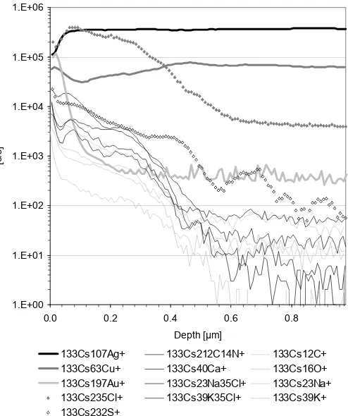
SIMS positive ion depth profiles have been obtained from all metal thread exterior sides and one interior side only. Figure 4 shows a typical depth profile of the exterior of a gilt metal thread. The initial intensities for contaminants such as C, K, Cl and Na containing compounds is high but decreases rapidly as the sample surface is etched. In sample PNM5_24 the gold intensity reaches its maximum at a depth of approximately 20 nm, and its minimum, i.e. bulk intensity at 200 nm. Other samples had the gold layer interface between 100-500 nm (PNM2_19) and 40-300 nm (PNM1_38). These values are consistent with other recent studies on metal threads, which revealed lower gold layer thicknesses than previously proposed (Enguita et al. 2002; Tronner et al. 2002).
Chlorine and sulphur ions are probably derive from corrosion compounds and showed a lower rate of decrease than other contamination residues. The lower intensity of 133Cs232S+ compared to 133Cs235Cl+ was attributed to the lower sensitivity of positive ion SIMS to sulphur. Sulphur compounds showed higher intensities in negative ion SIMS mass spectra and XPS analysis indicated similar atomic concentrations of chlorine and sulphur.
1.E+00 1.E+01 1.E+02 1.E+03 1.E+04 1.E+05 1.E+06
0.0 0.2 0.4 0.6 0.8
Depth [µm]
[c
/s
]
133Cs107Ag+ 133Cs212C14N+ 133Cs12C+
133Cs63Cu+ 133Cs40Ca+ 133Cs16O+
133Cs197Au+ 133Cs23Na35Cl+ 133Cs23Na+
133Cs235Cl+ 133Cs39K35Cl+ 133Cs39K+
133Cs232S+
Figure 4. SIMS positive ion depth profile of the exterior of metal thread PNM5_24
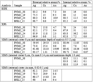
The SIMS, XPS and EDX analyses present values in relative atomic % of metallic components only, Table 2. EDX, at 25 kV accelerating voltage, has a depth analysis of > 0.2 µm, while XPS probes the surface to a depth of < 10 nm and SIMS depth profiles in the range of 0 to 2 µm. All PNM samples showed gilding on the exterior side only, with the highest Au concentrations in the PNM5_24 sample. The concentrations of copper and silver exceeded that of gold on the surface and throughout the gold layer of all samples indicating that the surfaces were not gilt with pure gold but with silver – copper – gold alloy or the alloy formed during gilding or the silver and copper ions precipitated through the gold layer. Precipitation of Ag and Cu ions was also supported by the XPS results, which showed low gold concentrations at the outer surface. The SIMS depth profile of the interior of PNM1_38 showed the highest copper concentration near the surface and decreased with increasing depth to the bulk value determined with the exterior face analysis. Previous SIMS depth profile analyses of the interior of single sided gilt metal threads showed similar results (unpublished analyses of MODHT samples), suggesting copper ion migration towards the surfaces of the metal filament.
Table 2. EDX, XPS and SIMS positive ion depth profiling analyses of the alloy compositions of exterior and interior surfaces of metal threads.
External relative atomic% Internal relative atomic %
Analysis Sample Ag Cu Au Ag Cu Au
SIMS (external crater 0 µm and maximum depth of analysis 0.5-2 µm)
PNM1_38 59.95 29.53 10.52 88.94 11.05 0.01
PNM2_19 73.8 25.4 0.8 92 7.8 0.2
PNM5_24 61.68 18.33 19.99 89.61 10.36 0.03 BXL2_15 93.17 1.31 5.52 98.71 0.42 0.87 SIMS (internal crater 0 µm, Cu max 0.14 µm and maximum depth 1.8 µm)
PNM1_38 88.8 11.1 0.0
83.8 16.1 0.1
88.0 11.9 0.0
SIMS (external crater Au max, 0.02-0.1 µm)
PNM1_38 61.1 24.4 14.5
PNM2_19 70.3 20.9 8.8
PNM5_24 60.17 15.83 24
BXL2_15 90 0.6 9.4
3.2 Accelerated tarnishing and corrosion on metal threads
Table 3. Colour variation of tarnish on Light Oddy tested silver coupons.
* number for dominant tarnish colour is stated first
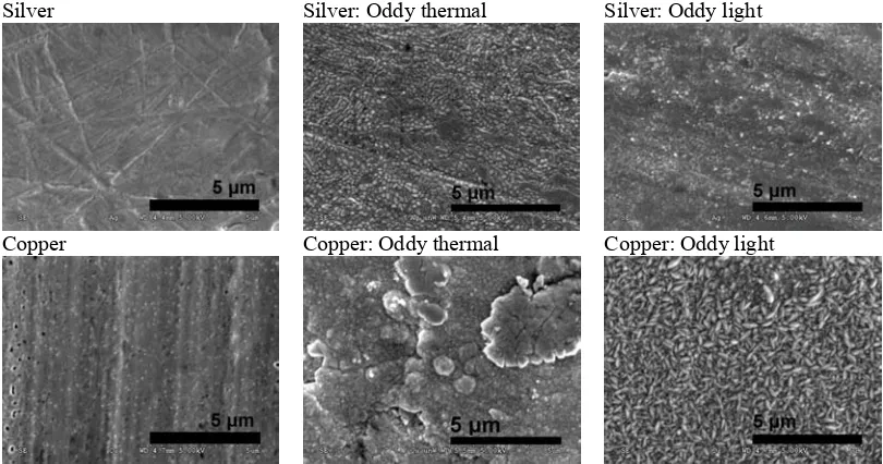
SEM micrographs of the corrosion morphologies on silver and copper are shown in Figure 5. In areas with little corrosion the initial growths were found to develop along scratches and pits on the surface. Generally the corrosion growths on copper were more rounded and accumulated than corrosion on silver, which formed a more evenly distributed layer with uniformly sized corrosion crystals and little differences in height. In addition, some of the copper coupons associated with photodegradation of wool showed regular longitudinal crystals.
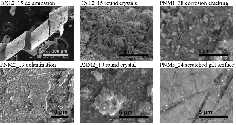
The morphological appearance copper sulfide corrosion on metal threads has been described as “trees”, “bushes” or “black spot disease”, while silver sulfide crystals have been described as “dendrites” breaking the gold layer (Hoke and Petrascheck-Heim 1977), “whiskers”, “black fuzzies”, “flower-or tree-shaped” and “mushroom-shaped bubbles” (Jaro and Toth 1997). While these descriptions are subjective, there does seem to be a clear distinction between sharp and rounded crystals. The cause for the different growth behaviour of the sulphide crystals is unknown. Corrosion morphologies on metal threads in this study were found to be rounded crystals, Figure 6. Corrosion cracking, flaking and delamination were observed on all samples, particularly BXL2_15.
Delamination and differences in the cracking structure have previously been attributed to either stress-corrosion causes, or the manufacturing technique, for example folding and stretching of a metal foil or strip prior to cutting into narrower strips for winding (Jaro and Toth 1997), or joining of thin foils by hammering and subsequent cutting into strips (Hardin and Duffield 1986). However, corrosion growth in layers and cracking of corrosion crystals has been observed on heavily corroded samples of the modern Oddy tested coupons, Figure 5.
Silver Silver: Oddy thermal Silver: Oddy light
Copper Copper: Oddy thermal Copper: Oddy light
Figure 5. Corrosion crystal morphology on Thermal and Light Oddy tested silver and copper coupons. Textile material Side A* Side B* Legend
Control 0 0 0 0 0 = no tarnish undyed 5 4 1 4 1 = yellow
weld 3 5 1 3 2 = green
woad 5 1 1 5 3 = red
madder 3 1 1 5 4 = purple Silk
brazil 3 3 1 3 5 = blue undyed 0 6 2 2 6 = black
weld 2 2 2 2 woad 5 1 4 1 madder 5 4 3 6 Wool
BXL2_15 delamination BXL2_15 round crystals PNM1_38 corrosion cracking
PNM2_19 delamination PNM2_19 round crystal PNM5_24 scratched gilt surface
Figure 6. Corrosion on metal threads.
XPS analysis of the coupons showed that the relative levels of oxygen and carbon were significantly higher on copper coupons than on silver coupons, with the atomic ratio O/Cu being ~ 2.5 and C/Cu ~ 5, while O/Ag was ~ 0.5 and C/Ag ~ 1.3. These ratios did not change significantly due to the accelerated tarnishing tests. Other low level contaminants were chlorine and nitrogen. Only the sulphur to metal atomic ratios showed significant increases due to accelerated tarnishing, Table 4. Both the sulphur atomic % and the atomic ratios of sulphur to metal were higher on silver than on copper, and were also higher due to the degradation of wool than silk. This agreed with the visual assessment of levels of tarnishing. The sulphur levels were higher on those metal coupons exposed to light irradiated wool and silk compared to thermally degraded wool and silk. This could be due to the relative exposure periods or the greater degradation of the light.
XPS high resolution scans of the S (2p) peaks showed that sulphur was mainly present in its S2+ form; only small amounts of S6+ were detected on copper coupons, probably as sulfates, Figure 7. The maximum atomic sulphur to metal ratios were > 0.5 indicating that sulphur was present not only as Ag2S and Cu2S, which would yield maximum ratios of 0.5 if the whole metal surface was converted to sulfide. The nature of this additional sulphur concentration is at present uncertain. However, future research will focus on establishing the nature of the corrosion layer, its durability to aqueous washing and possible detergent adsorption on clean and corroded surfaces.
XPS analysis of metal threads showed the presence of sulfide on all samples, and low levels of sulfate on PNM2_19 only. Chlorine was also present on all samples, usually in similar concentrations to the sulphur. The highest ratios of sulphur to metals were detected on the interior of BXL2_15, where atomic % S/(Ag+Cu) = 0.34.
Table 4. Relative sulphur levels on tarnished Oddy tested silver and copper coupons. Atomic % ratio
Oddy test set
Test material
S/Cu S/Ag
Control 0.05 0.07
Silk 0.04-0.09 0.16-0.22
Thermal
Wool 0.13-0.39 0.35-0.51
Control 0.09 0.09
Silk 0.19-0.33 0.39-0.59
Light
Sulfate
Sulfide
x 10 2 40
50
60
70
80
90
10 0
CS
17 4 17 2 17 0 16 8 16 6 16 4 16 2 16 0 Bi nd in g Ene rgy (e V)
Figure 7. XPS high resolution scans of S (2p) on Light Oddy tested copper coupons incubated with undyed silk – and undyed wool –.
4. Conclusions
Measurements of gilt metal threads from Renaissance tapestries indicate a correlation between metal thread diameter, metal strip width and the number of coils per unit length of thread. Those threads with the smallest diameter (< 350 µm) and most coils (> 10 per 5 mm) appeared to be of “superior” quality. Rare triple wrapped metal threads were reported for the first time observed in tapestries belonging to the set of the “Fables of Ovid” of the Royal Spanish collection, manufactured in Brussels, 1545. SEM micrographs of silver and copper coupons incubated with wool or silk and exposed to heat or light irradiation showed variations in corrosion morphologies, particularly of the copper and silver sulfides. XPS analysis of accelerated tarnished coupons and corrosion products on metal threads confirmed the presence of mainly sulfide corrosion with some sulfate and chlorine residues also on the metal threads.
SIMS analysis indicated the presence of thin surface gilt layers with gold concentration maxima at depths of 20 – 100 µm. Combined EDX, SIMS and XPS analyses of silver and copper suggested metal ion migration and/or application of a gold alloy rather than pure gold to the silver - copper alloy substrate.
Acknowledgments
The authors thank Trevor Jones (UMIST) and Tanya Moran (CSMA) for expert SEM and XPS analytical work.
We also gratefully acknowledge the European Commission (FP5) for funding the MODHT project.
References
Braun-Ronsdorf (1961) Gold and Silver Fabrics from Medieval to Modern Times, C.I.B.A. Review 3, 2-16.
Budney, M. and Tweedle, D. (1985) The Early Medieval Textiles at Maaseik, Belgium, The Antiquaries Journal LXV Pt II, 353-389.
Enguita, O., Climent-Font, A., Garcia, G., Montero, I., Fedi, M. E., Chiari, M. and Lucarelli, F. (2002) Characterization of Metal Threads Using Differential PIXE Analysis, Nuclear Instruments and Methods in Physics Research Section B: Beam Interactions with Materials and Atoms 189, 1-4, 328-333.
Garside, P. (2002). Investigations of Analytical Techniques for the Characterisation of Natural Textile Fibres Towards Informed Conservation, (Ph.D.), University of Southampton, Southampton: 1-232. Geijer, A. and Thomas, E. B. (1964-65). The Viminacium Gold Tapestry, in: Meddelanden fran Lunds. Lund, Universitets Historika Museum: 223-236.
Hacke, A. M., Carr, C. M., Brown, A. and Howell, D. (2003) Investigation into the Nature of Meta Threads in a Renaissance Tapestry and the Cleaning of Tarnished Silver by UV/Ozone (UVO) treatment, Journal of Materials Science 38, 3307-3314.
Hardin, I. R. and Duffield, F. J. (1986) Characterization of Metallic Yarns in Historic Persian Textiles by Microanalysis. Historic Textile and Paper Materials, American Chemical Society.
Hoke, E. and Petrascheck-Heim, I. (1977) Microprobe Analysis of Gilded Silver Threads from Mediaeval Textiles, Studies in Conservation 22, 49-62.
Howell, D., Mitchell, R., Carr, C. M. and Walton, J. (1999) X-ray Photoelectron Spectroscopy (XPS) and Time-of-Flight Secondary Ion Mass Spectrometry (ToF-SIMS) Study of the Tarnishing of Metal-Coated Textiles, Journal of the Textile Institute 90, 3, 50-59.
Indictor, N. and Blair, C. (1990) The Examination of Metal from Historic Indian Textiles Using Scanning Electron Microscope-Energy Dispersive X-Ray Spectrometry, Textile History 21, 2, 149-163. Indictor, N., Koestler, R. J., Blair, C. and Wardwell, A. E. (1988) The Evaluation of Metal Wrappings from Medieval Textiles Using Scanning Electron Microscopy-Energy DispersiveX-Ray Spectrometry, Textile History 19, 1, 3-19.
Jaro, M. (1990) Gold Embroidery and Fabrics in Europe: XI-XIV Centuries, Gold Bulletin 23, 2, 40-57.
Jaro, M., Gal, T. and Toth, A. (2000) The Characterization and Deterioration of Modern Metallic Threads, Studies in Conservation 45, 2, 95-105.
Jaro, M., Gondar, E. and Toth, A. (1990) Reconstruction of Gilding Techniques Used for Medieval Membrane Threads in Museum Textiles, Archeometry '90, Birkhaeuser Verlag Basel 317-325. Jaro, M. and Toth, A. (1994) Possibilities of Reconstruction of Metal Coating Techniques Used for Solid Metal Strips or Wires in Museum Textiles. Proceedings of the 4th International Conference on Non-Destructive Testing of Works of Art, Berlin, Deutsche Gesellschaft für Zerstörungsfreie Prüfung e.V., Berlin.
Jaro, M. and Toth, A. (1997) Deterioration of Metal Threads and Other Metallic Decorations Made of Gold, Silver or Gilt Silver on Museums Textiles. Problems of their Conservation, Met. 95, Actes Conf. Int. Conserv. Met. 201-208.
King, E. (2003). Digging up the Romans, (webpage) Museum of London
http://www.museumoflondon.org.uk/MOLsite/learning/features_facts/digging/people/o1.html.
Oddy, W. A. (1977) The Production of Gold Wire in Antiquity. Hand-Making Methods before the Introduction of the Draw-Plate, Gold Bulletin 10, 3, 79-87.
Reyer de, D., Jeantet, A. Y., Pilbout, S., Anglo, A. and Monnerot, M. (2002) Les Lamelles desFils Metallique Organiques dans les Textiles Medievaux: ApprcheMethodologique de leur Origine Biologique, Studies in Conservation 47, 122-133.
Schreier, B. A. and Bresee, R. R. (1979) History of Decorative Metallic Yarns. AATCC 1979 National Technical Conference, Hyatt House, Cherry Hill, NJ, Host Delaware Valley Section.
Skals, I. (1991) Technical and Analytical Notes: Metal Thread with Animal-Hair Core, Studies in Conservation 36, 240-242.