letters to nature
NATURE | VOL 390 | 4 DECEMBER 1997
509
A role for oestrogens in the
male reproductive system
Rex A. Hess., David Bunick, Ki-Ho Lee, Janice Bahr, Julia A. Taylor, Kenneth S. Korach & Dennis B. Lubahn
(Hess, Bunick, Lee) Department of Veterinary Biosciences, University of Illinois, 2001 S. Lincoln, Urbana, Illinois 61802, USA. (Bahr) Department of Animal Sciences, University of Illinois, Urbana, Illinois 61801, USA. (Taylor, Lubahn) Departments of Biochemistry and Child Health, University of Missouri, Columbia, Missouri 65211, USA. (Korach) National Institute of Environmental Health Sciences, NIH, RTP, North Carolina 27709, USA.
Oestrogen is considered to be the 'female' hormone, whereas testosterone is considered the 'male' hormone.However, both hormones are present in both sexes. Thus sexual distinctions are not qualitative differences, but rather result from quantitative divergence in hormone concentrations and differential expressions of steroid hormone receptors. In males, oestrogen is present in low concentrations in blood, but can be extraordinarily high in semen, and as high as 250 pg ml-1 in rete testis fluids [1,2], which is higher than serum oestradiol in the female [3]. It is well known that male reproductive tissues express oestrogen receptors [4-7], but the role of oestrogen in male reproduction has remained unclear. Here we provide evidence of a physiological role for oestrogen in male reproductive organs. We show that oestrogen regulates the reabsorption of luminal fluid in the head of the
epididymis. Disruption of this essential function causes sperm to enter the epididymis diluted, rather than concentrated, resulting in infertility. This finding raises further concern over the potential direct effects of environmental oestrogens on male reproduction and reported declines in human sperm counts [8,9].
Classic cellular responses to the hormone oestrogen are mediated through nuclear oestrogen receptors (ER), which function as ligand-dependent transcription factors. Efferent ductules of the testis are known to express high amounts of ER-alpha [10,11], higher even than uterine tissue, and both the alpha and beta forms of ER are present in efferent ductules and the epididymis [10]. These ductules form a series of small tubules that transport sperm from the testis to the epididymis
[12]. In humans, one third of the epididymal head consists of efferent ductules [13]. In addition to ciliated cells that stir the luminal fluid, their epithelia contain non-ciliated cells that resemble proximal tubule cells in the kidney. The non-ciliated cells have a reabsorptive function that results in the uptake of water, ions and proteins from the ductal lumen [12,14]. Ductules in the rat reabsorb nearly 90% of the rete testis fluid, coupling water and active ion transport in an electroneutral environment, in which Na sup + and water are reabsorbed at equal rates, thereby increasing the concentration of sperm as they enter the epididymis [15,16]. This method of concentrating sperm improves their survival and maturation during epididymal storage and ensures that a large number of sperm are released upon ejaculation, increasing the randomness of fertilization and providing genetic variation [14]. These data and the observation that efferent ductules contain the highest concentrations of ER in the male led us to hypothesize that oestrogen participates in the regulation of fluid reabsorption in the male reproductive tract.
To test this hypothesis, we used the ER-alpha gene knockout mouse (ERKO) [17,18]. The ERKO male is infertile [18], but its testes appear normal until puberty, when they begin to degenerate as early as 20-40 days of age. By 150 days, the testes are atrophic [19]. Sperm from the ERKO male are abnormal and sperm concentrations are significantly reduced in the epididmysis [19]. The reproductive tract in ERKO males contains a dilated rete protruding into the testis (Figure 1a,
Figure 1b). Downstream from the rete, the efferent ductules are also swollen (Figure 1c, Figure 1d), with luminal areas more than twice the size of those of wild-type males at 90 days of age. From these observations, it appears that luminal fluid is not being removed by the ductal epithelium or that there is an excess of fluid secreted by the testis, causing fluids to accumulate in seminiferous tubules, rete and efferent ductules. Epithelial cells of the wild-type (Figure 1e) contain endocytotic vesicles and large PAS+ lysosomal granules, organelles common to cells active in the uptake of luminal fluids. However, these structures are greatly reduced or missing in the epithelium of ERKO (Figure 1f), which is decreased in height by 45%. Based on these data, we hypothesized that an increase in testis weight would occur as testicular secretions accumulated in the lumen of the seminiferous tubule. As postulated, a transient increase in testis weight in ERKO males is seen between 32 and 81 days of age and a decrease by 185 days
(Figure 2), suggesting that long-term atrophy of testes from
ERKO is caused by back-pressure of the luminal fluids [20,21]
letters to nature
NATURE | VOL 390 | 4 DECEMBER 1997
510
Figure 2. Testicular mass between 32 and 185 days of age (mean +/-s.em.). ERKO mass increases at 32-45 and 70-80 days. By 185 days, the ERKO testes are atrophic. Differences were determined by the unpaired Student's t-test (P < 0.05); N = 5-8 for wild-type mice (WT) and N = 7-10 for ERKO.
Figure 1. Seminiferous tubules, rete testis and efferent ductules in ERKO and wild-type mice. a, Wild-type seminiferous tubules exhibit normal spermatogenesis and a small rete testis (RT). b, ERKO rete testis and seminiferous tubules (ST) are dilated. Spermatogenesis appears abnormal in several tubules. c, Wild-type efferent ductules (DE) have narrow lumen. d, ERKO efferent ductules have dilated lumen. e, Wild-type efferent-ductule epithelium contains endocytotic vesicles (EV) and numerous PAS+ lysosomes (L). f, ERKO efferent-ductule epithelium is reduced in height and non-ciliated cells (N) contain fewer lysosomes and endocytotic vesicles; C, cilia. Scale bars: a, b, 100 µm; c, d, 50 µm; e, f, 10 µm.
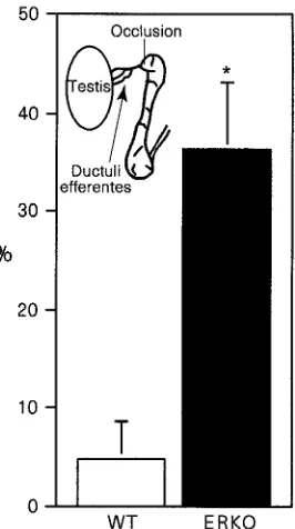
Figure 3. Change in testis mass 48h after occlusion of the initial-segment epididymis. Values shown are mean (+/- s.e.m.) percentage difference between the occluded and sham-operated side. Differences between wild-type (WT) and ERKO means were determined by the unpaired Student's t-test (P < 0.05).
letters to nature
NATURE | VOL 390 | 4 DECEMBER 1997
511
ductules. The contralateral side served as the control. Testes were removed 48 h after surgery and the difference in weight between the occluded and sham-operated side represented the build-up of luminal fluid caused by a lack of reabsorption in the efferent ductules. After ductal occlusion, testes of ERKO weigh 30% more than testes of wild-type mice (Figure 3). However, these data do not remove the possibility that the increase in weight results from an abnormal rate of fluid secretion by the seminiferous epithelium in the testis of ERKO. Therefore, we also unilaterally occluded the rete testis in a group of ERKO and wild-type males and compared differences in testicular weight changes at 24 h. Instead of showing increases in fluid secretion, the testis in ERKO secretes significantly less fluid (P < 0.05) in 24 h than does the wild-type: 22.4 +/- 3.0% increase versus 37.9 +/- 5.3%, respectively. In the ERKO male, efferent ductules do not appear capable of reabsorbing luminal fluids received from the testis. Thus the ERKO mouse provides evidence that oestrogens may be responsible for the regulation of fluid transport, which increases the concentration of sperm before entering the epididymis.
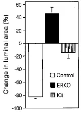
To assess the function of oestrogen in adult tissues without developmental influence, we treated wild-type mice for 3 days with an anti-oestrogen compound and used in vitro methods of analysis to determine fluid reabsorption. ICI 182,780 (ICI Pharmaceuticals) is a pure anti-oestrogen that inhibits increases in uterine weight stimulated by 17 beta-oestradiol and binds both ER-alpha and ER-beta [22,23]. Small segments of adult efferent ductules were isolated for organ culture and the tubular ends were ligated with fine suture, preventing the inflow of culture medium. Using this in vitro model, we compared the function of ductules in wild-type, ERKO and ICI-treated males over a 24-h period. In ductules from wild-type mice, the epithelium is capable of rapidly removing luminal fluid, which results in the collapse of the ductule walls (Figure 4). After 24 h of ligation, the luminal area of ductules from wild-type decreased 82% (Figure 5). In contrast, the luminal area of ductules from ERKO increased 46%, and ductules from wild-type mice treated with ICI showed only a slight decrease of 14%. Thus efferent ductules from ERKO are incapable of reabsorbing luminal fluid, while wild-type ductules remove most of the fluid within 3 h. Blockage of ER function, by treating ductules from wild-type with an anti-oestrogen, significantly inhibits fluid reabsorption.
Thus ER-alpha is important both for normal growth of the male reproductive tract and for adult function of the efferent ductules. However, because wild-type ICI-treated ductules in vitro did not swell like the tissues from ERKO mice, the mechanisms of oestrogen action are not fully understood. For example, it is not known whether the role of ER-beta is the same as that of ER-alpha in efferent ductules. Recent data in HeLa cells have shown that anti-oestrogens and 17 beta-oestradiol produce opposite effects on ER-beta, depending upon the type of response element complex
Figure 5 The effects of in vitro ligation on changes in luminal area of efferent
ductule segments 24h after ligation. Mean (± s.e.m.) percentage difference between 0h and 24 h in luminal areas for isolated ductules from wild-type controls, ERKO and ICI-treated mice.
formed [24]. Thus ER-beta function in ductules of ERKO males could account for the in vitro observations. Regardless of the precise mechanisms, developmental disruption of ER-alpha in mice causes a total loss of net fluid reabsorption. In the human, mutational dysfunction in ER-alpha and P450 aromatase (the enzyme that converts androgens to oestrogens) decreases sperm counts and results in poor sperm viability [25-27]. Our data suggest that oestrogen deficiency or oestrogen insensitivity in man might also result in the accumulation of fluid in efferent ductules and subsequent atrophy of the testis. Tamoxifen, which is commonly known to be a mixed oestrogen agonist/antagonist in different tissues, has been used to increase sperm counts in oligospermic men [28]. In conclusion, our data describe a physiological endpoint of oestrogen action in the male reproductive system. This finding is important given recent concerns over reported declines in human sperm counts and speculation that exposure to environmental oestrogens may be a cause of this [8,9,29]. The extensive presence of ER (both alpha and beta) at other sites in the male reproductive system and throughout the body [10,11,23] makes it possible that new and unexpected functions may be found for the 'female' hormone in
men.
………
Methods
Histology and morphometry. Testis and mid-proximal efferent ductules were fixed and processed for light microscopy [30]. Luminal area, ductal circumference and epithelial height were taken in 5 areas per ductule cross-section in 5 ductules per mouse.
Initial-segment epididymal occlusions and rete testis ligation. Under surgical anaesthesia, initial-segment epididymidis was occluded by cauterization or rete testis was ligated with 0000 suture. The contralateral testis was sham operated. The animals were allowed to recover and testes were taken at either 48 h (initial-segment occlusions) or 24 h (rete ligation) and weighed. For the initial segment occlusions, N = 7 for both wild-type and ERKO. For rete testis occlusions, N = 14 for wild-type mice and N = 12 for ERKO.
In vitro ligation of efferent ductules. One group of wild-type mice was given single daily injections of ICI 182,780 (1 mg per kg) for 3 days before removal of the ductules. Efferent ductules were microdissected into 1.5-mm lengths and incubated for 24 h in M199 culture medium containing
dihydrotestosterone (4 x 10⁻⁷ M), 17 beta-oestradiol (1 x 10⁻⁹ M), bovine
lipoprotein (0.16 mg ml-1) or all components plus ICI 182,780 (1 x 10⁻⁶ M) at
34°C in humidified 95% air/5% CO2. After 24 h, segments were ligated on both
ends to prevent entry and exit of fluids. Digital images of the ductules were analysed at 0, 3 or 12 and 24 h after ligation (Figure 4, Figure 5). Ductal segments damaged by microdissection or stretching were discarded. Only segments with rapid ciliary beat and clear lumens were ligated. N = 3 mice for each treatment group, and 3-12 ductal segments per animal were analysed. Differences between means were determined by a one-way analysis of variance (P < 0.0001) followed by Bonferroni multiple comparisons test (P < 0.001).
Received 29 July; accepted 17 September 1997.
---1. Ganjam, V. K. Amann, R. P. Steroid content of fluids and sperm entering and leaving the bovine epididymis, in epididymal tissue, and in accessory sex gland secretions. Endocrinology 99, 1618-1630 (1976).
2. Free, M. J. & Jaffe, R. A. Collection of rete testis fluid from rats without previous efferent duct ligation. Biol. Reprod. 20, 269-278 (1979).
3. Smith, M. S., Freeman, M. E. & Neill, J. D. The control of progesterone secretion during the estrous cycle and early pseudopregnancy in the rat: prolactin, gonadotropin and steroid levels associated with rescue of the corpus luteum of pseudopregnancy. Endocrinology 96, 219-226 (1975).
4. Cooke, P. S., Young, P., Hess, R. A. & Cunha, G. R. Estrogen receptor expression in developing epididymis, efferent ductules, and other male reproductive organs. Endocrinology 128, 2874-2879 (1991).
letters to nature
NATURE | VOL 390 | 4 DECEMBER 1997
512
6. Schleicher, G., Drews, U., Stumpf, W. E. & Sar, M. Differential distribution of dihydrotestosterone and estradiol binding sites in the epididymis of the mouse. An autoradiographic study. Histochemistry 81, 139-147 (1984).
7. West, N. & Brenner, R. Estrogen receptor in the ductuli efferentes epididymis and testis of rhesus and cynomologus macaques. Biol. Reprod. 42, 533-538 (1990). 8. Sharpe, R. M. & Skakkebaek, N. E. Are oestrogens involved in falling sperm counts and disorders of the male reproductive tract? Lancet 341, 1392-1395 (1993). 9. Auger, J., Kunstmann, J. M., Czyglik, F. & Jouannet, P. Decline in semen quality among fertile men in Paris during the past 20 years. N. Engl. J. Med. 332, 281-285 (1995).
10. Hess, R. A. et al. Estrogen receptor (alpha & beta) expression in the excurrent ducts of the adult male rat reproductive tract. J. Androl. 18, 602-611 (1997). 11. Fisher, J. S. et al. Immunolocalisation of oestrogen receptor-alpha within the testis and excurrent ducts of the rat and marmoset monkey from perinatal life to adulthood. J. Endocrinol. 153, 485-495 (1997).
12. Ilio, K. & Hess, R. Structure and function of the ductuli efferentes: A review. Microsc. Res. Tech. 29, 432-467 (1994).
13. Yeung, C. H., Cooper, T. G., Bergmann, M. & Schulze, H. Organization of tubules in the human caput epididymidis and the ultrastructure of their epithelia. Am. J. Anat. 191, 261-279 (1991).
14. Robaire, B. & Hermo, L. in The Physiology of Reproduction (eds Knobil, E. & Neill, J.) 999-1080 (Raven, New York, 1988).
15. Chan, H. C., Zhou, W. L., Fu, W. O., Ko, W. H. & Wong, P. Y. Different regulatory pathways involved in ATP-stimulated chloride secretion in rat epididymal epithelium. J. Cell. Physiol. 164, 271-276 (1995).
16. Clulow, J., Jones, R. & Hansen, L. Micropuncture and cannulation studies of fluid composition and transport in the ductuli efferentes testis of the rat: comparisons with the homologous metanephric proximal tubule. Exp. Physiol. 79, 915-928 (1994).
17. Lubahn, D. B. et al. Alteration of reproductive function but not prenatal sexual development after insertional disruption of the mouse estrogen receptor gene. Proc. Natl Acad. Sci. USA 90, 11162-11166 (1993).
18. Korach, K. S. et al. Estrogen receptor gene disruption: molecular characterization and experimental and clinical phenotypes. Recent Prog. Horm. Res. 51, 159-186 (1996).
19. Eddy, E. M. et al. Targeted disruption of the estrogen receptor gene in male mice causes alteration of spermatogenesis and infertility. Endocrinology 137, 4796-4805 (1996).
20. Smith, G. The effects of ligation and the vasa efferentia and vasectomy on testicular function in the adult rat. Endocrinology 23, 385-399 (1962).
21. Hess, R., Moore, B., Forrer, J., Linder, R. & Abuel-Atta, A. The fungicide benomyl [methyl 1-butylcarbamoyl-2-benzimidazole carbamate] causes testicular dysfunction by inducing the sloughing of germ cells and occlusion of efferent ductules. Fundam. Appl. Toxicol. 17, 733-745 (1991).
22. Wakeling, A., Dukes, M. & Bowler, J. A potent specific pure antiestrogen with clinical potential. Cancer Res. 51, 3867-3873 (1991).
23. Kuiper, G. G. et al. Comparison of the ligand binding specificity and transcript tissue distribution of estrogen receptors alpha and beta. Endocrinology 138, 863-870 (1997).
24. Paech, K. et al. Differential ligand activation of estrogen receptors ER alpha and ER beta at AP1 sites. Science 277, 1508-1510 (1997).
25. Carani, C. et al. Effect of testosterone and estradiol in a man with aromatase deficiency. N. Engl. J. Med. 337, 91-95 (1997).
26. Morishima, A., Grumbach, M. M., Simpson, E. R., Fisher, C. & Qin, K. Aromatase deficiency in male and female siblings caused by a novel mutation and the physiological role of estrogens. J. Clin. Endocrinol. Metab. 80, 3689-3698 (1995).
27. Smith, E. P. et al. Estrogen resistance caused by a mutation in the estrogen-receptor gene in a man. N. Engl. J. Med. 331, 1056-1061 (1994). Erratum, N. Engl. J. Med. 332, 131 (1995).
28. Krause, W., Holland-Moritz, H. & Schramm, P. Treatment of idiopathic oligozoospermia with tamoxifen-a randomized controlled study. Int. J. Androl. 15, 14-18 (1992).
29. Sharpe, R. M. Declining sperm counts in men-is there an endocrine cause? J. Endocrinol. 136, 357-360 (1993).
30. Hess, R. A. & Moore, B. J. in Methods in Reproductive Toxicology (eds Chapin, R. E. & Heindel, J. J.) 52-85 (Academic, San Diego, 1993).
Acknowledgements. We thank E. Jassim and C. Finnigan-Bunick for technical assistance, P. Cooke, V. K. Ganjam and D. J. Miller for reviews of the manuscript and A. Wakeling and Zeneca Pharmaceuticals for providing ICI 182, 780.