Development of Candidate Antigens for Rapid Test Kit to Detect
Autoantibodies in Patients with Systemic Lupus Erythematosus
Wisnu Barlianto1, Hani Susianti2, Singgih Wahono3, Nelly Ismayasih2,
Rossy Meilani2, Kusworini Handono2
1Department of Pediatrics, Faculty of Medicine Brawijaya University/Dr. Saiful Anwar General Hospital,
Malang, Indonesia
2Department of Clinical Pathology, Faculty of Medicine Brawijaya University/Dr. Saiful Anwar General
Hospital, Malang, Indonesia
3Department of Internal Medicine, Faculty of Medicine Brawijaya University/Dr. Saiful Anwar General
Hospital, Malang, Indonesia Email: [email protected]
ABSTRACT
Systemic Lupus Erythematosus (LES) is an autoimmune inflammatory disease characterized by the formation of anti-nuclear antibodies (ANA) and anti-double stranded DNA (anti-dsDNA) antibodies as diagnostic markers. Detection of such autoantibodies requires advanced equipment and trained personnel.
This study was conducted to acquire candidate antigens that can be used for rapid test kit for practical and accurate detection of ANA and anti-dsDNA to speed up SLE diagnosis.
Nuclear proteins and DNA derived from cell lines, hair follicles, and leukocytes of SLE patients and healthy individuals were isolated using QiaGEN kit and modified-manual procedure. Antigen-antibody bonds were tested by dot blot assay.
The strongest binding between DNA antigens of a healthy individual and antibodies occurred at dilution factors of 1:5,120 for the antigen and 1:2,560 for the antibody. The strongest binding between nuclear protein antigens from the cell line and antibodies occurred at dilution factors of 1:512 for the antigen and 1:1,600 for the antibody.
Nuclear antigens derived from cell line and DNA antigens of healthy individuals were antigen candidates for the
development of ANA and anti-dsDNA rapid detection tests.
Keywords: ANA; anti-dsDNA; diagnostic test; rapid test; SLE.
INTRODUCTION
Systemic lupus erythematosus (SLE) is an autoimmune inflammatory disease with diverse clinical symptoms and high mortality rates (Zhu & Mohan, 2007). Lately, SLE is more frequently encountered, with women make up 80% of the patients. This disease is thought to be the fourth leading cause of disability in women. Most patients are between adolescence and fourth decade of life, which is the productive age so the impact becomes more apparent (Mok & Lau, 2003). The pathogenesis is still not clearly understood, but immune system dysregulation that causes the secretion of pathogenic autoantibodies triggering tissue damage has been reported by expert (Lauwerys & Houssiau, 2003). Antinuclear antibody (ANA) and anti-double stranded DNA (anti-dsDNA) are commonly present in SLE and considered as the laboratory markers of the disease (Petri et al, 2012; Krishnamurthy & Mahadevan, 2011).
are highly sensitive and specific for SLE, found in 40-90% of patients with active disease (Pagana & Pagana, 2013). At present, commercial kits manufactured by foreign companies for the detection of ANA and anti-dsDNA are available in Indonesia. However, expensive price, the need for a trained and experienced personnel and a laboratory with full facilities such as that on the tertiary or referral hospital, limit the use of these kits. Such examinations are difficult to be done in primary and secondary facilities such as in the laboratory of the public healthcare center, necessitating an effort to develop a SLE diagnostic kit that can easily be applied, particularly at the primary healthcare center. One application of the diagnostic tools that is quick and easy to perform is a rapid test kit method, such as that applied to the pregnancy test or malaria immunochromatographic test (ICT). Therefore, early diagnosis and management can be established, which will improve the life expectancy of SLE patients.
Based on above descriptions, the purpose of this study is to acquire nuclear and DNA antigens as candidates to develop a new easy-to-use diagnostic kit in strip format, based on the principle of ANA and anti-dsDNA autoantibodies detection, which would allow an earlier diagnosis of SLE in Indonesia.
MATERIALS AND METHODS
This study was conducted from January 2016 to October 2016 in three locations: The Biomedical Central Laboratory-Medical Faculty of Brawijaya University, the Central Laboratory of Dr. Saiful Anwar General Hospital Malang, and the Bioscience Laboratory of Brawijaya University.
Subjects and study design.
This study was a laboratory experimental research. Isolation and purification of nuclear proteins and DNA
were performed on various samples: cell lines, hair follicles, and leukocytes of SLE patients/healthy individuals/donated blood obtained from the Indonesian Red Cross Society. Acquired antigens were tested against antibodies derived from sera of ANA-positive and anti-dsDNA-positive SLE patients. Antigen immunogenicity obtained from various samples were then compared and set as candidate antigens for the rapid test.
Samples.
HeLa cells, hair follicles, and leukocytes of SLE patients and healthy individuals were used as samples in this study. Patients with SLE who became the subjects of this study were those who visited the Internal Medicine Polyclinic at the Dr. Saiful Anwar General Hospital Malang. The 2012 SLICC criteria for SLE were used to establish the diagnosis and all of the patients were tested for ANA and anti-dsDNA. Healthy individuals were those without SLE and history of chronic inflammatory disease and were illness-free during the sample collection period. Freshly donated blood obtained from donors at the Malang branch of the Indonesian Red Cross Society were also used as the source of samples.
Isolation of Nuclear Proteins.
mL ice cold PBS and resuspended. After another centrifugation at 6000 rpm for 5 minutes in cold room, all supernatants were removed. The tubes were put on ice and 5X more CE buffer was added to the pellets (i.e., if the pellet is 20 µL, add 100 µL of CE buffer). The pellets were resuspended in the CE buffer and incubated for 5 minutes on ice. The tubes were then vortexed from time to time and centrifuged at 3000 rpm for 5 minutes. After the supernatant (the cytoplasmic extract. Note that the pellet is not dense) was harvested, the pellets were resuspended in 1 µL CE buffer without NP-40 and centrifuged again at 3000 rpm for 5 minutes in cold room. The supernatant was removed after the centrifugation and an equal volume of NE buffer was added to the pellets (i.e., if the pellet is 40 µL, add 40 µL of NE buffer). The pellets were resuspended and incubated for 10 minutes on ice and vortexed from time to time. After another centrifugation at 14,000 rpm for 5 minutes at 4oC, the supernatant (the nuclear extract) was harvested. The extracts can be stored at -80oC.
DNA extraction from cell culture (QiaGen). Hela cell culture: A certain amount of cell (max. 5 x10⁶ cells) was centrifuged at 300 g for 5 minutes. The pellets were then resuspended in 20 µL proteinase K. Optional: if DNA with free-RNA genomics were needed, 4 µL of Rnase A (100 mg/ml) can be added, mixed with vortex, and incubated for 2 minutes in room temperature. 200 µL of buffer AL was added (without ethanol addition) and mix thoroughly by vortexing, then incubated for 10 minutes at 56oC. 200 µL of ethanol (96 – 100%) was added to the sample and was mixed thoroughly by vortexing. The mixture was pipetted into the DNeasy Mini spin column and placed in a 2 mL collection tube. After centrifugation at 6000 g (8000 rpm) for 1 minute, the
flow-through and the collection tube were discarded. The DNeasy Mini spin column was placed in a new 2 mL collection tube, 500 µL Buffer AW1 was added and centrifuged at 6000 g (8000 rpm) for 1 minute. Flow-through and collection tube were discarded. The DNeasy Mini spin column was placed in a new 2 mL collection tube, 500 µL Buffer AW2 was added and centrifuged at 20,000 g (14,000 rpm) for 3 minutes to dry the DNeasy membrane. Flow-through and collection tube were discarded. The DNeasy Mini spin column was placed in a clean 2 mL collection tube and 100 µL Buffer AE was pipetted directly onto the DNeasy membrane. After incubation for 1 minute at room temperature and centrifugation at 6000 g (8000 rpm) for 1 minute, the final were inserted into a 1.5 mL collection tube. 100 µL of K-lysed buffer was added to the tube and incubated for at least 1 hour at 55ºC, then vortexed and centrifuged at the end of the incubation period. The reaction was stopped by heating the tube at 70°C for 10 minutes. After centrifugation at 12,000 rpm for 10 minutes at 4°C, the supernatant was removed. The extract can be stored at -20°C.
Wizard® Genomic DNA Purification Kit (whole blood & cell culture).
are then removed by a salt precipitation step, which precipitates the proteins but leaves the high molecular weight genomic DNA in solution. Finally, the genomic DNA is concentrated and desalted by isopropanol precipitation.
Procedure: 300 μL blood collected in EDTA, heparin or citrate anticoagulant tubes was used as the sample. 900 μL Cell Lysis Solution was added in a sterile 1.5 mL microcentrifuge tube. The tube containing blood was gently rocked until thoroughly mixed. The blood was then transferred to the tube containing the Cell Lysis Solution and the tube was inverted 5 to 6 times. The mixture was incubated for 10 minutes at room temperature. After centrifugation at 13,000 to 16,000 g for 20 seconds, the supernatant was removed. 10 to 20 µL of residual liquid that remain in the tube was vortexed vigorously for 10 to 15 seconds. 300 μL Nuclei Lysis Solution was added to the tube containing resuspended cells. The solution was then pipetted 5 to 6 times to lyse the leukocytes. 1.5 µL RNase Solution was added to the nuclear lysate and the sample was mixed by inverting the tube 2 to 5 times. The mixture was incubated for 15 minutes at 37°C and then cooled to room temperature. 100 μL Protein Precipitation Solution was added to the nuclear lysate and vortexed vigorously for 10–20 seconds. After centrifugation at 13,000 to 16,000 g for 3 minutes at room temperature, the supernatant was transferred into a clean 1.5 mL microcentrifuge tube containing 300 μl of room temperature isopropanol. The solution was mixed by inversion until the white thread-like strands of DNA visible. The mixture was centrifuged at 13,000 to 16,000 g for 1 minute at room temperature. DNA will be visible as a small white pellet. The supernatant was decanted and 1 sample volume of 70% ethanol was added to the
DNA. The tube was inverted several times to wash the DNA pellet and the sides of the microcentrifuge tube before centrifuged again at 13,000 to 16,000 g for 1 minute at room temperature. The ethanol was carefully aspirated using a drawn Pasteur pipette. The tube was inverted on a clean absorbent paper and the pellet was allowed to dry for 10 to 15 minutes. 100 μL of DNA Rehydration Solution was added to the tube and the DNA was rehydrated by incubating the tube at 65°C for 1 hour. Alternatively, the DNA can be rehydrated by incubating the solution overnight at room temperatur or 4°C. The DNA can be stored at 2 to 8°C.
Binding test between nuclear proteins and DNA with the serum of SLE patient (Dot blot Assay).
Dot blot assay is a simple immunodetection method to identify specific proteins in sample. If a protein has been immobilized on the protein-binding membrane such as PVDF (polyvinylidene fluoride) or nitrocellulose, it can be reacted with specific antibody directed against the protein. After binding between protein and primary antibody (autoantibody derived from the patient serum) has been formed, the binding between primary antibody and labeled secondary antibody will follow. This will be visualized by changes in the label color or substrate which level can be measured with a densitometer.
μl TBS skim milk was added to each well. The plate was covered with aluminium foil and incubated for 2 hours. Each well was washed three times for 3 minutes each with 50 μl TBS tween. After the plate was dried using absorbent paper, 50 μL of primary antibody was added. The plate was then covered with aluminium foil and incubated overnight at 4ºC. Each well was again washed three times for 3 minutes each with 50 μl TBS tween. 50 µL of secondary antibody was added to each well, covered with aluminium foil, and incubated for 1 hour at room temperature. Each well was again washed three times for 3 minutes each with 50 μl TBS tween. 50 μl substrate was added to each well in a dark room and incubated for 20 minutes. Color changes were then observed and measured using densitometer.
Antigen immunogenicity test using dipstick. 3 µL of antigen was dropped on the dipstick tip. The dipstick was incubated for 1 hour at 370C and fixed by dipping in absolute methanol, then dried for 1 hour. The dipstick was soaked in 10% sucrose solution for 10 minutes, and then dried. The dipstick was placed into a serum-containing well for 30 minutes, and then transferred into a protein A solution containing human Ig for 15 minutes. The dipstick was read immediately.
RESULTS AND DISCUSSION
Role of autoantibodies in SLE diagnosis. SLE is a chronic and complex autoimmune disease. It is often called “the great imitator” or disease with thousand faces, because the clinical symptoms are very broad and difficult to recognize (Zhu & Mohan, 2007). The characteristic of SLE is the production of autoantibodies directed against the core antigens, such as ds-DNA and chromatin, resulting in immune complex-mediated organ damage [Zhu & Mohan, 2007; Lauwerys & Houssiau, 2003).
The diagnosis of most autoimmune diseases including SLE is complicated by the absence of specific signs and symptoms and the lack of diagnostic criteria and validated standard biomarkers for identifying early disease (Wallace, 2007). Delay in diagnosis and treatment can lead to increased morbidity and healthcare costs.
Various autoantibodies can be found in a patient with SLE, but at the early stage, the most important autoantibody is ANA, an antibody that reacts against nuclear, nucleoli or perinuclear antigens such as nucleic acids, histones, chromatins, and ribonuclear proteins (Castro & Gourley, 2010). ANA titer has a good validity for diagnosis and is included in the criteria to diagnose SLE (Kavanaugh et al, 2000). Another specific autoantibody is anti-dsDNA autoantibody, an important marker used in the diagnosis and evaluation of SLE. This antibody is found in 40% - 90% of SLE patients with active disease (Pagana & Pagana, 2013).
Development of SLE diagnostic kit.
easy-to-use, inexpensive, practical diagnostic test that can be applied at all primary healthcare centers.
Several foreign companies have successfully developed diagnostic kits for other autoimmune diseases such as rheumatoid arthritis (RA) that is quick and easy to perform. Some of these kits use rheumatoid factor (RF) and cyclic citrullinated peptide (CCP) which will bind to the autoantibodies produced by RA patients. These kits have been proved to have quite high sensitivity and specificity (De Rycke et al, 2004). Meanwhile, rapid diagnostic kit for SLE itself has not been widely developed due to various types of autoantibodies that can be found in SLE patients which make it difficult to determine the specific antibody for diagnosis. There is no data on the dominant type of autoantibodies produced by SLE patients in Indonesia because autoantibody profiling has never been done in this country. The most frequently reported autoantibodies in Indonesian patients with SLE are ANA and anti-dsDNA (Kalim et al, 2012).
SLE diagnostic kit has been produced in 2001 using a suspension of polystyrene latex particles coated with deoxyribonucleoprotein (DNP). Autoantibodies produced by SLE patient will bind to the latex material which can be evaluated using agglutination reaction. However, this anti-DNP kit is not specific to SLE only. Positive results can also be found in RA, scleroderma, chronic discoid lupus, polyarteritis nodosa, and dermatomyositis. Therefore, identification of a specific antigen in SLE patients is important to determine the dominant type of antigens that can be developed into a SLE diagnostic kit.
Utilization of the diagnostic kit to be developed.
There are a lot of signs and complaints expressed by the patients that should be
able to raise the physician’s suspicion of the presence of SLE. These include reproductive-age women that come with fever of unknown cause, hair loss, skin rash, joint pain, oral ulcer, etc (Petri et al, 2012). Such changes will prompt the patient to seek treatment from doctors at the public healthcare center or general physicians, in accordance with the national healthcare program. The diagnostic kit that will be developed will be utilized in reproductive-age women suspected of having SLE. General physicians and doctors in public healthcare centers who find at least 2 signs or symptoms in reproductive-age women will be asked to be alert to the possibility of SLE and to perform autoantibody test using the kit that will be developed and other appropriate laboratory tests. Establishing SLE diagnosis in women with above manifestations will greatly help them to achieve successful treatment, increase life expectancy, and improve functional qualities. The diagnostic kit that will be developed is also important to diagnose SLE in several autoimmune diseases that can mimic the disease or to establish a differential diagnosis.
immunogenicity test by mixing the antigens with the serum of SLE patients containing high levels of ANA and anti-dsDNA.
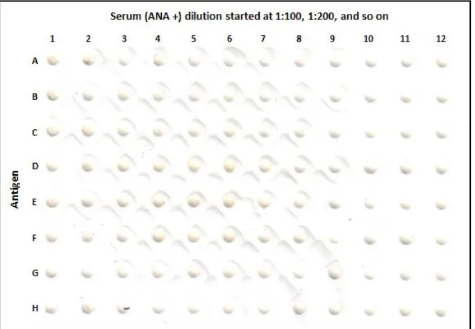
Checkerboard test results of nuclear protein antigens derived from leukocytes and antibodies obtained from the serum of a SLE patient with high ANA levels.
In this checkerboard test, samples of nuclear protein antigens were isolated from
a SLE patient and primary antibodies were also obtained from the serum of a SLE patient with high ANA levels. Antigen dilutions started at 1:16, 1:32, 1:64, and so on, whereas antibody dilutions started at 1:100, 1:200, 1:400, and so on (Figure 1). No color changes were observed after the addition of substrate, suggesting that the isolated nuclear protein antigens can not bind the antibodies (ANA) of SLE patients.
Figure 1. Checkerboard test results of nuclear protein antigens derived from leukocytes and antibodies obtained from the serum of a SLE patient with high ANA levels.
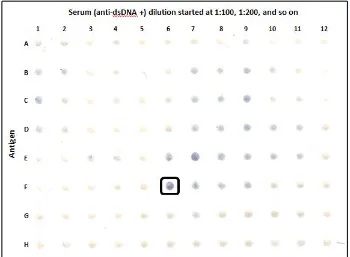
Checkerboard test results of DNA antigens derived from leukocytes and antibodies obtained from the serum of a SLE patient with high anti-dsDNA levels.
In this checkerboard test, samples of DNA antigens were obtained from the leukocytes of a SLE patient and primary antibodies were also obtained from the serum of a SLE patient with high anti ds-DNA levels. Antigen dilutions started at 1:20, 1:40, 1:80, and so on, whereas antibody dilutions started at 1:100, 1:200, 1:400, and so on. The most prominent color change after the substrate addition was observed on position F6 (antigen dilution 1:640 and
Figure 2. Checkerboard test results of DNA derived from leukocytes (as antigens) and serum (as antibodies) of SLE patients.
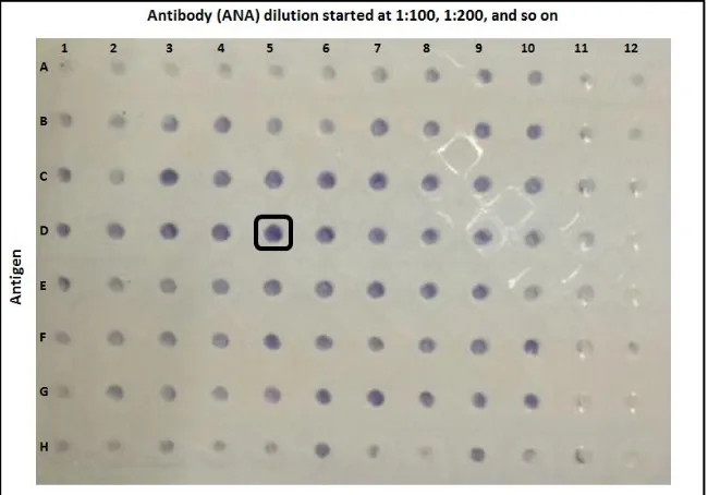
Checkerboard test results of DNA derived from leukocytes of a healthy individual and antibodies obtained from the serum of a SLE patient with high anti-dsDNA levels.
In this checkerboard test, samples of DNA antigens were obtained from the leukocytes of a healthy individual using method that was introduced by Kotsimbos et al (DNA extraction method performed on the hair follicles). The primary antibodies were obtained from the serum of a SLE patient with high anti ds-DNA levels. Antigen dilutions started at 1:20, 1:40, 1:80, and so on, whereas antibody dilutions started at 1:20, 1:40, and so on. The most prominent color change after the substrate addition was observed on position G8 (antigen dilution 1:5120 and antibody dilution 1:2560) (Figure 4). Based on this results, it can be concluded that the DNA antigens isolated from the leukocytes of healthy individuals can recognize anti-dsDNA.
Figure 4. Checkerboard test results of DNA derived from leukocytes of a healthy individual and antibodies obtained from the serum of a SLE patient with high anti-dsDNA levels.
Checkerboard test results of nuclear antigens derived from cell lines and antibodies obtained from the serum of a SLE patient with high ANA levels.
In this checkerboard test, samples of antigens were obtained from the culture of HeLa cell line, whereas the primary antibodies were obtained from the serum of a SLE patient with high ANA levels. Antigen dilutions started at 1:64, 1:128, 1:256, and so on and antibody dilutions started at 1:100, 1:200, 1:400, and so on. The most prominent color change after the substrate addition was observed on position D5 (antigen dilution 1:512 and antibody dilution 1:1600) (Figure 5). Based on this results, it can be concluded that the DNA antigens isolated from the leukocytes of healthy individuals can recognize anti-dsDNA.
The limitation of this study is that the binding between the nuclear proteins and DNA and serum of SLE patients does not produce noticeable differences in color on all strips. This is likely caused by the low specificity of the isolated antigens (poor antigen purity) or the antigens are not attached perfectly to the strip, so that they cannot bind the antibody proteins from the patients’ serum.
CONCLUSION
The checkerboard test results of nuclear protein and DNA antigens derived from cell line and healthy controls (with several extraction methods) show good antigen-antibody binding. Therefore, it can be used as prototypes for ANA and anti-dsDNA rapid tests which will be helpful for initial screening of patients suspected of having SLE in primary healthcare services.
ACKNOWLEDGEMENT
The authors would like to acknowledge the financial support from the Ministry of Health of the Republic of Indonesia.
REFERENCES
Castro, C., & Gourley, M. (2010). Diagnostic testing and interpretation of tests for autoimmunity. Journal of Allergy and Clinical Immunology, 125 (2), S238-S247.
De Rycke, L., Peene, I., Hoffman, I., Kruithof, E., Union, A., Meheus, L., Lebeer, K., Wyns, B., Vincent, C., Mielants, H., Boullart, L., Serre, G., Veys, E. & De Keyser, F. (2004). Rheumatoid factor and anticitrullinated protein antibodies in rheumatoid arthritis: diagnostic value, associations with radiological progression rate, and extra-articular manifestations. Annals
of Rheumatic Diseases, 63 (12): 1587-1593.
Hsieh, S. C. & Yu, C. L. (2013). Autoantibody profiling in systemic lupus erythematosus. Current Biomarker Findings, 3: 55-65.
Kalim, H., Wahono, S., Suryana, B. P. P., Puspitasari, L., Wijayanto, F. H., & Handono, K. (2012). Association between serum level of Vitamin D with autoantibodies expression, disease activity (SLEDAI) and bone mineral density (BMD) in patients with Systemic Lupus Erythematosus (SLE).
Arthritis Research and Therapy, 14 (suppl 1): P23.
Kavanaugh, A., Tomar, R., Reveille, J., Solomon, D. H., & Homburger, H. A. (2000). Guidelines for clinical use of the antinuclear antibody test and tests for specific autoantibodies to nuclear antigens. Archives of pathology & laboratory medicine, 124 (1), 71-81. Krishnamurthy, S., & Mahadevan, S. (2011).
Systemic lupus erythematosus: recent concepts in genomics, pathogenetic mechanisms, and therapies. ISRN Immunology, 2011.
Lauwerys, B. R., & Houssiau, F. A. (2003). Involvement of cytokines in the pathogenesis of systemic lupus erythematosus. Cytokines and Chemokines in Autoimmune Disease,
237-251.
Mok, C. C., & Lau, C. S. (2003). Pathogenesis of systemic lupus erythematosus.
Journal of Clinical Pathology, 56: 481-90.
Pagana, K. D., & Pagana, T. J. (2013). Mosby’s Manual of Diagnostic and Laboratory Tests (3rd ed.). Philadelphia: Mosby Elsevier.
... & Sturfelt, G. (2012). Derivation and validation of the Systemic Lupus International Collaborating Clinics classification criteria for systemic lupus erythematosus. Arthritis & Rheumatism, 64(8), 2677-2686 Wallace, D. J. (2007). The Clinical
Presentation of Systemic Lupus Erythematosus. In: D. J. Wallace, & B.
H. Hahn (Eds.), Dubois’ Lupus
Erythematosus (7th ed.) (pp. 638-646). Philadelphia: Lippincott Williams & Wilkins.
Zhu, J., & Mohan, C. (2007). SLE 1,2,3… genetic dissection of lupus. Advances in Experimental Medicine and Biology,