Limited evidence for white spot syndrome virus susceptibility associated
with expression of
Pm
VRP15 in local population of giant tiger shrimp
(
Penaeus monodon
)
Aushia Tanzih Al Haq
1*, Murwantoko
2, Trijoko
1, Nastiti Wijayanti
1,
Ch. Retna Handayani
3, Rarastoeti Pratiwi
11Department of Biology, Faculty of Biology, Universitas Gadjah Mada, Yogyakarta, Indonesia 2Department of Fisheries, Faculty of Agriculture, Universitas Gadjah Mada, Yogyakarta, Indonesia
3Main Center for Brackishwater Aquaculture and Fisheries, Jepara, Indonesia
Abstract
White spot syndrome virus (WSSV) is a devastating viral disease in shrimp aquaculture. Infection of WSSV in penaeid shrimps affects immune defense and changes gene expression. PmVRP15 has been reported as a part of the WSSV propagation pathway that is highly up-regulated in hemocytes at the acute phase of WSSV infection. This study analyzed the expression of PmVRP15 in local populations of giant tiger shrimp (Penaeus monodon) to be associated with susceptibility to WSSV. Tested populations consisted of an inbreeding population (G8) and outbreeding population (G8iA) from Jepara, Indonesia. Susceptibility was determined by cumulative mortality, median lethal time (LT50), and severity of infection at time of death. Though all populations were susceptible to WSSV, the irst mortality in G8 occurred at 18 hours post-infection (hpi) with mild infection,
while irst mortality of G8iA occurred at 30 hpi with severe infection. The LT50 of G8 was signiicantly lower than that of G8iA, indicating that G8iA was less susceptible to WSSV than G8. Relative PmVRP15 transcripts of G8iA were insigniicantly down-regulated, whereas relative PmVRP15 transcripts of G8 were insigniicantly up-regulated. Although it’s still not conclusive, the results of this study suggest that PmVRP15 has weak potential as a WSSV susceptibility marker in G8 and G8iA broodstock selection.
Keywords: Disease susceptibility, genetic marker, Penaeus monodon, PmVRP15, WSSV susceptibility
Introduction
White spot syndrome virus (WSSV) is one of the pathogens causing high mortality in giant tiger shrimp (Penaeus monodon) aquaculture. WSSV has been reported to cause harvest failure with a 100% morbidity and mortality level in three to ten days (Jeswin et al., 2013; Reddy et al., 2013). This virus is an enveloped, double-stranded DNA virus that is classiied into genus Whispovirus and family Nimaviridae (Tonganunt et al.,
2009). White spot disease can be detected by the appearance of white lesions inside the carapace due to abnormal calcium deposition in the cuticle (Alifuddin et al., 2003; Reddy et al., 2013). Either natural or experimental WSSV susceptibility in penaeid shrimps has been reported in many studies (Alifuddin et al., 2003; Corteel et al., 2012; Escobedo-Bonilla et al., 2006; Hayes et al., 2010; Lo et al., 1996; Lo et al., 1997; Mathew et al., 2007).
Hemocytes play an essential role in the immune system of crustaceans through their molecules which are able to recognize the structure of invading organisms and trigger the removal mechanism (Johansson et al., 2000). A Penaeus monodon viral responsive protein, PmVRP15, has been found as a gene that is responsive to the infection of WSSV in
*Corresponding author:
Aushia Tanzih Al Haq
hemocytes by mediating WSSV propagation at the acute phase (Vatanavicharn et al., 2014). Vatanavicharn et al. (2014) also conducted RNA interference to alter PmVRP15 expression and elucidated that PmVRP15 was associated with WSSV susceptibility in a P. monodon population in Thailand. This signifies the importance of determining the change in expression of PmVRP15 in association with susceptibility to WSSV infection. At the commercial level, change in gene expression, which equates to the effect on itness, helps to predict and decide the potentially best population or family of P. monodon to be used as the broodstock in the next selective breeding program.
I n 2 0 0 5 , t h e M a i n C e n t e r f o r Brackishwater Aquaculture and Fisheries (MCBAF) Jepara, Central Java, Indonesia, started a selective breeding program to improve the growth rate and survival of P. monodon. Regarding the outbreak in 1990, producing a WSSV-tolerant P. monodon population was one of the focuses of the MCBAF selective breeding program. However, this existing breeding program does not consider the use of genetic markers to select broodstocks with desired traits. While this study was being conducted, the selective breeding program in MCBAF Jepara reached an eighth generation of an inbreeding (domesticated) population called G8 and produced an outbreeding population called G8iA. The aims of this study were to determine whether the populations had the potential to adapt to a WSSV infection and to evaluate whether the expression of PmVRP15 could provide a genetic basis to mark WSSV susceptibility in the G8 and G8iA populations.
Materials and methods
Maintenance of experimental shrimp
Penaeus monodon used in the experiment consisted of 1) an eighth generation of inbreeding (domesticated) population called G8, and 2) an outbreeding population called G8iA. G8iA was the offspring from introgression of wild type males of an Aceh
population to the females of the seventh generation of the inbreeding population (G7). These shrimps were transferred from NSBC (National Shrimp Broodstock Center) ponds to the Laboratory of Health Management of Aquatic Organism, MCBAF Jepara, Central Java, Indonesia. The mean body weight (MBW) of the experimental P. monodon was 20–25 g. Shrimps were fed twice a day with commercial pelleted shrimp feed at a rate of 3% MBW. They were allowed to acclimatize for a week in iberglass tanks and randomly chosen for WSSV detection using one-step PCR to conirm the absence of WSSV. The WSSV-free P. monodon were then randomly maintained in 100-l plastic containers illed with disinfected seawater. Each container was fitted with air-stone aeration and natural photoperiod. Water exchange (50%) was performed daily throughout the trial. Salinity ranged between 20 and 30%, water temperature ranged between 26.3 and 28.5°C, dissolved oxygen ranged between 5 and 7.17 g ml-1, and water pH ranged from 7.34 to 8.
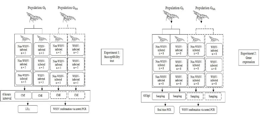
This study consisted of two experiments. The first experiment aimed to determine cumulative mortality in order to examine susceptibility of the population to WSSV exposure, while the second experiment aimed to quantify PmVRP15 expression. Each experiment was conducted in both G8 and G8iA populations. Each population was divided into two groups, a non-infected group (control group) and WSSV-infected group. All experiments were conducted in triplicates. A total of 60 shrimps were used in the irst experiment (susceptibility test), while in the second experiment (gene expression assay) 96 shrimps were used (Fig. 1). Each population was maintained in different containers.
Preparation of viral inoculums
4°C. The supernatant was iltered through a 0.45 μm membrane ilter. The extract was a 10-1 virus extract. This viral inoculum
was prepared according to Hameed et al. (2006). The median lethal dose (LD50) was determined based on the method described in Gopikrishna et al. (2012). The diluted 10-2
virus extract was subsequently used for a challenge test in G8 and G8iA.
Challenge experiment and sampling
Two independent challenge tests were conducted in triplicates to determine the cumulative mortality and to quantify gene expression in sampled hemocytes. Treated shrimps were injected intramuscularly at a rate of 0.02 ml of WSSV extract g-1 of body
weight into the dorsolateral part of the fourth abdominal segment of the shrimp between the tergal plates into the muscle of the third abdominal segment using a 1 ml syringe with a 20 G 0.9 x 25 mm needle (Escobedo-Bonilla et al., 2006). Control shrimps were injected with PBS 1x at the same rate. Mortality was recorded at 6 hour intervals.
Dead shrimps, which showed no physical response to mechanical stimulation, were removed and the tissues from gills and/ or pleopods were preserved in 95% alcohol for WSSV detection. Cumulative mortality was used to estimate the median lethal time (LT50). For the expression proile, random sampling of hemocytes was conducted at 48 hpi by collecting 0.2 ml of hemolymph from the ventral sinus in the second abdominal segment using a 1 ml syringe provided with a 20 G 0.9 x 25 mm needle previously illed with 0.2 ml of 10% tri-sodium citrate (Andrade, 2011; Peraza-Gomez et al., 2014). Hemolymph was then poured into a microtube and kept on ice. The hemolymph (100 µl) sample was immediately centrifuged at 5,000 x g for 5 minutes at 4°C to separate the hemocyte (pellet) from the hemolymph (Vatanavicharn et al., 2014). The supernatant was discarded whereas the pellet (the hemocyte) was used in RNA isolation. The remaining 100 µl of the hemolymph sample was stored at -20°C to be used in WSSV detection.
Fig. 1. Experimental design. The irst experiment aimed to examine susceptibility within populations (left), whereas
RNA extraction and cDNA synthesis
Total RNA was extracted from isolated hemocytes with TRIzol reagent (Invitrogen, USA), quantified using a NanoDrop 1000 spectrophotometer (Thermo Fisher Scientific Inc., USA), and treated with RNase free DNase I (Invitrogen, USA) to remove the contaminating DNA. Thermal cycling of cDNA synthesis was performed at 75°C for 5 minutes, chilled on ice for 5 minutes, 60°C for 15 minutes, and 85°C for 5 minutes with 1–160 ng RNA, 1 μM cocktails of antisense gene-speciic primers, 1 μM dNTP, 0.2 U/μl RNasin (Promega, USA), 5 mM MgCl2 (Promega, USA), 0.05 U/μl AMV RT-ase, and 5 μl of 5x AMV RT-buffer (Promega, USA).
Real-time PCR
The PmVRP15 transcript levels from isolated RNA were assayed with 10 μl of 1x KAPA SYBR Fast qPCR Master Mix (Kapa Biosystems, USA), 0.1 μM of PmVRP15-RTF/R and β-actin-F/R primers, and 10 x diluted cDNA in 7500 Fast Real-time PCR System (Applied Biosystems, USA). The sequences of primer set for β-actin (5’ GAACCTCTCGTTGCCGATGGTG 3’ [forward] and 5’ GAAGCTGTGCTACGTG GCTCTG 3’ [reverse]) and PmVRP15 (5’ CGTCCTTCAGTGCGCTTCCATA 3’ [forward] and 5’ ACAGCGACTCCAAGGTCT ACGA 3’ [reverse]) used in this study were based on those described in Vatanavicharn et al. (2014). Thermal cycling was performed at 95°C for 3 minutes, 40 cycles of 95°C for 30 seconds, 60°C for 30 seconds, and 72°C for 45 seconds. A melt curve stage was performed at 95°C for 15 seconds, 60°C for a minute, and 95°C for 30 seconds, and 60°C for 15 seconds. The results were presented as average relative expression ratio of PmVRP15 transcript levels of the WSSV-infected shrimps compared with the non-infected shrimps after normalization to the reference gene β-actin. The relative expression ratios were calculated as 2-∆CT (Schmittgen and Livak, 2008).
Rapid DNA extraction and conirmation of
WSSV infection
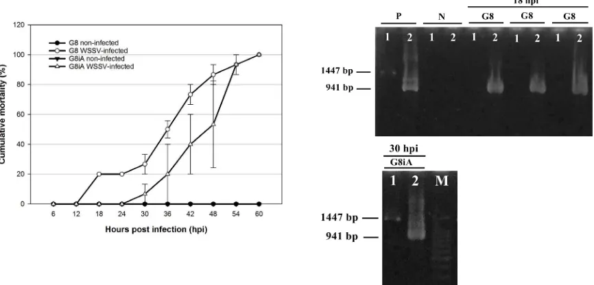
DNA extraction was conducted by homogenizing 50–100 µl of hemolymph or 0.2 g of tissue (gills/pleopods) with 500 µl of lysis buffer (10% SDS pH 7.2, 5M NaCl, 0.5M EDTA pH 8, and 1M Tris-HCl pH 8) and 1 µl of 10 μg/ml proteinase K until the tissues were inely dispersed. The mixture was then incubated at 95°C for 10 minutes using a heating block before centrifugation at 12,000 x g for 10 minutes. Supernatant (approximately 100 µl) was transferred to a new tube containing 400 µl of absolute ethanol. The tube was vortexed briely and centrifuged at 12,000 x g for 5 minutes. Supernatant was removed and the DNA pellet was air-dried on the bench. The DNA pellet was then dissolved by adding 100 µl of dH2O to the tube (Wang and Chang 2000). Before being used in PCR, the DNA was diluted 100 times by combining 1 µl of DNA isolate with 99 µl dH2O. Detection of WSSV using nested PCR was conducted according to the Manual of Diagnostic Tests for Aquatic Animals ( http://www.oie.int/international- standard-setting/aquatic-manual/accesss-online/). Lightly WSSV-infected shrimps showed a positive result only in the second step of PCR, yielding a 941 bp PCR fragment, while severely WSSV-infected shrimps showed 1447 bp and 941 bp PCR fragments both in irst step and second step of PCR (Lo et al. 1997).
Data Analysis
Cumulative mortality data were used to estimate median lethal time (LT50) using 3-Parameter-Weibull survival distribution analysis in Minitab 16 software at the P < 0.05 level. Threshold cycle (CT)data from ABI 7500 software was copied into Microsoft Excel to measure the relative expression change of PmVRP15 transcript levels of the WSSV-infected shrimps compared with the non-infected shrimps after normalization to the reference gene β-actin. The statistical signiicance of differences among 2-∆C
of each population was calculated by a two-sample t-test in SigmaPlot 13.0 where the signiicance was accepted at the P < 0.05 level.
Results and Discussion
The irst mortality in G8 was recorded at 18 hpi whereas the irst mortality in G8iA was recorded at 30 hpi, indicating that the inbred population (G8) died sooner than the outbred population (G8iA) in responding to the WSSV infection. However, 100% mortality of both G8 and G8iA occurred at 60 hpi (Table 1). This demonstrates that all populations were susceptible to white spot disease. Nevertheless, the large standard error bar showed that G8iA had more variable mortality time than G8, suggesting that G8iA showed a highly variable degree of susceptibility to WSSV within individuals in the population (Fig. 2 left). Based on conirmation of WSSV-infection using nested PCR, P. monodon from G8 that were dead at 18 hpi (irst mortality) showed a positive WSSV result only after the second step of amplification, which yielded a 941 bp PCR fragment (Fig. 2 right), indicating that the shrimps from G8 died with a light infection of WSSV. Compared
with the light infection at early death in G8, G8iA individuals that died at 30 hpi (first mortality) gave WSSV-positive results in both steps of PCR and yielded 1447 bp and 941 bp PCR fragments (Fig. 2 right); thereby indicating that G8iA shrimps were able to bear more severe infections in considerably longer periods than G8. All control P. monodon from G8 and G8iA that were alive and terminated at 66 hpi were WSSV-negative.
Table 1. Median lethal time (LT50) of WSSV-infected shrimp in the population of P. monodon Fabricius estimated with 3-Parameter-Weibull distribution analysis. Data were derived from three replicates. Supercripted letters indicate statistical signifance.
Population
Time of irst mortality
(hpi)
Time of 100% mortality
(hpi)
LT50 (hpi)
G8 18 60 45.17 ± 1.28
a
G8iA 30 60 50.09 ± 1.04b
Cumulative mortality G8 and G8iA was analyzed using 3-Parameter-Weibull survival distribution analysis and the LT50 of the two populations were compared to examine the degree of susceptibility. As Table 1 shows,
the median lethal time (LT50) of WSSV-infected G8iA (50.09 ± 1.04 hpi; 95% conidence interval: 48.04–52.13) was significantly higher than that of G8 (45.17 ± 1.28 hpi; 95% conidence interval: 42.67–47.68) (P < 0.05), indicating that G8iA had a higher survival time than G8. These results would suggest that the outbred population (G8iA) was less susceptible to the WSSV infection than the inbred population (G8). This result was consistent with previous studies reporting that wild type penaeid shrimps and outbred penaeid shrimps showed higher survival times than inbred populations in response to WSSC infection (Gopikrishna et al., 2012; Hayes et al., 2010). There is also evidence that genetic polymorphism between two different populations of L. vannamei affects their WSSV resistance/susceptibility level (Liu et al., 2014).
The significant difference in median lethal time and distinct capability to bear infection suggested there was little genetic variation between G8 and G8iA involving in WSSV susceptibility. The genetic variation may promote different immune responses to WSSV infection. According to the MCBAF Jepara breeding history, G8 was produced from a limited broodstock that had undergone decreased genetic variation since G1 (Prastowo et al., 2009). G8iA also revealed higher genetic variation compared with G8 (Purnamaningrum 2015). Moreover, wild type male P. monodon from Aceh, the parental population of G8iA, showed higher heterozygosity compared with the other populations of male P. monodon in Indonesia (Prastowo et al., 2010). Thus, the less WSSV susceptible G8iA likely emerged from genetic variation resulting from breeding with the wild type Aceh population. A limited broodstock, which is often caused by harvest activities, and domestication by inbreeding could reduce genetic variation in apopulation, accumulate recessive alleles, and eliminate resistant alleles (Brieuc et al., 2009; Cock et al., 2009), thereby affecting the G8 and G8iA populations’ ability to adapt to
the viral infection and contributing to their susceptibility.
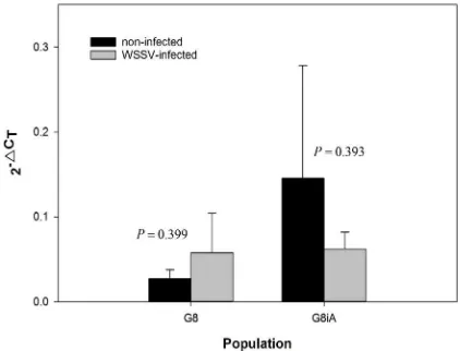
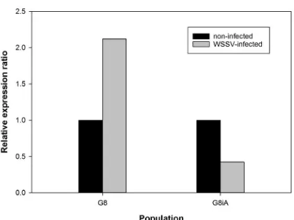
PmVRP15 has been found as a nuclear membrane-like protein acting to mediate replication of WSSV in hemocytes in early infection (Vatanavicharn et al., 2014). A linear form of ∆CT (2-∆CT) showed that baseline expression (non-infected) of PmVRP15 in G8iA was higher than G8. WSSV infection induced an increased expression of PmVRP15 in G8 (Fig. 3). Contrarily, PmVRP15 inducible expression (WSSV-infected) of G8iA decreased (Fig. 3). The relative expression ratio showed that the PmVRP15 transcripts level in G8 was up-regulated to 2.12 fold, while G8iA showed a down-regulation of PmVRP15 transcripts level to 0.42 fold (Fig. 4). The melt curve which showed a single peak at 83–84°C (data not shown) indicated that ampliication of PmVRP15 and β-actin was speciic. However, both the up-regulation of PmVRP15 in G8 (P = 0.399) and down-regulation in G8iA (P = 0.393) were not statistically significant (Fig. 3), contradicting a previous study that showed that PmVRP15 expression in the Thai population was signiicantly up-regulated in hemocytes at 48 hpi (Vatanavicharn et al., 2014).
Fig. 4. PmVRP15 relative expression ratios in G8 and G8iA in response to WSSV infection. The data represent the fold change of each population calculated from nine different biological replicates. Relative expression ratio >1, 1, and <1 indicate that the gene expression level was up-regulated, remained the same, or was down-regulated compared with the non-infected P. monodon.
The disparity between this present study and Vatanavicharn et al. (2014) can be explained in two ways. Firstly, as Vatanavicharn et al. (2014) hypothesized, severity of WSSV infection may affect stimulation of PmVRP15 expression in hemocytes. WSSV propagation of experimental P. monodon among these two local populations and the Thai population in the prior study were not monitored. Therefore, at present, we cannot determine how severe WSSV infection may regulate the expression of PmVRP15. Refinement in the infection protocol by prior quantification of viral loads in the challenge test may also help to observe the impact of WSSV severity on PmVRP15 expression. Secondly, PmVRP15 expression in this present study was measured from individual challenged shrimps, while PmVRP15 expression in Vatanavicharn et al. (2014) was measured from pooled challenged shrimps. As shown in Fig. 3, the large SEM bar of the PmVRP15 transcripts level shows a high variability of PmVRP15 expression within individuals. This indicates that the signiicant up-regulation of PmVRP15 in Vatanavicharn et al. (2014) tends to represent the up-regulated
gene expression within the Thai population. Glass et al. (2005) argue that even though the gene expression increased by a treatment, the quality of representation of individuals does not improve the representation of the pool, implying that the gene expression information obtained by the pooled sample and individuals are not comparable. However, since the inferences of this study are needed for estimation in population, we assume that pooled sampling of hemocytes would be more representative to compare baseline expression and inducible expression within population. Number of individuals in pooling strategy should be examined carefully to consider population characteristics in expressing genes (Muniesa et al., 2014; Peng et al., 2003; Taylor et al., 2010).
in G8 and G8iA can be marked with other considerable immune-related gene expression rather than PmVRP15. As suggested by Cock et al. (2009), it will be helpful to screen a large number of individuals from the G8 and G8iA populations that demonstrate either resistance or susceptibility to natural WSSV infection, thus exposing a representative WSSV susceptibility genetic marker to be used in MCBAF’s Jepara selective breeding program. Despite this possibility, future research will still be needed to study the association of PmVRP15 expression and WSSV susceptibility in another independent population of P. monodon to reveal the possibility of PmVRP15 as a universal genetic marker for WSSV susceptibility.
Acknowledgements
The authors would like to thank Rahayu Rahardianti, Evi Maftuti Nur, and Amri Yudhistira for their technical guidance during the research. We are also indebted to Salim Arrokhman and Dr. Budi Setiadi Daryono for their helpful evaluation to the manuscript. This work was a joint research project between MCBAF Jepara, Indonesia, and the Faculty of Biology of Universitas Gadjah Mada in part of a giant tiger shrimp breeding program for broodstock selection.
References
Alifuddin, M., Dana, D., Eidman, M., Malole, M.B. and Pasaribu F.H. 2003. Patogenesis Infeksi Virus White Spot (WSV) pada Udang Windu (Penaeus monodon Fab.) [Pathogenesis of White Spot Virus Infection (WSV) on Black Tiger Shrimp (Penaeus monodon Fab.)]. Jurnal Akuakultur Indonesia 2(2): 82-92.
Andrade, A.J. 2011. Shrimp immunological r e a c t i o n s a g a i n s t W S S V : r o l e o f haemocytes on WSSV fate. Master’s dissertation, Universiteit Gent.
Brieuc, M.S.O, Purcell, M.K., Palmer, A.D. and Naish K.A. 2015. Genetic variation underlying resistance to infectious hematopoietic necrosis virus in a steelhead
trout (Oncorhynchus mykiss) population. Dis Aquat Org 117: 77-83.
Cock, J., Gitterle, T., Salazar, M. and Rye, M. 2009. Breeding for disease resistance of Penaeid shrimps. Aquaculture 286: 1-11. Corteel, M., Dantas-Lima, J.J., Tuan, V.V.,
Thuong, K.V., Wille, M., Alday-Sanz, V., Pensaert, M.B., Sorgeloos, P. and Nauwynck H.J. 2012. Susceptibility of juvenile Macrobrachium rosenbergii to different doses of high and low virulence strains of white spot syndrome virus (WSSV). Dis Aquat Org 100: 211-218. Dutta ,S., Chakrabarty, U., Mallik, A. and
Mandal, N. 2013. Experimental evidence for white spot syndrome virus (WSSV) suceptibility linked to a mictosatellite DNA marker in giant black tiger shrimp, Penaues monodon (Fabricius) J Fish Dis 36(6): 293-597.
Escobedo-Bonilla, C.M., Audoorn, L., Wille, M., Alday-Sanz, V., Sorgeloos, P., Pensaert, M.B. and Nauwynck H.J. 2006. Standardized white spot syndrome virus (WSSV) inoculation procedures for intramuscular or oral routes. Dis Aquat Org 68: 181-188.
Ghosh, J., Lun, C.M., Majeske, A.J., Sacchi, S., Schrankel, C. and Smith L.C. 2011. Invertebrate immune diversity. Dev Comp Immunol 35: 959-974.
Glass, A., Henning, J., Karopka, T., Scheel, T., Bansemer, S., Koczan, D., Gierl, L., Rolfs, A. and Gimsa, U. 2005. Representation of Individual Gene Expression in Completely Pooled mRNA Sample. Biosci Biotechnol Biochem 69(6): 1098-1103. Gopikrishna, G., Gopal, C., Shekhar, M.,
Kumar, K.V., Kannappan, S. and Ponniah, A.G. 2012. Growth performance and white spot syndrome virus resistance in families of kuruma shrimp (Marsupenaeus japonicus). Indian J of Geo Mar Sci 41(1): 56-60.
virus-infected penaeid shrimp. Penaeus monodon and Penaeus indicus, by low cytometric analysis. Aquaculture 256: 111-120. Hayes ,B.J., Gitterle, T., Gopikrishna, G.,
Gopal, C., Krishna, G., Jahageerdar, S., Lozano, C., Alavandi, S., Paulpandi, S., Ravichandran, P. and Rye, M. 2010. Limited evidence for genetic variation for resistance to the white spot syndrome virus in Indian populations of Penaeus monodon. Aquacult Res 41: e872 - e877. Jeswin, J., Anju, A., Thomas, P.C., Paulton,
M. and Vijayan, K. 2013. Survivability of Penaeus monodon during white spot syndrome virus infection and its correlation with immune related genes. Aquaculture 380-383: 84-90.
Johansson ,M.W., Keyser, P., Sritunyalucksana, K. and Soderhall, K. 2000. Crustacean haemocytes and haematopoiesis. Aquaculture 191(13): 45-52.
Liu, J., Yu, Y., Li, F., Zhang, X. and Xiang, J. 2014. A new anti-lipopolysaccharide factor (ALF) gene with its SNP polymorphisms related to WSSV-resistance of Litopenaeus vannamei. Fish Shellish Immunol 39: 24-33. Lo, C., Chen, C., Peng, S., Chen, Y., Chou,
C., Yeh, P., Huang, C., Chou, H., Wang, C. and Kou, G. 1996. Detection of baculovirus associated with white spot syndrome (WSBV) in penaeid shrimps using polymerase chain reaction. Dis Aquat Org 25: 133-141.
Lo, C., Ho, C., Chen, C., Liu, K., Chiu, Y., Yeh, P., Peng, S., Hsu, H., Liu, H., Chang, C., Su, M., Wang, C. and Kou. G. 1997. Detection and tissue tropism of white spot syndrome baculovirus (WSBV) in captured brooders of Penaeus monodon with a special emphasis on reproductive organs. Dis Aquat Org 30: 53-72.
Luo, T., Zhang, X., Shao, Z. and Xu, X. 2003. PmAV, a novel gene involved in virus resistance of shrimp Penaeus monodon. FEBS Lett 551: 53-57.
Mathew, S., Kumar, K.A., Anandan, R., Nair, P.G.V. and Devadasan, K. 2007. Changes in tissue defence system in white spot
syndrome virus (WSSV) infected Penaeus monodon. Comp Biochem Phys C 145: 315-320.
Muniesa, A., Ferreira, C., Fuertes, H., Halaihel, N. and de Blas, I. 2014. Estimation of the Relative Sensitivity of qPCR Analysis Using Pooled Samples. PLoS ONE 9(4): e93491. Peraza-Gomez, V., Luna-Gonsalwz, A.,
Gonzales-Prieto, J., Fierro-Coronado, A. and Gomzalez-Ocampo, H. 2014. Protective effect of microbial immunostimulants and antiviral plants against WSSV in Litopenaeus vannamei cultured under laboratory conditions. Aquaculture 420-421: 160-164. Peng, X., Wood, C.L., Blalock, E.M., Chen, K.C.,
Landield, P.W. and Stromberg, A.J. 2003 Statistical implications of pooling RNA samples for microarray experiments. BMC Bioinformatics 4 : 1-9.
Prastowo, B.W., Rahardianti, R., Nur, E.M. and Taslihan, A. 2009 Proil heterogenitas genetik induk udang windu (Penaeus monodon) turunan F1 melalui analisis DNA mitokondria-RFLP dan RAPD [Genetic heterogeneity proile of giant tiger prawn (Penaeus monodon) broodstock F1 revealed by mitochondria DNA-RFLP and RAPD]. J Fish Sci XI(1): 25-30.
Prastowo, B.W., Pancoro, A., Susanto, A., Nur, E.M. and Rahardianti, R. 2010. Pure line Selection of Black Tiger Shrimp Broodstock through Microsatellite Marker. Ministry of Marine and Fisheries, Jakarta.
Purnamaningrum, A. 2015. Variasi Genetik Morfologis dan Molekular Udang Windu (Penaeus monodon Fabricius, 1798) Hasil Inbreeding G7 dan Outcrossing dengan Induk Alam [Morphological and Molecular Variation of Giant Tiger Prawn (Penaeus monodon Fabricius, 1798) from Inbreeding of G7 and Outcrossing of G7 with Natural Broodstock]. Master’s dissertation, Universitas Gadjah Mada.
Schmittgen, T.D. and Livak, K.J. 2008. Analyzing real-time data by the comparative Ct method. Nat Protoc 3(6): 1101-1108.
Taylor, S.M., Juliano, J.J., Trottman, P.A., Grifin, J.B., Landis, S.H., Kitsa, P., Tshefu, A.K. and Meshnick, S.R. 2010. High-Throughput Pooling and Real Time PCR-Based Strategy for Malaria Detection. J Clin Microbiol 48(2): 512-519.
Tonganunt M., Saelee M., Chotigeat, M. and Ponghdara, A. 2009. Identiication of a receptor for activated protein kinase C1 (Pm-RACK1), a cellular gene product from black tiger shrimp (Penaeus monodon) interacts with a protein, VP9 from the white spot syndrome virus. Fish Shellish Immunol 26: 509-514.
Vatanavicharn, T., Prapavorarat, A., Jaree, P., Somboonwiwat, K. and Tassanakajon, A. 2014. PmVRP15, a novel responsive protein from the black tiger shrimp, Penaeus monodon, promoted white spot syndrome virus replication. PLoS ONE 9(3): e91930.
Wang, Y. and Chang, P. 2000. Yellow head virus infection in the giant tiger prawn Penaeus monodon cultured in Taiwan. Fish Pathol 35(1): 1-10.