Histopathological changes in broiler chickens fed afl
atoxin and
cyclopiazonic acid
Raja Kumar1, and Chidambaram Balachandran2*
1Department of Veterinary Pathology, Rajiv Gandhi College of Veterinary and Animal Sciences, Puducherry, India
2Department of Veterinary Pathology, Veterinary College and Research Institute, Namakkal, Tamilnadu, India
KUMAR, R., C. BALACHANDRAN: Histopathological changes in broiler chickens fed afl atoxin and cyclopiazonic acid. Vet. arhiv 79, 31-40, 2009.
ABSTRACT
Experimental mycotoxicoses was induced into broiler chickens by feeding 1 ppm afl atoxin (AF) and 20
ppm cyclopiazonic acid (CPA) from 0 to 28 days of age to evaluate the gross and histopathological changes. Grossly, AF and AF-CPA fed birds showed enlargement, yellowish discoloration of the liver while the CPA fed birds showed enlargement and congestion. The CPA and AF-CPA fed birds showed thickening of crop and necrosis and thickening of proventricular mucosa. Histopathologically, degenerative and necrotic changes were observed in the liver, kidneys, intestine, pancreas, heart, pectoral muscle, spleen and bursa of Fabricius of all toxin fed birds. Besides, hyperplastic changes were also observed in the crop, proventriculus and gizzard in the CPA fed birds. The lesions were more marked in the AF-CPA group. The study revealed that AF and CPA in combination could act cumulatively and adversely affect the health of broiler chicken.
Key words:broiler chicken, afl atoxin, cyclopiazonic acid, pathology
Introduction
In nature, mycotoxins rarely occur as a single contaminant since many fungal species that produce mycotoxins grow and produce their toxic metabolites under similar conditions. The co-occurrence of mycotoxins can also arise through a single mould producing more than one toxin and simultaneous contamination by two or more moulds, from the same or different species. Furthermore, a typical poultry ration is made up of several grain sources, each of which may be contaminated with a different mycotoxins or more than one mycotoxin.
Afl atoxins (AF) are a group of heterocyclic toxic metabolites of toxigenic fungi
Aspergillus fl avus and A. parasiticus. The mechanisms of action of afl atoxins involve their *Corresponding author:
metabolism to reactive intermediates, which bind to macromolecules with consequent disruption of transcriptional and translational processes (DIAZ, 2005). Cyclopiazonic acid (CPA), an indole tetramic acid, is a mycotoxin produced by several species of Aspergillus
and Penicillium fungi. CPA is a potent, specifi c and reversible inhibitor of the sarcoplasmic
and endoplasmic reticulum Ca2+ ATPase (KARON et al., 1994).
Co-occurrence of the most potent mycotoxin, the afl atoxin and cyclopiazonic acid, an emerging important mycotoxin, has been reported in various feed and feedstuffs
(GALLAGHER et al., 1978; BALACHANDRAN and PARTHASARATHY, 1996). Besides,
certain strains of Aspergillus fl avus produce both AF and CPA (GALLAGHER et al., 1978). It has been suggested that a combination of mycotoxins at low concentration may have negative effects, even though the concentrations of individual mycotoxins are well below the concentrations reported to cause negative effects. The above facts and the limited literature on the subject prompted the authors to study the pathological changes in broiler chickens following oral consumption of sublethal doses of AF and CPA singly and in combination.
Materials and methods
Production and estimation of afl atoxin. Afl atoxin was produced on rice (SHOTWELL
et al., 1966). Fifty grams of rice was soaked in 25 mL of water for two hours with frequent
shaking. The fl asks were autoclaved, cooled and inoculated with one mL of spore suspension (containing about 106 spores) of A. parasiticus NRRL 2999. The fl asks were kept at room temperature in slant position and hand shaken vigorously six to ten times a day to avoid clump formation. On the tenth day, mouldy rice was briefl y steamed for
fi ve minutes to kill the spores and dried overnight at 60ºC in a hot air oven. The mouldy rice was powdered and the afl atoxin content was estimated by the ROMER method (1975). A known quantity of mouldy rice powder was blended with water and extracted using acetone and fi ltered. The extract was purifi ed by adding cupric carbonate and mixed with ferric chloride gel (ferric chloride sodium hydroxide) and fi ltered. The fi ltrate was mixed with sulphuric acid and extracted with chloroform in a separating funnel. The extracted product was mixed with potassium hydroxide and potassium chloride mixture in another separating funnel. The lower layer was collected and run over anhydrous sodium sulphate. The fi nal extract was dried. A known quantity of chloroform was added to dilute the extract and taken to a thin layer chromatograph (TLC) for quantifi cation. The samples and standard (M/s Sigma Chemicals, USA) were spotted on the TLC plates and developed in a chloroform:acetone mixture for 45 minutes. The plates were compared under a UV lamp in a chromatoview cabinet. The afl atoxin content was calculated according to ANONYM. (1980) specifi cations.
Production and estimation of cyclopiazonic acid. One hundred grams of rice was
taken in a 500 mL conical fl ask. After adding 20 mL of distilled water to each fl ask, they were autoclaved, cooled and inoculated with one mL of spore suspension (containing about 106 spores) of P. griseofulvum NRRL 3523 and kept at room temperature in slant position (RATHINAVELU and SHANMUGASUNDARAM, 1984). The fl asks were shaken once daily to avoid clump formation. After 15 days, the culture was steamed for 5 minutes to kill the spores and was dried at 50ºC in an oven for about 3 hours and powdered. The mouldy rice was powdered and mixed with chloroform and fi ltered. The extract was dissolved in chloroform and concentrated. The concentrate was spotted on to a TLC plate. The plate was developed in chloroform-ethyl acetate-formic acid-toluene. The plate was dried and sprayed with one percent p-dimethylaminobenzaldehyde in n-butanol, dried and exposed to HCL vapour. A bright, purple colour confi rmed the presence of CPA and the corresponding unreacted areas containing the toxin were scrapped off., extracted with methanol and the extract was mixed p-dimethylaminobenzaldehyde and 5N HCl and read in an Erma photoelectric colorimeter using green fi lter. The amount of CPA was determined from the standard curve prepared (RATHINAVELU and SHANMUGASUNDARAM, 1984).
Experimental design. Known amounts of powdered rice cultures containing AF and
CPA were incorporated into AF and CPA free broiler starter mash to yield 1 ppm AF, 20 ppm CPA and 1 ppm AF +20 ppm CPA. One control diet was also prepared. The diets contained 23 percent crude protein. Forty commercial broiler chickens (Vencob, India) were weighed, wing banded and randomly allotted to four treatment groups. Each treatment consisted of two replicates of fi ve chickens. The birds were raised on battery brooders with ad libitum supply of feed and water. The diets were fed from 0 to 28
days of age. The trial was repeated with another 40 birds. The birds were sacrifi ced at the termination of the experiments following ethical procedures. Detailed necropsy was conducted and gross lesions observed were recorded. Representative pieces of tissues from the liver, kidneys, crop, proventriculus, gizzard, intestine, heart, skeletal muscle, pancreas, spleen and bursa of Fabricius were collected and fi xed in 10% buffered formol saline for histopathological examination. Paraffi n embedded tissues were sectioned to 5-6 μm thickness and stained with haematoxylin and eosin following standard procedures
(BANCROFT and STEVENS, 1996).
Results
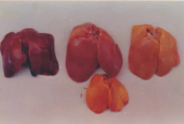
Grossly, the livers from the AF fed birds showed enlargement, pallor or yellowish discoloration (Fig. 1), occasional mottling and gall bladder distension. Kidneys were enlarged, pale or congested with a few petechiae. CPA fed birds showed congestion, enlargement and occasional yellow depressed foci in the liver. The crop and proventricular
mucosa showed thickening (70%). The lesions were observed in all the AF-CPA fed birds (100%).
Fig. 1. Liver CPA toxicosis. Congestion (upper left), afl atoxicosis-paleness and yellow discoloration (middle and upper right), AF-CPA toxicosis - yellow discoloration (bottom).
Fig. 2. Afl atoxicosis. Liver showing acinar arrangement of regenerating hepatocytes.
H&E, scale bar = 40 μm.
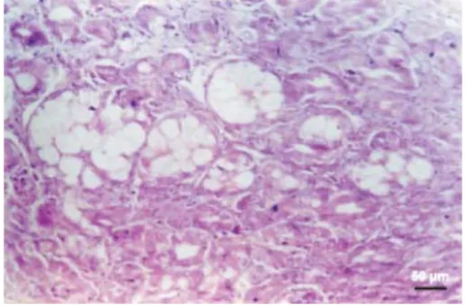
Fig. 3. CPA toxicosis. Liver showing microvesicular fatty degeneration of hepatocytes. H&E, scale bar = 100 μm.
Fig. 4. AF-CPA toxicosis. Liver showing macrovesicular fatty degeneration and fatty cyst formation in the regenerating hepatocytes.
H&E, scale bar = 50 μm.
Fig. 5. AF-CPA toxicosis. Kidney showing thickening of glomerular basement membrane
and collapse of glomerulus . H&E, scale bar = 5 μm.
Fig. 6. AF-CPA toxicosis. Crop mucosa showing epithelial hyperplasia and vacuolar
degeneration. H&E, scale bar = 50 μm.
Fig. 7. Afl atoxicosis. Proventriculus showing partial necrosis of mucosa, dilated crypts and submucosal edema. H&E, scale bar = 50 μm.
Fig. 8. CPA toxicosis. Proventriculus
Microscopically, the livers of the AF fed birds revealed vacuolar degeneration, macrovesicular fatty degeneration and ballooning degeneration of the hepatocytes. The hepatocytes showing fatty changes coalesced to form fatty cysts. The architecture of the liver was completely altered and the regenerating hepatocytes were arranged in acinar or ductular patterns (Fig. 2). The acinar hepatocytes also showed fatty degeneration and fatty cysts in some areas. Congestion, bile duct hyperplasia, focal infi ltration of heterophils and mononuclear cells, perivascular infi ltration of mononuclear cells and heterophils and
fi brosis were also noticed in the livers.
Livers from CPA fed birds showed congestion, sinusoidal distension, microvesicular fatty degeneration (Fig. 3), focal necrosis of hepatocytes, biliary hyperplasia, Kupffer cell hypertrophy, periductular lymphocytic infi ltration and thickening of capsules. In the AF-CPA fed birds, the livers revealed congestion, macrovesicular fatty degeneration and extensive fatty cyst formation (Fig. 4). The regenerated hepatic cells were arranged in an acinar pattern and these cells also showed fatty degeneration and fatty cysts. Hepatocellular necrosis, marked bile duct hyperplasia, perivascular fi brosis, focal collections of lymphocytes, periportal heterophilic and lymphocytic infi ltration, necrosis, large collections of lymphocytes around bile ducts, marked thickening of capsule and Kupffer cell hypertrophy were also seen.
Kidneys revealed congestion, focal haemorrhages and degeneration of tubular epithelium in all toxin fed groups. In addition, occasional thickening of basement membrane in the AF fed birds, focal necrosis and mononuclear cell infi ltration in the CPA fed birds and thickening of glomerular basement membrane and collapse of glomerular tuft (Fig. 5) in the AF-CPA fed group were noticed.
Consistent changes observed in the mucosa of the crop of the AF and AF-CPA fed birds were epithelial hyperplasia, vacuolar degeneration (Fig. 6), keratinization, parakeratosis, necrosis and fi brosis. The lesions were more marked in the AF-CPA fed birds and in addition, the crop muscles showed fatty degeneration and hyaline degeneration. In the AF fed birds, the proventriculus showed partial to full thickness necrosis (Fig. 7) and diffuse lymphocytic and heterophilic infi ltrations in the lamina propria. In the CPA fed birds, hyperplasia of the proventricular epithelium characterized by an increase in the height of the villi (Fig. 8), vacuolar degeneration, moderate lymphocytic infi ltration, disruption and fi brosis of the lamina propria, focal areas of erosion and full thickness necrosis were noticed. In the AF-CPA fed birds, vacuolar and hyaline degenerations of the proventricular muscles were also seen. The CPA fed birds showed dilatation of the glands of the gizzard. Besides, the AF-CPA fed birds showed hyperplasia and vacuolar degeneration of the epithelium, defective keratinoid membrane formation (Fig. 9) and lymphocytic infi ltration in the submucosa of the gizzard.
All toxin fed groups showed catarrhal enteritis with lymphocytic or mononuclear cell infi ltrations. Congestion, a decrease in the volume of the zymogen granules, dissociation of acinar cells, necrosis and mononuclear cell infi ltration were the lesions observed in the pancreas of the toxin treated groups. Granular degeneration of sarcoplasm, necrosis and collection of mononuclear cells in the heart and hyaline degeneration, necrosis and mononuclear cell infi ltration of the pectoral muscle were also observed.
The spleens showed lymphoid depletion, an increase in the number of germinal centres and reticulum cell hyperplasia in all toxin treated birds. The bursa of the Fabricius of the AF fed group revealed a lack of cortico-medullary differentiation, generalized lymphoid depletion and heterophilic infi ltration. Glandular transformation of isolated follicles and plical epithelial hyperplasia with cystic changes were also observed in the CPA and AF-CPA fed birds. The lesions were more marked in the AF and AF-AF-CPA fed group.
Discussion
Mycotoxicoses were induced in broiler chicken by feeding AF (1 ppm) and CPA (20 ppm) singly and in combination, to study the pathological changes in different organs. The gross pathological changes in the liver, kidneys, crop and proventriculus observed in AF and CPA fed birds agreed with those of earlier studies (ASPLIN and CARNAGHAN,
1966; BALACHANDRAN and RAMAKRISHNAN, 1987;DORNER et al., 1983; CULLEN et al.,
1988; BALACHANDRAN et al., 1998). Ulceration of the proventriculus and nodular lesions
in CPA fed birds have also been described in literature (DORNER et al., 1983; CULLEN
et al., 1988; BALACHANDRAN et al., 1998). Probably higher doses of toxin (25 ppm and
above) are required to produce such effects. In AF-CPA fed birds, the lesions were more marked and consisted of both the lesions seen in AF and CPA fed birds. SMITH et al. (1992)
reported a hard fi brotic spleen and atrophy of the gizzard at the higher dose levels of 3.5 ppm of AF and 50 ppm of CPA.
Histopathological changes observed in the livers of AF and CPA fed birds was similar to earlier reports (CARNAGHAN et al., 1966; BALACHANDRAN and RAMAKRISHNAN,
1987; DORNER et al., 1983; CULLEN et al., 1988; BALACHANDRAN et al., 1998) but occurred
at a lower dose level in CPA toxicosis in the present study. The marked cytoplasmic vaculoation in the AF-CPA fed birds in the present study is perhaps the result of inhibition of protein synthesis in the hepatocytes by both the toxins, involving different pathways, whereby the apparent intracellular fatty degeneration was magnifi ed (PIER et al., 1989). Further fatty degeneration and fatty cyst formations in the acinar hepatocytes have not been reported previously. These changes suggest the possible cumulative effect AF and CPA.
BALACHANDRAN et al., 1998). However, glomerular basement membrane thickening in the AF fed birds was not reported in the earlier studies. The glomerular basement membrane thickening, karyomegaly and collapse of the glomerular tuft in the AF-CPA fed birds also suggest the cumulative interaction of these toxins in inducing severe changes in the kidneys. The lesions observed in the crop, proventriculus, gizzard and intestines concurred with the fi ndings of earlier studies (DORNER et al., 1983; CULLEN et al., 1988;
BALACHANDRAN et al, 1998). However, the lesions observed in these organs in AF fed
birds and parakeratosis of the crop mucosa in CPA toxicosis have not been reported previously. Hyperplasia, degeneration of glandular epithelium and defective keratinoid formation in the gizzard and degenerative and necrotic changes in the muscles of the crop and proventriculus observed in the present study indicated the possible cumulative effects of AF and CPA in combination.
Degenerative and necrotic changes were observed in the myocardium of all toxin treated birds. However, in AF fed birds, haemorrhages were noticed in the epicardium and myocardium (BALACHANDRAN and RAMAKRISHNAN, 1987) whilst in CPA fed birds subacute to chronic infl ammation of the myocardium (DORNER et al., 1983), absence of lesions except granulocytic infi ltration (CULLEN et al., 1988) and fatty degeneration and necrosis (BALACHANDRAN et al., 1998) were reported earlier. Lesions observed in the skeletal muscle, pancreas, spleen and the bursa of the Fabricius of all toxin treated birds were similar to earlier reports (BALACHANDRAN and RAMAKRISHNAN, 1987; DORNER
et al., 1983; CULLEN et al., 1988; BALACHANDRAN et al., 1998).
The effects of exposure to several combinations of mycotoxins in poultry have been reported to be cumulative. However, synergistic interactions have been observed in AF and ochratoxin (HUFF and DOERR, 1981) and CPA and -2 toxin (KAMALAVENKATESH
et al., 2005) whereas the interaction was cumulative or less than cumulative in CPA and
T-2 Toxin (KUBENA et al., 1994) and in CPA and ochratoxin (GENTLES et al., 1999). It has also been reported that the effect of AF and CPA was less than cumulative for certain parameters, such as haemoglobin, albumin and glucose in poultry (SMITH et al., 1992). Histopathological changes in the liver, digestive tract and kidneys were more pronounced in the birds fed with AF-CPA than in animals fed single toxins, suggesting that AF and CPA have at least additive effect. These fi ndings are of substantial importance in view of the known co-production by Aspergillus fl avus and natural co-existence of these two
toxins in nature (GALLAGHER et al., 1978).
References
ANONYMOUS (1980): AOAC. Offi cial Methods Of Analysis. 13th ed., Association of Offi cial
Analytical Chemists, Washinton, D.C.
ASPLIN, F. D., R. B. A. CARNAGHAN (1961): The toxicity of certain groundnut meal for poultry with special reference to their effects on ducklings and chicken. Vet. Rec. 73, 1215- 1219.
BALACHANDRAN, C., R. RAMAKRISHNAN (1987): An experimental study on the pathology of afl atoxicosis in broiler chicken. Indian Vet. J. 64, 911-914.
BALACHANDRAN, C., K. R. PARTHASARATHY (1996): Occurrence of cyclopiazonic acid id feed and feed stuffs in Tamil Nadu, India. Mycopathologia 133,159-162.
BALACHANDRAN, C., K. R. PARTHASARATHY, A. SUNDARARAJ (1998): Experimental study on pathology of cyclopiazonic acid mycotoxicosis in broiler chicken. Indian Vet. J. 75, 693-697.
BANCROFT, J. D., A. STEVENS (1996): Theory and Practice of Histological Techniques. 4 ed., Churchill Livingstone, London.
CARNAGHAN, R. B. A., G. LEWIS, D. S. P. PATTERSON, R. ALLCROFT (1966): Biochemical and pathological effects of groundnut poisoning in chickens. Path. Vet. 3, 601-615.
CULLEN, J. M., M. WILSON, W. M. HAGLER, J. F. ORT, R. J. COLE (1988): Histologic lesions in broiler chicks given cyclopiazonic acid orally. Am. J. Vet. Res. 49, 728-732.
DIAZ, D. E. (2005): The Mycotoxin Blue Book. Nottingham University Press, Nottingham. DORNER, J. W., R. J. COLE, L. G. LOMAX, H. S. GOSSER, U. L. DIENER (1983): Cyclopiazonic
acid production by Aspergillus fl avus and its effects on broiler chickens. Appl. Environ.
Microbiol. 46, 698-703.
GALLAGHER, R. T., J. L. RICHARD, H. M. STAHR, R. J. COLE (1978): Cyclopiazonic acid production by afl atoxigenic and non-afl atoxigenic strains of Aspergillus fl avus. Mycopathologia
66, 31-36.
GENTLES, A., E. E. SMITH, L. F. KUBENA, E. DUFFUS, P. JOHNSON, J. THOMPSON, R. B. HARVEY, T. S. EDRINGTON (1999): Toxicological evaluations of cyclopiazonic acid and ocratoxin A in broilers. Poultry Sci. 78, 1380-1384.
HUFF, W. E., J. A. DOERR (1981): Synergism between afl atoxin and ochratoxin A in broiler
chickens. Poultry Sci. 60, 550-555.
KAMALAVENKATESH, P., S. VAIRAMUTHU, C. BALACHANDRAN, B. MURALIMANOHAR, G. DHINAKARAJ (2005): Immunopathological effect of the mycotoxins cyclopiazonic acid and T-2 toxin on broiler chicken. Mycopathologia 159, 273-279.
KARON, B. S., J. E. MAHANEY, D. D. THOMAS (1994): Halothane and cycloiazonic acid modulate Ca-ATPase oligomeric state and function in sarcoplsmic reticulum. Biochemistry 33, 13928-13937.
KUBENA, L. F., E. E. SMITH, A. GENTLES, R. B. HARVEY, T. S. EDRINGTON, T. D. PHILLIPS, G. E. ROTTINGHAUS (1994): Individual and combined toxicity of T-2 toxin and cyclopiazonic acid in broiler chicks. Poultry Sci. 73, 1390-1397.
PIER, A. C., E. L. BELDEN, J. A. ELLIS, E. W. NELSON, L. R. MAKI (1989): Effect of cyclopiazonic acid and afl atoxin singly and in combination on selected clinical, pathological and immunological responses of guinea pigs. Mycopathologia, 105, 135-142.
ROMER, R. T. (1975): Qualitaitve/quantitiatve analysis for detection and estimation of afl atoxin (Romer mini column method). J. Assoc. Off. Anal. Chem. 58, 500-506.
SHOTWELL, O. L., C. W. HESSELTINE, R. D. STUBBLEFIELD, G. W. SORENSON (1966): Production of afl atoxin on rice. Appl. Microbiol. 14, 425-428.
SMITH, E. E., L. F. KUBENA, C. E. BRAITHEWAITE, R. B. HARVEY, T. D. PHILLIPS, A.
H. REINE (1992): Toxicological evaluation of afl atoxin and cyclopiazonic acid in broilers
chickens. Poultry Sci. 71, 1136-1144.
KUMAR, R., C. BALACHANDRAN: Patohistološke promjene u tovnih pilića koji su u hrani dobivali afl atoksin i ciklopiazonsku kiselinu. Vet. arhiv 79, 31-40, 2009.
SAŽETAK
Pokusna mikotoksikoza u tovnih pilića bila je izazvana davanjem 1 ppm afl atoksina (AF) i 20 ppm
ciklopiazonske kiseline (CPK) od dana valjenja do 28. dana života, u svrhu promatranja patoanatomskih i
patohistoloških promjena. Patoanatomski je u pilića koji su dobivali AF i AF-CPK ustanovljena povećana
žućkasta jetra, a u pilića koji su dobivali CPK povećana jetra s kongestijom. U pilića koji su dobivali CPK i AF-CPK ustanovljeno je zadebljanje voljke te nekroza i zadebljanje sluznice žljezdanoga želuca. Patohistološki su ustanovljene degenerativne i nekrotične promjene u jetri, bubrezima, crijevima, gušterači, srcu, prsnom mišićju, slezeni i Fabricijevoj burzi u svih pilića koji su dobivali toksin. Povrh toga, hiperplastične promjene bile su ustanovljene u voljki, predželucu i želucu u pilića koji su dobivali CPK. Oštećenja su bila mnogo jače izražena u skupini pilića koja je istodobno dobivala AF i CPK. Istraživanje je pokazalo da AF i CPK djeluju kumulativno i nepovoljno na zdravlje tovnih pilića.
Ključne riječi:tovni pilići, afl atoksin, ciklopiazonska kiselina, patologija
Received: 30 May 2007 Accepted: 21 December 2008