INDONESIAN FISHERIES RESEARCH JOURNAL
Manuscript send to the publisher:
Indonesian Fisheries Research Journal
Research Center for Capture Fisheries
Jl. Pasir Putih I Ancol Timur Jakarta 14430 Indonesia
Phone (021) 64711940
Fax: (021) 6402640
Email:
[email protected]
Indonesian Fisheries Research Journal is published by Research Center for Capture
Fisheries. Budgeting F.Y. 2008.
ISSN 0853 - 8980
Published by:
Agency for Marine and Fisheries Research
Volume 14 Number 2 December 2008
Acreditation Number: 101/Akred-LIPI/P2MBI/10/2007
(Period: November 2007-November 2010)
Indonesian Fisheries Research Journal is the English edition of
fisheries research journal. The first published in 1994 with publishing frequently is
once a year. Since 2005, this journal published twice on JUNE and DECEMBER.
Head of Editor Board:
Prof. Dr. Ir. Endi Setiadi Kartamihardja, M.Sc.
Members of Editor Board:
Prof. Dr. John Haluan Dr. Ir. Subhat Nurhakim, M.S.
Dr. Ir. Wudianto, M.Sc. Dr. Ir. Indra Jaya
Refrees for this Number:
Prof. Dr. Ir. Ari Purbayanto, M.Sc. (Bogor Agriculture Institute) Dr. Ir. Setyo Budi Susilo, M.Sc. (Bogor Agriculture Institute)
Dr. Ir. Augy Syahailatua (Research Center for Oceanography-Indonesian Institute for of Science)
Managing Editors:
Dra. Endang Sriyati
i
PREFACE
Indonesian Fisheries Research Journal Volume 14 No.2 December 2008 is the second publising English Journal of Research Center for Capture Fisheries in 2008. This journal is one of media for communication of fisheries scientiest. This publication is supported by the financial budget of Research Center for Capture Fisheries, fy 2008.
This number contains six scientific papers concerning one article of inland waters, three papers of marine resources, and two papers of post harvest technology fisheries.
We deeply thank to the editorial boards for their highly contribution in evaluating the papers before published. Their effort had increased the quality of the journal. Finally, we hope this journal would contribute to fisheries science as well as development of fisheries in Indonesia.
ISSN 0853 - 8980
INDONESIAN FISHERIES RESEARCH JOURNAL
Vol.14 No.2 - December 2008
CONTENTS
Page
i
iii
51-65
67-74
75-82
83-89
91-95
97-101
iii PREFACE ………...
CONTENTS ………..…...………..
Fish Community Structure in Relation to Water Quality of the Down Stream of Musi River, South Sumatera, Indonesia
By: Husnah, Eko Prianto, Makri, and Hilda Z. Dahlan ………...…………..…… Demersal Fish Resources Result of MV. SEAFDEC 2 Survey in the South China Sea of Indonesia By: Wudianto and Bambang Sumiono ………...………….. Biological Aspects, Density, and Distribution of the Alfonsino (Beryx splendens) in the Indian Ocean Ekslusive Economic Zone of Indonesia
By: Fayakun Satria, John Haluan, Eko Sri Wiyono, and Wudianto ………...……….……. Additional Valid Record of Marbled Hawkfish, Cirrhites pinnulatus (Bloch & Schneider, 1801) (Perciformes: Cirrhitidae) from North Sulawesi
By: Teguh Peristiwady ………...……….
Antibacterial Potency of Chitosan Oligomer Produced by Bacillus Licheniformis MB-2 Chitosanase By: Ekowati Chasanah, Meidina, and Maggy T. Suhartono ………...……….. Use of Aspergillus repens in the Moulding Process of Dried Fish Stick Made from Little Tuna
(Euthynnusaffinis)
91
Antibacterial Potency of Chitosan ... by Bacillus Licheniformis MB-2 Chitosanase (Chasanah, E., et al.)
ANTIBACTERIAL POTENCY OF CHITOSAN OLIGOMER PRODUCED
BY
Bacillus licheniformis
MB-2 CHITOSANASE
Ekowati Chasanah1), Meidina2), and Maggy T. Suhartono3) 1) Marine Biotechnology Lab, Center for Marine and Fisheries Product Processing and
Biotechnology Research-Petamburan, Jakarta
2) Food Science Program, Graduate School, Bogor Agricultural University-Bogor, Indonesia
3) Department of Food Science Technology, Faculty of Agricultural Technology, Bogor Agricultural University-Bogor, Indonesia Received September 26-2007; Received in revised form March 25-2008;
Accepted October 24-2008
ABSTRACT
Bacillus licheniformis MB-2 chitosanase isolated from hot spring water in Manado, Indonesia,
was used to produce chitosan oligomers. Both crude and pure Bacillus licheniformis MB-2 chitosanase were used to prepare chitosan oligomers, and the antibacterial activity of the resulting oligomers were tested towards 6 pathogenic bacteria, including Pseudomonas aerugenusa, Salmonella
typhimurium, Escherichiacoli, Staphylococcusaureus, Listeriamonocytogenes, and Bacilluscereus.
By contact method, the oligomers, at the MIC value (Salmonella typhymurium of 321 ppm, Pseudomonas
aeruginosa, Staphylococcus aureus, and Escherichia coli (MIC of 402 ppm) and the contact time of 24
h, were able to reduce all pathogenic bacteria tested by 2 to 5 log cycles. Using Pseudomonas
aeruginosa protease, the oligomers were capable of reducing the protease activity by 64%, indicating
that antiprotease might be involved in the antibacterial mechanism by these oligomers.
KEYWORDS: Bacillus licheniformis MB-2 chitosanase, oligomer, antibacterial agent, protease inhibitor
INTRODUCTION
Chitosan is a natural polymer containing N
-acetyl-D-glucosamine and D-glucosamine residues. Having biodegradable and biocompatible properties, this deacetylated form of chitin polymer has been used widely at various industries such as food, agricultural, pharmaceutical, and waste water treatments. To improve its application, i.e enhancing the water solubility properties, the polymer is usually modified by substituting the functional groups with chemical substances or partially hydrolyzed chemically or enzymatically. Application of enzyme for partially degrading chitosan was more preferable because of the mild and safe process as well as specific reaction, resulting in high quality oligomer products. Specific size oligomer was reported to maintain various prominent biological activities such as antimicrobial properties.
Study on antimicrobial properties of chitosan has been reported by a number of scientists (Sagoo et al., 2002; Helander, 2001; Rhoades & Roller, 2000; Muzarelli et al., 1990; El-Ghaouth et al., 1992). At low pH (<6.3), chitosan polymers showed antifungal activity (Roller & Covill, 1999; Rhoades & Roller, 2000). Partially degraded chitosan or chitosan oligomer was reported to have antibacterial properties against some pathogenic bacteria compared to the native one (Kendra & Hadwiger, 1984). The antibacterial mechanism has been proposed as loosing of barrier function of bacterial cell wall caused by chitosan
binding to the outer membrane of bacteria, and Chung
et al. (2004) reported that there was a positive relationship between antibacterial activity of chitosan and surface characteristics of bacterial cell wall. Another possible mechanism through inactivation of important enzymes such as protease has never been reported yet.
Bacillus licheniformis MB-2 has been isolated from hot spring water of Manado and be able to produce thermostable chitosanase. In pure form, the enzyme hydrolyzes specifically chitosan, producing pentamer and hexamer of chitooligosaccharide (Chasanah, 2004). Preliminary study on antibacterial properties of oligomers from the crude enzyme using 4 concentration (0.005 tp 0.170 U per mg chitosan) showed that oligomer produced by applying 0.1 U per mg chitosan and reaction time of 1 to 3 hours was able to reduce the 6 pathogenic bacteria tested (Meidina et al., 2004). Further study on the sensitivity and mechanism of antibacterial through their potency as anti protease was assesed in this study using P. aeruginosa protease. Six pathogenic bacteria frequently associated with food borne infection and intoxication was used.
MATERIALS AND METHODS
Chemicals
Chitin and chitosan (85% deactylated) were purchased from Sigma. Colloidal chitosan (about 76%
Corresponding author:
92
deacetylated) was chemically prepared from chitosan by method of Trudel and Asselin (199). All other reagents were of analytical grade. Pseudomonas aeruginosa, Staphylococcus aureus, Salmonella typhimurium, Escherichia coli were obtained from Pertamina Hospital, Jakarta, while Listeria monocytogenes and Bacillus cereus was from Veterinery Research Institute, Bogor. Commercial antibiotics, i.e kanamycin, amphicillin, and cephotaxim was used as control, obtained in 5 mg vial.
Microorganism and Enzyme Production
Bacillus licheniformis MB-2 was cultivated in medium consisted of 0.24% chitosan, 0.25% casiton, 1% MgSO4, 1.4% K2HPO4, 0.02% CaCl2.2H2O, 0.002% FeSO4.7H2O (pH7.0), and incubated at 55°C, 120 rpm shaker waterbath. A seed culture (15%) (pH7.0) of the bacteria (18 h) was used. The supernatant containing the enzyme was collected after centrifuging the fermentation broth at 8,000 g for 20 min.The enzyme was further concentrated by addition of 80% saturated ammonium sulphate. Purification
Cell free supernatant containing 30% saturated ammonium sulphate (75 ml) was applied onto hydrophobic Butyl Sepharose 4 fast flow matrix, which had been previously equilibrated with 30% saturated ammonium sulphate to 0.05 m phosphate bufer (pH6). After sample application, the column was washed with the same buffer, and the eluate was obtained by washing with linear gradient of 15% saturated ammonium sulphate phosphate buffer (0.05 m, pH 6) and 0.05 m phosphate buffer (without ammonium sulphate) with elution rate of 2 ml per h. Each fraction (3 ml) was collected, and the protein and chitosanase activity were measured.
Chitosanase Assay
Chitosanase assay was conducted according to Yoon et al. (33), with modifications. The reaction mixture consisting of 100 ìl of 1% colloidal chitosan, 100 ìl 0.05 m phosphate buffer (pH 6) and 100 ìl of the enzyme solution was incubated at optimum temperature for 30 min. The reaction was stopped by incubating the mixture at -10°C for 15 min. The amount of reducing sugar in the mixture was determined by a modified method of Schales (27). An amount of 200 ìl of the solution was further mixed with 1 ml schales reagent and 800 ìl aquadest, and are further heated in boiling water for 15 minutes, centrifuged for 10 min at 3,000 g and the absorbance was read at ë=420 nm. A
blank was prepared using aquadest. One unit of chitosanase activity was defined as the amount of the enzyme which produces 1 ìmol of reducing sugar (glucosamine) per minute.
Protein Determination
Protein content was determined based on Bradford method using bovine serum albumin as the protein standard at 0.2 to 1.2 mg protein per ml. The reaction mixture contained 100 ìl of sample, 1 ml of aquadest and 1 ml Bradford reagent. After vortexing the mixture, the absorbance was read at 595 nm. A blank was prepared by substituting sample solution with 100 ìl of aquadest.
Chitosan Oligomer Preparation
Oligomer chitosan was made by reacting the enzyme (0.1 unit per mg chitosan), both crude and pure, to 1 of 85% deacetylated chitosan for 1 hour (Meidina et al., 2004). Oligomer was harvested by boiling the mixture to inactivate the enzyme followed by sentrifugation (10,000 rpm, 10 minutes). The oligomer (supernatant) was sterilized at 121°C, 15 minutes before applied for antibacterial test.
Viability Study of Pathogenic Bacteria
Bacterial viability was analyzed by contact method (Carson & Riley, 1995). Bacterial culture amounting to 104 CFU per ml were incubated along with chitosan oligomer, and incubated in medium broth at 37°C. The number of bacteria at 0, 1, 3, 11, and 24 hours were plated and counted as colony forming unit per ml The chitosan oligomer concentration used was 1 and 1.5 MIC value (Meidina et al., 2004)
Identification of Chitosan Oligomer
Chitosan oligomer formed was identified using dual lambda 440 mode HPLC, with 60% acetonitrile solvent in water as the moving phase. Detection was based on retention time. The flow rate speed used was 1 ml per min. The oligosaccharides standard (monomer to hexamer of glucosamine) was used at 1% (w/w). Chitosan oligomer as anti protease
Protease enzyme was produced by inoculating 1 ose of Pseudomonas aeruginosa into LB and incubated for 24 h in 37°C shaker water bath. Extracellular protease was harvested by centrifuging the broth at 10,000 g for 15 min. Protease was assayed using 2% casein according to Bergmeyer et al. (1983).
93 Inhibitor protease was analysed according to Anson
In Imada et al. (1989).
RESULTS AND DISCUSSION
Enzyme and Oligomer Production
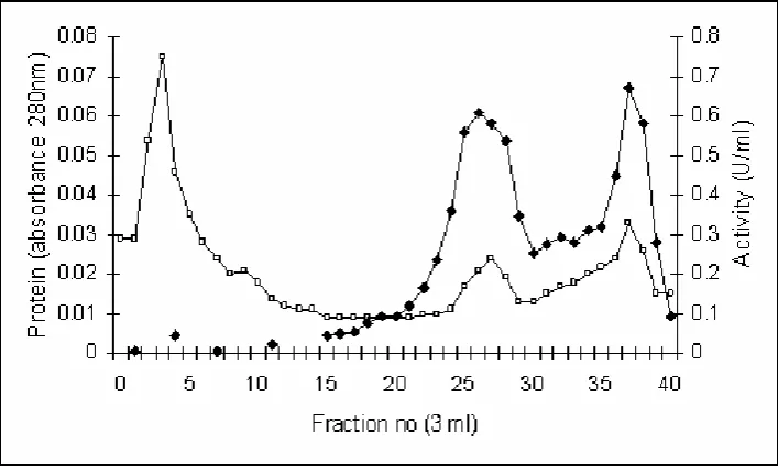
Bacillus licheniformis MB-2 chitosanase was harvested at the seventh days of fermentation using 15% starter, at 55°C 120 rpm shaker bath. Crude enzyme was obtained by concentrating the cell free supernatan with 80% saturated ammonium sulphate. Purification was done by hydrophobic interaction colomn chromatography as previously mention, and the relatively pure enzyme (Figure 1, Fraction 2) was collected.
Oligomer chitosan was made by reacting the enzyme, both crude, and pure, to 1 of 85% deacetylated chitosan. Optimal concentration of the crude enzyme effectively retarded the 6 pathogenic bacteria tested was 0.1 IU per mg chitosan with reaction time of 1 to 3 h (Meidina etal., 2004). The diameter of a clear zone as antibacterial indication was about 10 to 19 mm for the 6 pathogenic bacteria. This concentration was used in this experiment with reaction time of 1 h. For pure enzyme, the same concentration was used. In this experiment, oligomer possesing antibacterial properties was identified based on HPLC analysis. It was found that oligomer mixture were in the from of 2 to 6 unit oligomer.
Figure 1. Elution profile of chitosanase and protein during hydrophobic interaction chromatography using Butyl Sepharose 4 FF matrix.
Remarks: = chitosanase activity; = absorbance 280 nm•
Antibacterial Study
Previous study by Meidina resulted that MIC value of oligomer produced by the crude chitosanase, was lower (321 to 562 ppm) compared to the polymer chitosan (7,000 to 10,000 ppm), but it was much higher compared to the commercial antibiotics (5 to 100 ppm). The MIC value of Salmonella typhymurium was 321 ppm, while the MIC of Pseudomonas aeruginosa,
Staphylococcus aureus, and Escherichia coli were 402 ppm, and the MIC of Listeria monocytogenes and
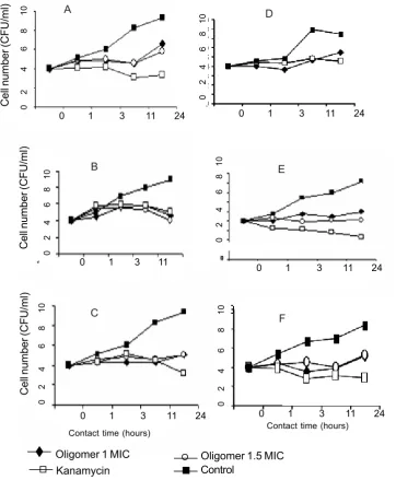
Bacillus cereus were 482 and 562 ppm, respectively. In this study, antibacterial potency of the oligomer was assesed by viability study. The concentration of oligomer used was at the MIC and 1.5 MIC, and the number of bacteria survived was counted during certain
time as indicated at Figure 2 below. Results showed that, in general, the number of all bacteria tested was reduced during 11 hours contact time. It was assumed that during that time all oligomer were already bound to the cells, and increasing bacterial number after that time may indicated the survived bacteria were capable to regain replication. The 1.5 MIC used was not significantly shown different effect to the number of bacterial reduction. Among bacteria tested,
Pseudomonas aeruginosa was the most sensitive. The number of the Pseudomonas aeruginosa was significantly reduced during the first 3 hours contact, while Escherichia coli and Salmonella typhimurium
were significantly reduced by 11 and 24 hours contact. On the other hand, gram positive pathogenic bacteria tested were reduced not as much as those gram negative bacteria. The results was in agree with
94
previous research results (Chung et al., 2004; Helander et al., 2001). It was reported that there was a positive relationship between antibacterial activity of chitosan and surface characteristics of bacteria cell wall. Gram negative bacteria which has more negative
charged and more hydrophilic were more sensitive to oligomer chitosan compared to the gram positive bacteria. Chitosan oligomer produced by pure chitosanase did not give different inhibition results as the crude one.
Contact time (hours)
Oligomer 1 MIC Oligomer 1.5 MIC
Kanamycin Control
Figure 2. Effect of chitosan oligomer concentration and contact time on to cell viability (A = Pseudomonas aeruginosa; B = Salmonella typhimurium; C = Escherichia coli; D = Staphylococcus aureus; E = Listeria monocytogenes; F = Bacillus cereus).
A
Anti Protease Activity of the Oligomer
Protease enzymes presented in bacterial cell surface has important role in defensive mechanism. It was hypothized that protease secreted by pathogenic bacteria was known as toxin, and some other were hydrolytic enzymes capable of degrading
extracellular matrix compounds of host polymer, for their carbon and energy sources.
In this experiment, chitosan oligomer produced by Bacillus licheniformis MB-2 was assessed for antiprotease. Results showed that chitosan oligomer at their MIC were able to retard 64% of Pseudomonas
aeruginosa protease having activity of 0.09 U per ml, while 25% retardation was obtained when using chitosan oligomer at 0.5 MIC. From the result, it can be deduced that the ability of chitosan oligomer, produced by Bacillus licheniformis MB-2 chitosanase, to inhibit pathogenic bacteria might through antiprotease mechanism.
CONCLUSIONS
1. Chitosan oligomers produced by Bacillus licheniformis MB-2 chitosanase has capability of reducing the 6 pathogenic bacterial tested by 2 to 5 log CFU per ml during 24 h contact.
2. Gram negative bacteria were more sensitive to the chitosan oligomer than the gram positive one. 3. The oligomers, at MIC value, was able to inhibit
Pseudomonas aeruginosa protease by 64%, suggesting that the ability to inactivate bacterial protease might be one of the antimicrobial mechanisms performed by chitosan oligomer. ACKNOWLEDGMENT
We acknowledged research support from Program B, Department of Food Science Technology, Faculty of Agricultural Technology, Bogor Agricultural University. The initial work was started through the cooperation between Research Center for Biotechnology, Bogor Agriculture University, Bogor, Indonesia and Bioproduct Research Center, Yonsei University, Seoul, Korea.
REFERENCES
Bergmeyer, H. U., J. Bergmeyer, & M. Grafi. 1983. Methods of enzymatic analysis. Vol.2. Weinheim. Verlag Chemie. Hal.1.007-1.009.
Carson, C. F. & T. V. Riley. 1995. Antimicrobial activity of the major components of the essential oil of
Melalueca alternifolia. Journal Appl Bacteriol. 78. 264-269.
Chasanah, E. 2004. Characterization of chitosanase of Bacillus licheniformis MB-2 from Manado hot spring water. Dessertation. Institut Pertanian Bogor. Bogor. Indonesia.
Chung, Y. C., Y. P. Su, C. C. Chen, G. Jia, H. I. Wang, J. C. G. Wu, & J. G. Lin. 2004. Relationship between antibacterial activity of chitosan and surface characteristics of cell wall. Acta Pharmacol Sin. 25. 932-936.
El-Ghouth A., R. Ponnampalam, F. Castaigne, & J. Arul. 1992. Chitosan coating to extent the storage life of tomatoes. Hortsci. 27. 1.016-1.018. Helander, I. M., E. L. Nurmiaho, R. Ahvenainen, J.
Rhoades, & S. Roller. 2001. Chitosan disrupts the barrier properties of the outer membrane of Gram-negative bacteria. Int Journal Food Microbiol. 71. 235-244.
Imada, C., U. Simidu, & N. Taga. 1989. Isolation and characterization of marine bacteria producing alkaline protease inhibitor. Bulletin Japanasse Soc Scientific Fishery. 51. 799-803.
Kendra, D. F. & L. A. Hadwiger. 1984. Characterization of the smallest chitosan oligomer that is maximally antifungal to Fusarium solani and elicits pisatin formation in Pisum sativum. Exp Mycol. 8. 276-281.
Meidina, B. S. L. Jenie, Sugiyono, & M. T. Suhartono. 2004. Aktivitas antibakteri oligomer kitosan yang diproduksi menggunakan kitosanase dari isolat
Bacillus licheniformis MB-2. Seminar Nasional PATPI. Jakarta.
Muzarelli, R. A. A., R. Farsi, O. Filippini, E. Giovanetti, G. Biagini, & P. E. Varaldo. 1990. Antimicrobial properties of N-Carboxybutyl chitosan. Antimicr Agents Chemoth.34. 2.019-2.023.
Roller, S. & N. Covill. 1999. The antifungal properties of chitosan in laboratory media and apple juice.
Int. Journal Food Microbiol. 47. 67-77.
Rhoades, J. & S. Roller. 2000. Antimicrobial actions of degraded and native chitosan against spoilage organism in laboratory media and foods. Appl. and Enviroment Microbiology. Jan. 80-86.
Sagoo, S., R. Board, & S. Roller. 2002. Chitosan inhibits growth of spoilage microorganisms in chilled pork products. Food Microbiol.19. 175-182.
Antibacterial Potency of Chitosan ... by Bacillus Licheniformis MB-2 Chitosanase (Chasanah, E., et al.)
INDONESIAN FISHERIES RESEARCH JOURNAL Instructions for Authors
Scope
Indonesian Fisheries Research Journal publishes research results on resources, oceanography and limnology for fisheries, fisheries biology, management, socio-economic and enhancement, resource utilization, aquaculture, post harvest, of marine, coastal and inland waters.
Manuscripts submission
Manuscripts submitted to the Journal must be original with clear definition of objective, materials used and methods applied and should not have been published of offered for publication elsewhere. The manuscripts should be written in English, in
double-spaced typing on one side A4-size white papers. Three copies of the manuscripts and a disk containing the manuscript file (s)
produced using MS Word must be submitted to the Managing Editor, Jl. Pasir Putih I Ancol Timur, Jakarta 14430, Indonesia,
E-mail: [email protected]. There are no page charges for the manuscripts accepted for publications, unless otherwise
indicated below.
Manuscripts preparation
- A title page should be provided which shows the title and the name(s) of the author(s). The author(s)’ position and affiliation
should be written as footnotes at the bottom of the title page.
- Title should not be more than 15 words.
- Abstract summarizes the study is not more than 250 words. Keywords (3-5) must be provided and conform with Agrovocs.
- Introduction should concisely establish the relate to which the article concerns, the purpose and the importance of the work.
Do not use any sub-headings.
- Materials and Methods should clearly and concisely describe the experiment with sufficient detail for independent repetition.
- Results are presented with optimum clarity with unnecessary detail. No same results should be presented in both tables and
figures. Tables (double spaced, same font size with text, no vertical line, lower case supercript letters to indicate, footnotes),
and illustration/figures should be printed on separate sheets and given arabic number consecutively. illustration/figures
should be submitted in original (1) and copied (2) forms and be sufficiently sharp for reproduction. 1 Photographs are
preferably black and white on glossy papers with good contrast. Color photographs of good quality are acceptable but the cost of reproduction must be paid for by the author(s). Exemption for bearing the cost can be sought from the publisher by written request.
- Conclusion should be short with regard to the title, objectives, and discussion of results.
- Acknowledgement, if necessary, should be kept minimum (less than 40 words).
- References should be cited in the text by the author(s)’ family or last name and date in one or two forms: Wang (1985) or
(Wang, 1985). For references with more than two authors, cite the first author plus et al. Full citation in alphabetical order is
required for the references list in the following style:
Sunarno, M.T.D., A. Wibowo & Subagja. 2007. Identifikasi tiga kelompok ikan belida (Chitala lopis) di Sungai Tulang Bawang,
Kampar dan Kapuas dengan pendekatan biometrik. Jurnal Penelitian Perikanan Indonesia. 13 (3): 1-14.
Sadhotomo, B. 2006. Review of environmental features of the Java Sea. Indonesia Fisheries Research Journal. 12 (2): 129-157.
Collins, A. 1977. Process in acquiring knowledge. In Anderson, R.C., Spiro, R.J. and Montaque, W.E. (eds.). Schooling and the
Acquisition of Knowledge. Lawrence Erlbaum, Hillsdale, New Jersey. p.339-363
Bose, A.N., Ghosh, S.N., Yang, C.T. and Mitra, A. 1991. Coastal Aquaculture Engineering. Oxford & IBH Pub. Co. Prt. Ltd., New
Delhi. 365 pp.
Smith, T.I.J. and Sandifer, P.O. 1983. Development of prawn (Macrobrachium rosenbergil) farms in temperate climates:
Prospects and problems in the United States. In Roger, G.L., Day, D. and Um, A. (eds.) Proceedings of the First International
Conference on Warm Water Aquaculture - Crustacea. Brigham Young University, Laie, Hawaii, USA. p 109-126.
Unpublished materials should not be used, except for thesis which is cited as follows:
Simpson, B.K. 1984. Isolation, Characterization and Some Applications of Trypsin from Greenland Cod (Gadus morhua).
PhD Thesis. Memorial University of New Foundland, St. John’s, New Foundland, Canada. 179 pp.
- Acronyms or uncommon abbreviations must be given in full with the first mentions; new abbreviations should be coined
only for unwieldy name and should not be used at all unless the names occur frequently.
- Latin name and family of the species should be given besides its common name at first mention in the manuscript, and the
common name only for the next mentions.
- SI Systems should be used for all measurements.