Journal of The Korean Wood Science and Technology
2016
∙
3
pISSN 1017-0715
eISSN 2233-7180
CONTENTS -
Ⅱ
Vol. 44 No. 2
March 2016
CONTENTS -
Ⅰ
Anatomical Characteristics of Paulownia tomentosaRoot Wood
Yue Qi, Jaehyuk Jang, Wahyu Hidayat, Aehee Lee, Sehwi Park, Seunghwan Lee, Namhun Kim …157
Size Reduction Characterteristics of Yellow Poplar in a Laboratory Knife Mill
Hyoung-Woo Lee………166
Quality Improvement of Oil Palm Trunk Properties by Close System Compression Method
Rudi Hartono, Imam Wahyudi, Fauzi Febrianto, Wahyu Dwianto, Wahyu Hidayat, Jae-Hyuk Jang, Seung-Hwan Lee, Se-Hwi Park, Nam-Hun Kim ………172
A Study on Dimensional Stability and Thermal Performance of Superheated Steam Treated and Thermal Compressed Wood
Hyunwoo Chung, Yeonjung Han, Jun-Ho Park, Yoon-Seong Chang, Yonggun Park,
Sang-Yun Yang, Hwanmyeong Yeo ………184
Comparison of Dynamic Sorption and Hygroexpansion of Wood by Different Cyclic Hygrothermal Changing Effects
Tiantian Yang, Erni Ma………191
Optimization of L-shaped Corner Dowel Joint in Modified Poplar using Finite Element Analysis with Taguchi Method
Qing Ke, Fan Zhang, Yachi Zhang………204
Enhancing Enzymatic Digestibility of Miscanthus sinensisusing Steam Explosion Coupled with Chemicals
Ji Young Jung, Jae-Kyung Yang ………218
Vol.
44
N
o.
2
2 0 1 6 ∙
3
(
(
��
March 2016
………
………
………
………
………
1. INTRODUCTION
The wood structure of woody plant species is
mainly studied on stems (Kim et al., 2012;
2013) and branches (Jacobsen et al., 2007a;
Poorter et al., 2010; Qi et al., 2014), mostly because of their obvious function in supporting
of plant’s life. Roots also support important
functions in plants. For example, they anchor the plants in the substratum, and facilitate the
uptake of water and minerals from the
environment. Furthermore, roots are important for the storage of reserves. As a consequence,
the root wood structure can significantly affect
Original Article
Anatomical Characteristics of
Paulownia tomentosa
Root Wood
1Yue Qi2⋅Jaehyuk Jang2,3⋅
Wahyu Hidayat2,4⋅Aehee Lee2⋅
Sehwi Park2,5⋅Seunghwan Lee2⋅
Namhun Kim2,†
ABSTRACT
This study investigated several anatomical characteristics of Paulownia tomentosa roots. The root wood was separated into three parts from stem base (top, middle, and base) at different positions below ground. Qualitative anatomical data suggested that the growth rings in earlywood and latewood were structurally different. Furthermore, the root wood vessels were found having 2 to 3 radial multiples and they were appeared in the form of clusters. In addition, some sheath cells and septate axial parenchyma were observed. Regarding the quantitative anatomical characteristics, vessel and ray numbers per mm2, as well as ray width and height
differed significantly among the top, middle, and base rood wood parts. However, there were no significant differences in vessel diameters, cell wall thickness, and width and length of wood fibers among those parts. The crystallinity of the root top part was slightly higher than that of the middle and base parts. Furthermore, the vessel numbers, ray numbers, and ray width and height in the near pith (NP) area were higher compared to those in the near bark (NB) area. However, the fiber width and fiber length at NP were lower than those at NB. Overall, this study demonstrated some significant differences in the anatomical characteristics of the top, middle, and base parts of root wood from Paulownia tomentosa.
Keywords : Paulownia tomentosa, root wood, wood anatomy
1 Date Received January 15, 2016, Date Accepted February 25, 2016
2 College of Forest and Environmental Sciences, Kangwon National University, Chuncheon 24341, Republic of Korea 3 The Institute of Forest Science, Kangwon National University, Chuncheon 24341, Republic of Korea
4 Faculty of Agriculture, Lampung University, Bandar Lampung 35145, Indonesia 5 Faculty of Forestry, Bogor Agricultural University, Bogor 16680, Indonesia
†
Yue Qi⋅Jaehyuk Jang⋅Wahyu Hidayat⋅Aehee Lee⋅Sehwi Park⋅Seunghwan Lee⋅Namhun Kim
the wood quality of the rest of the tree.
A few previous studies have investigated the
anatomy of root wood (Patel, 1970; Matsumura
and Butterfield, 2001). Schuldt et al. (2013) in-vestigated the density, anatomy, and hydraulic
properties of root wood of tropical rainforest
trees. Furthermore, several studies compared root wood with stem wood or branch wood.
Rao et al. (1989) claimed that the average
ves-sel tangential diameters, and vesves-sel length of root wood were substantially smaller than those
of stem wood. Stokke and Manwiller (1994)
showed that for black ash (Quercus velutina)
the vessel elements, fibers, rays, and axial pa-renchyma differed among stem, branch, and
root woods. In addition, Palhares et al. (2007)
found that the amount of parenchyma cells and thin-walled fibers differed between root wood
and stem wood. Lee and Eom (2011) identified
the wood anatomy of yellow-poplar grown in Korea, and compared stem and root wood for
their vessel and ray elements, and quantitative
anatomical features. Furthermore, Fortunel et al.
(2014) investigated differences of wood specific gravity and anatomy between branches and
roots in 113 Amazonian rainforest wood
species. They demonstrated that the fiber
properties were the major contributors to wood
specific gravity, independent of vessel
prop-erties, across branches and small woody root.
In Korea, Paulownia tomentosa is a
fast-growing tree species, and its wood can be
used for high-value applications, such as making
Korean traditional musical instruments and furniture. In order to optimally utilize Paulownia
tomantosa wood as a bioresource, and to gain more knowledge of its wood qualities, the
ana-tomical characteristics of stem, branch, and root
wood of this species should be studied. In our
previous study (Qi et al., 2014), we reported the anatomical and physical properties of branch
wood. In this study, we examined the root wood
anatomical characteristics, and compared the dif-ferences among its top, middle, and base parts.
2. MATERIALS and METHODS
2.1. Materials
The eleven and thirteen years old Paulownia
tomentosa trees were obtained from research
forest (N 37°51'/ E 127°48') of Kangwon na-tional university in South Korea (Table 1). The
experimental samples for investigating
anatomi-cal properties were cut from root parts of
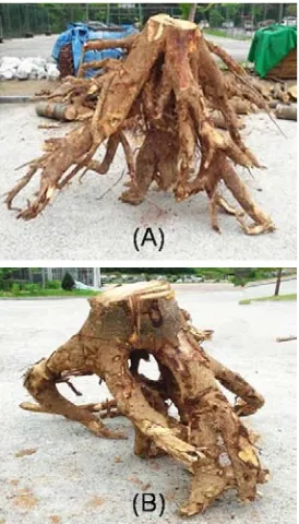
Paulownia tomentosa trees (Fig. 1). The rooting
depth of the studied root wood was
approx-imately 2.0 m. Therefore, samples were
col-lected as follows: the root top part consisted of root wood with a diameter of 17.5 cm and was
sampled 0.5 m below ground; the middle part
with a diameter of 12 cm and was sampled between 0.5 m and 1.0 m below ground; the
base part had a diameter of 8.5 cm and was
sampled between 1.0 and 1.5 m below ground.
Sample trees DBH (cm) Height (m) above ground
Paulownia tomentosa 1 30.9 15.0
Paulownia tomentosa 2 31.2 14.5
The obtained root were separated into near pith
(NP) and near bark (NB) parts before analyzing
their anatomical properties.
2.2. Anatomical characteristics
The blocks of NP and NB root wood were obtained from wood discs.
Fifteen-micro-meter-thick slices were cut from per root parts
by using a sliding microtome (Nippon Optical works, Japan). These sections were stained with
Safranin-Astra blue (Von, 1973; Lillie, 1977;
Jourez et al., 2001), dehydrated in a graded ser-ies of alcohol (50%, 70%, 90%, 95%, and
99%), mounted using Canada balsam, and
ob-served under an optical microscope (Nikon
Eclipse E600, Japan). The anatomical
character-istics, such as number and diameter of vessels,
height and width of rays, fiber length, and
structure were determined according to the IAWA hardwood feature list (IAWA Committee,
1989) using Total Imaging Solution software
(IMT, I-solution Lite, USA). For each charac-teristic, fifty random measurements were made
to determine a mean value.
2.3. Crystalline characteristics
An X-ray diffractometer (Rigaku DMAX2100V, Japan) equipped with a Cu target was used
for measuring the crystalline properties, at
40 kV and 40 mA. The root wood relative
crystallinity (%) and crystallite width (mm) were calculated by Segal’s equation (Segal et
al., 1959) and Scherrer’s equations (Alexander
et al., 1969), respectively.
2.4. Statistical analysis
Differences in the quantitative features among
the root wood top, middle, and base were
stat-istically examined with one-way ANOVA, and
post-hoc Duncan’s tests (IBM SPSS ver. 21, USA).
3. RESULTS and DISCUSSION
3.1. Qualitative anatomical characteristics
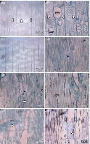
The anatomical characteristics are presented
in Fig. 2 A-H. The root wood showed rela-tively indistinct growth ring boundaries, because
Fig. 1. The samples: (A) the root obtained from
Yue Qi⋅Jaehyuk Jang⋅Wahyu Hidayat⋅Aehee Lee⋅Sehwi Park⋅Seunghwan Lee⋅Namhun Kim
of a less narrow zone of flattened axial
paren-chyma cells between the earlywood and
late-wood (Fig. 2 A and B). The root late-wood vessels
were solitary, occurred in radial multiples of 2 to 3, and were occationally clustered (Fig. 2 A
and B). Axial parenchyma was aliform and
confluent (Fig. 2 C). The wood rays consisted mostly of 2 to 4-seriate cells, along with
occa-sional uniseriate, and were surrounded by
sep-tate and nonsepsep-tate axial parenchyma (Fig. 2 D - F). Sheath cells were present, but only rarely
(Fig. 2 D). Ray cells displayed procumbent
forms (Fig. 2 G and H). Furthermore, vessel
perforation plates were simple, and the inter-vascular pits alternated (Fig. 2 E and H). As a
result, the growth rings were indistinct in the
root wood, whereas they were distinct in stem
and branch wood (Jeong et al., 2008; Lee,
1994; Park et al., 1981; Qi et al., 2014). These
results also agreed with Fayle (1968b), Patel (1971), Stokke and Manwiller (1994), and Lee
and Eom (2011).
3.2. Quantitative anatomical characteristics
Anatomical characteristics of Paulownia
to-mentosa root wood are summarized in Table 2. The vessel numbers were 5.8, 5.0, and 4.5 per
mm2 in top, middle, and base parts of root
wood, respectively. These differences were sig-nificant with the top part showing the highest
value among them. The vessel numbers in NP
and NB wood were similar, with no significant
difference between them. The average vessel number in root wood was less than compared
with that in branch wood, which was
approx-imately 10 per mm2 (Qi et al., 2014).
The tangential diameters of vessels in root
wood were 186.0, 182.2, and 182.5 µm, where-as the radial diameters were 249.6, 238.5, and
246.5 µm in top, middle, and base parts,
respectively. The vessel diameters did not dis-tinctly differ among the root parts, and no
sig-nificant difference were identified between NP
and NB wood. The average tangential vessel di-ameter in branch wood was 180.0 µm and the
radial dimeter 210.2 µm (Qi et al., 2014).
Overall, there were no distinct differences in
vessel diameter between root wood and branch wood.
The cell wall thickness of wood fibers in root
wood was 3.4, 3.4, and 3.6 µm in tangential di-rection, and 3.2, 4.2, and 4.0 µm in the radial
direction of top, middle, and base parts,
respectively. No significant statistical differ-ences were noted in cell wall thickness among
the different root parts. Furthermore, there was
no difference in the cell wall thickness between
NP and NB. The cell wall thickness was thick-er in branch wood than in root wood, because
of the gelatinous layer existing in cell walls of
the branch wood (Qi et al., 2014).
The width of wood fiber was 29.0, 28.8, and
28.3 µm, and the length of wood fibers was
725.2, 746.0, and 772.5 µm in top, middle and base parts, respectively. The fiber width and
length were not different among the root parts.
No significant differences were displayed in
Yue Qi⋅Jaehyuk Jang⋅Wahyu Hidayat⋅Aehee Lee⋅Sehwi Park⋅Seunghwan Lee⋅Namhun Kim
stem wood, whereas the root wood fiber length
was shorter than in stem wood (Jeong et al.,
2008; Lee, 1994). Furthermore, fiber length in
root wood was slightly shorter than in branch wood,which had an average value of 800 µm
(Qi et al., 2014).
The average ray numbers of root wood were 14.2, 12.1, and 10.9 in the top, middle and
base parts, respectively. Statistical analysis
showed that the ray numbers were significantly different among the root parts (Table 2), and
the ray numbers decreased from the top of the
root wood to the base. There were no differ-ences in ray numbers between NP and NB.
Furthermore, in our previous study, branch
wood ray numbers were ca. 18 per mm2 (Qi et
al., 2014), which makes the ray numbers of
root wood lower than those of branch wood. Wood ray width was 38.0, 46.8, and 52.3
µm, and ray height was 283.2, 311.2, and 325.1
µm in the top, middle, and base parts of root wood, respectively. The ray width and height
were significantly different among the root
parts, and increased from the top to the base part. The ray width and height in NP were a
little higher than those in NB. Interestingly, the
ray width and height in the root wood were higher than in the branch wood, which had a
Characteristics Top Root Base P-value
NP* NB* Mean NP* NB* Mean NP* NB* Mean
Notes: means within a row followed by the same capital letter are not significantly different at 5% significance level using Duncan’s test.
ray width of 34.5 µm, and a ray height of
227.7 µm (Qi et al., 2014).
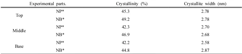
3.3. Crystalline characteristics
The crystalline characteristics of Paulownia tomentosa root wood are shown in Table 3.
The relative crystallinity of root wood was
slightly different in the top, middle, and base parts (Table 3). Furthermore, the relative
crys-tallinity in NB was a somewhat higher than
that in NP. The crystallinity in NB varied from
49.2% (top) to 44.8% (base), whereas the crys-tallinity in NP was 45.3% (top), 42.3%
(middle) and 42.2% (base), respectively.
However, there was no obvious trend for crys-tallite width differences among the different
root parts. The crystallinity of branch wood,
which was 61.9%, showed higher value than that of root wood (Qi et al., 2014). This
differ-ence in relative crystallinity, like the cell wall
thickness difference, can be explained by the
presence of the gelatinous layer in the branch wood cell walls (Goto et al., 1978; Harada and
Goto, 1982; Lee et al., 1997; Lee and Kim,
1993).
4. CONCLUSION
Anatomical characteristics of root wood from
Paulownia tomentosa were investigated.
Overall, the root wood growth rings were
indis-tinct in root parts. Furthermore, the vessels in the root wood appeared solitary, with radial
multiples of 2 to 3, and were occasionally
clustered. Some sheath cells and septate axial parenchyma were additionally observed in the
wood samples.
The vessel and ray numbers, as well as the ray width and height were significantly different
among the root wood top, middle and base
parts. The ray numbers decreased from the top
to base parts, while the ray width and height increased. In contrast, there were no significant
differences in vessel diameters, cell wall
thick-ness, and wood fiber width and length among the different root parts. Furthermore, the top
part crystallinity was slightly higher than in the
middle and base parts of root wood. There
were no big differences between the anatomical characteristics of the NB and NP. Overall,
the top, middle, and base parts of root
wood differed in some of their anatomical
Experimental parts. Crystallinity (%) Crystallite width (nm)
Top NP* 45.3 2.78
NB* 49.2 2.78
Middle NP* 42.3 2.70
NB* 46.9 2.68
Base NP* 42.2 2.58
NB* 44.8 2.87
Yue Qi⋅Jaehyuk Jang⋅Wahyu Hidayat⋅Aehee Lee⋅Sehwi Park⋅Seunghwan Lee⋅Namhun Kim
characteristics.
ACKNOWLEDGEMENT
This study is supported by 2015 Research Grant
from Kangwon National University (No. 520150258).
Yue Qi also sincerely thanks the ACES-KNU
schol-arship of Kangwon National University for financial
support from 2012.
REFERENCES
Alexander, L.E. 1969. X-ray diffraction in polymer
science. Wiley-Interscience, Amsterdam pp:
423∼424.
Fortunel, C., Ruelle, R., Beauchene, J., Fine, P.V.A.,
Baraloto, C. 2014. Wood specific gravity and
anatomy of branches and roots in 113
Amazonian rainforest tree species across
environmental gradients. New Phytologist 202:
79∼94.
Goto, T., Harada, H., Saiki, H. 1978. Fine structure
of cellulose microfibrils in poplar gelatinous
layer and valonia. Wood Science and
Technology 12: 223∼231.
Harada, H., Goto, T. 1982. The structure of cellulose
microfibrils in Volonia. “cellulose of other
natural polymer systems”. Malcolm Brown, Jr.,
Eds., Plenum press: 383∼401.
IAWA Committee. 1989. IAWA List of microscopic
features for hardwood identification. IAWA
Bulletin n.s. 10(3): 219∼332.
Jacobsen, A.L, Agenbag, L., Esler, K.J., Pratt, R.B.,
Ewers, F.W., Davis, S.D. 2007a. Xylem density,
biomechanics and anatomical traits correlate with
water stress in 17 evergreen shrub species of the
Mediterranean-type climate region of South
Africa. Journal of Ecology 95: 171∼183.
Jeong, S.H., Park, B.S. 2008. Wood properties of the
useful tree species grown in Korea. Korea Forest
Research Institute 29: 348∼368.
Jourez, B., Riboux, A., Leclercq, A. 2001.
Anatomical characteristics of tension and
opposite wood in young inclined stem of
Poplar (Populus euramericana cv ‘Ghjoy’).
IAWA Journal 22: 133∼157.
Kim, J.H., Jang, J.H., Ryu, J.Y., Hwang, W.H.,
Febraianto, F., Kim, N.H. 2013. Comparison of
anatomical characteristics of White Jabon and
Red Jabon grown in Indonesia. Journal of
Korean Wood science and technology 41(4):
327∼336.
Kim, J.H., Jang, J.H., Kwon, S.M., Febraianto, F.,
Kim, N.H. 2012. Anatomical properties of major
planted and promising species growing in
Indonesia. Journal of Korean Wood science and
technology 40(4): 244∼256.
Korean standards association. 2004. KS F 2198.
Lee, M.R., Eom, Y.G. 2011. Comparative wood
anatomy of stem and root in Korean-grown
Yellow-poplar (Liriodendron tulipipfera L.).
Journal of Korean Wood science and technology
39(5): 406∼419.
Lee, S.H., Hwang, W.J., Kim, N.H. 1997. Some
ana-tomical characteristics in tension and opposite
wood of Quercus mongolica Fisher. Journal of
Korean Wood science and technology 25(3):
43∼49.
Lee, W.Y., Kim, N.H. 1993. Crystal structure of
ten-sion wood by x-ray diffraction method. Journal
of Korean Wood Sciencen and Technology 21
(4): 65∼73.
Lee, P.W. 1994. The structures of Korean domestic
woods. -Microscopic anatomy- JeongMinSa,
Seoul, Republic of Korea.
Lillie, R.D. 1977. Conn’s biological stains. Williams
Matsumura, J., Butterfield, B.G. 2001. Microfibril
angles in the root wood of Pinus radiata and
Pinus nigra. IAWA Journal 22: 57∼62.
Palhares, D., de Paula, J.E., Rodringues Pereira,
L.A., dos Santos Silveira, C.E. 2007.
Comparative wood anatomy of stem, root and
xylopodium of Brosimum gaudichaudii
(Moraceae). IAWA Journal 28(1): 83∼94.
Patel, R.N. 1971. Anatomy of stem and root wood
of Pinus radiata D. Don. New Zealand Journal
of Forest Science 11: 37∼49.
Poorter, L., McDonaldM, I., Alarcon, A., Fichtler,
E., Licona, J.C., PenaClaros, M., Sterck, F.,
Villegas, Z., Sass-Klaassen, U. 2010. The
im-portance of wood traits and hydraulic
con-ductance for the performance and life history
strategies of 42 rainforest tree species. New
Phytologist 185: 481∼492.
Qi, Y., Jang, J.H., Park, S.H., Kim, N.H. 2014.
Anatomical and Physical Characteristics of
Korean Paulownia (Paulownia coreana) Branch
Wood. Journal of Korean Wood science and
technology 42(5): 510∼515.
Rao, R.V., Sharma, B., Dayal, R. 1989. Anatomy of
aerial rootwood of Sonneratia caseolaris (L.)
Engler (Sonneratioideae). IAWA Bull.n.s 10(4):
374∼378.
Schuldt, B., Leuschner, C., Brock, N., Horna, V.
2013. Changes in wood density, wood anatomy
and hydraulic properties of the xylem along the
root-to-shoot flow path in tropical.
rainforest trees. Tree Physiology 33: 161∼174.
Segal, L., Creely, J.J., Martin, A.E., Conrad, C.M.
1959. An empirical method for estimating the
degree of crystallinity of native cellulose using
the X-ray diffractometer. Textile research Journal
29: 786∼794.
Stokke, D.D., Manwiller, F.G. 1994. Pro-portions of
wood elements in stem, branch, and root wood
of black oak (Quercus velutina). IAWA Journal
15(3): 301∼310.
Von Aufsess, B.H. 1973. Microscopic scope of
lignifications by staining methods. European
Journal of Wood and Wood Products 31(1):