Pathomechanism of atherothrombotic disease
caused by
Streptococcus mutans
Purwanto, I Dewa Ayu Susilawati
Biomedic Division Dentistry Faculty University of Jember Indonesia
ABSTRACT
Background: Streptococcus mutans, the main bacteria of dental caries, can easily invade circulatory system and was reported to have the ability to survive and colonize on atherosclerotic plaque, therefore was suspected to contribute in pathomechanism of atherotrombotic disease. It was assumed that S. mutans play a role in atherotrombotic disease due to its potency to produce proteases which were directly or indirectly via (Matrix metalloproteinases, MMPs) causing subendothelial vascular collagen fragmentation leading to platelet aggregation. Purpose: This study purposed to demonstrate in vitro that S. mutans could (directly or indirectly) cut subendothelial vascular collagen (type IV collagen) to become fragments which subsequently stimulated platelet aggregation. Method: Collagen fragmentation was analyzed by means of Sodium Dodecyl Sulphatpolyacrylamid Gel Electrophoresis (SDS-PAGE) and Soluble Biotinylated Assay (SBA), while platelet aggregation was analyzed microscopically and spectrophotometrically. Result: Result showed that S. mutans (directly and indirectly), could fragmentate type IV subendothelial vascular collagen leading to platelets aggregation. Conclusion: The potency of S. mutans on subendothelial collagen degradation and platelet aggregation may suggest the molecular mechanism of S. mutans involvement in pathogenesis of atherothrombotic disease.
Key words: Streptococcus mutans, protease, MMPs, collagen, platelet.
Correspondence: Purwanto, Biomedic Division Dentistry Faculty University of Jember Indonesia. E-mail:[email protected]
INTRODUCTION
Studies related to S. mutans (the primary
bacterial cause of dental caries) are mostly reviewing its role in dental caries. This is due to the ability of S. mutans to metabolize glucose to produce acids that cause tooth demineralization. Meanwhile,
based on genome analysis, S. mutans can also
produce proteases.1 Unfortunately research that
concerned with its proteolytic potency of S. mutans
is very limited.
Streptococcus mutans, from deep carious lesion
can easily invade into systemic blood circulation2
and was reported to have ability to survive and colonize on endothelium3 and coronary atherosclerotic
plaque,4 therefore, it was suspected to contribute in
pathomechanism of atherotrombotic disease. It was reported that 40% S. mutans enter blood stream during tooth brushing and 88% after periodontal surgery.2 Our
previous study found that antibody anti-S. mutans exist in blood serum of acute myocardial infarction patients.5
coronary heart disease when the disease involving coronary arteria and causes stroke when involving the arteria that supply blood to the brain, i.e internal
carotid arteria. The potency of S. mutans role in
pathogenesis of atherotrombotic disease has not been studied yet.
New paradigm that emerged recently shown that atherothrombotic disease is caused by inflammation response against the invasion of bacteria into the circulatory blood vessel wall. Bacterial invasion induce activation of phagocytes to produce proteinases mainly matrix metalloproteinase (MMPs), that causes the degradation of vascular collagen matrix. Collagen vascular damage readily induce platelet aggregation followed by activation of the coagulation cascade resulting in thrombus formation.7-9
It was reported that some bacteriae could induce production of MMPs zymogen (pro-MMPs) in
phagocytes such as monocyte.10 Meanwhile there
were also reported that proteases of bacteria could
activate pro-MMPs to become active MMPs.11,12 Those
enzymes play a key role in the degradation of
extracellular matrix, including collagen. S. mutans
capable to produce proteases, therefore it was
assumed that S. mutans proteases can activate
monocyte pro-MMPs to become active MMPs, that can breake down collagen into fragments. In addition to its potency to activate MMPs, S. mutans proteases was thought to be able to degrade collagen directly. The aim of this study was analyzing the role of
S. mutans in pathogenesis of atherotrombotic disease,
in vitro, by proving that S. mutans directly or indirectly (by its interaction with monocyte) break down vascular type IV collagen to become fragments, and those fragments stimulated platelet aggregation.
MATERIALS AND METHODS
Peripheral venous whole blood from healthy
donor. Wild type S. mutans was obtained from
Microbiology Laboratory Faculty Dentistry University of Airlangga. TYC medium was purchased from Topley House UK, Type IV collagen (Sigma), BHIB medium (Sigma), Biotin-N-Hydroxysucci-nimide Ester (ICN Biomedicals, Inc). Biotin-N-Hydroxysuccinimide Esther (ICN Biomedicals, Inc.). Rabbit anti-human MMP-9 (Sigma); Anti-rabbit IgG-biotin (MP Biomedicals, Inc), Nitrocellulose membrane (NC, KPL), SA-HRP (streptavidin avidin-Horse Reddish peroxidase, Dako), TMB (Tetra methylbenzidine, KPL), high-range protein molecular weight marker (Bio-Rad).
Streptococcus mutans were suspensed in BHIB medium and incubated 2 x 24 hours at 37° C. Using spectroscopic methods, the concentration of S. mutans
was adjusted to 109 per ml. Protease of S. mutans
was prepared from culture medium after being incubated for 2x24 h, rt. The medium was centrifugated 3500 rpm, 10 min, rt. Supernatan contain proteases was then filtered using microfilter 0,2 µm (Sartorius)
Monocyte was isolated by means of ficoll hypaque centrifugation. Six ml heparinized whole blood was diluted in HBSS (1:3), then layered on ficoll (1:3) and centrifuged 30 min, 1400 rpm, at room temperature (rt). The buffy coat layer contain mononuclear cells are aspirated, put in falcon tube, washed 3 times and resuspensed with 2 cc HBSS. Suspension of mononuclear cells were then layered
on gelatin (0.5%) and incubated 1 h, 37°C, rt. Non
adherent cells were removed and rinsed 3 times. The adherent cell (monocytes) were gently scaped and suspensed in 3 cc HBSS, then washed 2 times.
Pellet cells were then resuspensed in 300 µl HBSS
and ready for further analysis.
MMPs was prepared from monocyte medium
after being incubated with S. mutans. A total of
250 µl monocytes suspension were exposed to
250 µl suspension of S. mutans and incubated for 18
h, 37°C. After centrifugated at 3500 rpm, 10 min, rt, supernatant contain active MMPs was aspirated and filtered (Sartorius 2µm). To confirm the presence of
MMPs products, we tested MMP-9 secretion by monocytes with imunocytochemistry assay.
Collagen type IV (5 mg) was dissolved in 2.5 ml of acetic acid 25%. For biotin labelling, 500 µl collagen
suspension was added to 500 µl d-biotin and stirred
4°C, 24 h. Dialysis was then performed to remove
excess biotin in sterile H2O, 24h, 4° C, followed by sterile PBS, 24 hours, 4° C.
Collagen fragmentation was analyzed by means of SDS-PAGE dan SBA (modified from methods by Romanelli et al, 1999).13 For direct fragmentation we
reacted biotinylated collagen with S. mutans proteases, while for indirect fragmentation with monocytes MMPs. Briefly 100 µl suspension containing S. mutans
protease or MMPs was reacted with 100 µl biotinylated
type IV collagen for 18 h, 37° C. This process caused collagen degradation into fragments. This reaction was
stopped by addition of 100 µl RSB and heating 100°C
SA-HRP for 2 h. After being washed 2 times, membranes were incubated with TMB substrate, stop reaction with aquades. Collagen fragments are visualized as a colored band on the membrane blot. The character (molecular weight) of collagen fragment were identified by comparing it with standard molecular weight proteins (Poncheau Red staining) . Platelet aggregation were analyzed using microscopic and spectrophotometric assay. Firstly, collagen fragments was prepared by SDS-PAGE, the identified fragments were isolated from gel using electroelusion methods. Eluent consist of collagen fragments were ready for further assay. For microscopic assay, 50 ul collagen fragments suspension were coated on cover slide in humid chamber, then, 500 ul of platelet suspension was layered on it and incubated for 18 h, 37° C. After removing the medium, cells was rinsed, fixed and stained with Giemsa. Platelet aggregation was observed under light microscope 400x magnification. Spectrophotometric assay was done to compare the optical density (334.4 nm) of platelet suspension before and after being reacted with suspension of collagen fragments.
RESULTS
Streptoccocus mutans produced proteases which can directly degrade type IV collagen into specific fragments. To perform fragmentation, biotinylated
collagen was reacted with medium culture of S.
mutans that was assumed to contain proteases, this was presented by SDS-PAGE (Figure 1A). To visualize collagen fragments only, SBA (biotinylated collagen) or ligand blotting method was used (Figure 1B).
A B
Figure 1. Fragmentation of type IV collagen by S. mutans
proteases.
collagen fragments and proteases-containing
culture medium of S. mutans).
B. SBA profile, line 1 protein marker, line 2, 3 and 4 type IV collagen fragments, comprises six fragments: 114, 70; 50; 40; 23 and 7 kDa.
This study also demonstrated that S. mutans
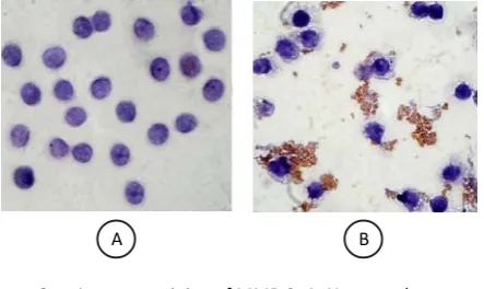
could induce monocyte to produce MMPs, one of them was MMP-9 (Figure 2). Furthermore, using SDS-PAGE method and SBA, it was proved that these MMPs can degrade type IV collagen to become fragments, they were fragments with molecular weigh of 70; 62 and 7 kDa (Figure 3)
A. SDS-PAGE profile, line 1 protein marker, line 2 untreated collagen, line 3 and 4 mixture of
A. SDS-PAGE profile, line 1 protein marker, line 2 & 3 untreated collagen, line 3 & 4 mixture of collagen fragments and MMPs.
B. SBA profile, line 1 protein marker, line 2, 3 and 4 type IV collagen fragments, comprises three fragments: 70; 62 and 7 kDa.
Figure 2. Immunostaining of MMP-9. A. Untreated monocyte did not express MMP-9; B. Monocytes induced by S. mutans produced MMP-9 (brown secrete)
A B
Figure 3. Fragmentation of type IV collagen by S. mutans -induced monocytes MMPs.
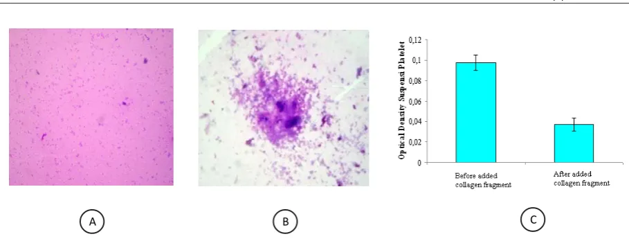
Formation of collagen fragments related to the the action of S. mutans were tested for its ability to induce platelet aggregation, Presentation of platelet aggregation test (interaction between platelets with fragments of collagen IV) were visualized in the form of microscopic picture and histograms of spectrophotometric data. Based on the microscopic image can be determined whether the aggregation of platelets occurred around the collagen fragments (Figure 4A, 4B). While spectrophotometric data presented the comparison of the optical density of platelet suspensions before and after being exposed to collagen fragments. Low optical density indicated that platelet aggregation occurs, and vice versa, a high optical density showed little platelet aggregation (Figure 4C).
DISCUSSION
S. mutans is well-known as an asidogenic bacterium, because they has enzymes for metabolizing carbohydrates that the final product are acids that causes a decrease in pH in the
environment surrounding the growth of S. mutans.
This acids production suggests that S. mutans
probably can lead to damage host proteins through a decrease of pH. However, in this study the hypothesis that collagen can be damaged by acidic
pH was not proven, because after culturing S.
mutans for 2 x 24 h, did not cause significantly changes in the pH to becomes acidic (data not shown). As an explanation for this is, the production
of acid by the glycolytic enzymes of S. mutans may
occur in cytoplasm, and it takes time to dispose acidic products into the extracellular space.
Therefore, the result of this study suggested that collagen degradation occurs not by acids but because of the activity of proteolysis enzymes.
Based on genomic analysis, S. mutans has a gene
that encodes the expression of several types of protease, i.e., serine protease HtrA, HtpX, Zn-dependent protease, two types of collagenase-like protease, serine protease RgpF and membrane proteases.1 These S. mutans proteases, however, has
not been characterized and identified its specificity. It is therefore important to do more research on the
S. mutans proteases.
In context of collagen degradation, in addition to
direct collagenolytic properties, S. mutans was
suggested to be able to induce monocyte to produce
MMPs,10 while other notions indicated that various
bacterial proteases may activate proMMPs.11,12
Therefore it is reasonably suspected protease S. mutans
can also enable to activate MMPs that cause collagen degradation. The results of this study proved, S. mutans
induced the expression of MMP-9 monocytes (evidenced by imunocytochemistry). Also shown that
S. mutans proteases can activate monocytes MMPs that cause degradation of collagen type IV. This study specifically analyzed the involvement of MMP-9, because it has been known that substrate specification of MMP-9 is type IV collagen.14
Type IV collagen is the principal constituent of subendothelial basement membrane, so it is a collagen of vascular wall which located at the most luminal surface. When there is invasion or attachment of foreign bacteria on vascular wall, there will be a vascular inflammatory response, in these circumstances, collagen type IV will be exposed firstly to the destructive effects of vascular inflammation. Therefore alleged that
Figure 4. Platelet aggregation assay. A. Platelets which incubated with HBSS seem to spread, B. Platelets that were exposed to collagen fragments appeare to aggregate (400 x). C. Optical density of platelet suspension before and after being incubated with collagen fragments
fragmentation of collagen type IV is the key step for platelet aggregation and thrombus formation.
Formation of collagen fragments related to the
action of S. mutans was proved to be able to induce
platelet aggregation. This results study supported the notion by Constantinides (1994), who stated that vascular collagen degradation is the most powerful stimulus for platelet aggregation and thrombus formation.15
Potency of S. mutans to induce vascular collagen degradation and platelet aggregation, concomitantly
with the ability of S. mutans to spread into blood
circulation, to envade endothelium and survive in coronary atherosclerotic plaque, provide better
understanding regarded the mechanism role of S.
mutans in pathogenesis of atherothrombotic diseases.
It can be concluded that Streptococcus mutans
produce proteases which directly or indirectly via activation of MMPs monocyte can be able to degrade type IV vascular collagen, and its fragments induce platelet aggregation. This mechanism may suggest the role of S. mutans cause atherothrombotic disease.
ACKNOWLEDGMENTS
We would like to thank “Hibah Pasca Project” of DP2M Dikti for funding this research. Special thank to Mbak Fitri, Mas Yuda and Mas Slamet for helping us in laboratory works.
REFERENCES
1. Ajdi D, Mc Shan WM, McLaughlin RE, Savic G, Chang J, Carson MB, Primeaux C, Tian R, Kenton S, Jia H, Lin S, Qian Y, Li S, Zhu H, Najar F, Lai H, White J, Roe BA, Ferritti JJ. Genome sequence of Streptococcus mutansUA159, a cariogenic dental pathogen. PNAS. 2002. Vol 99. No.2. p. 14434-9.
2. Vojdani A. A Look at infectious agent as posssible causative factor in cardiovascular disease. Part I. Science (microb. and virology/ immunologi). 2003 3. Stinson MW, Alder S, and Khumar S. Invasion and
killing of human endothelial cells by viridans group streptococci. J. Infect and Immun2003. p: 2365-72. 4. Nakano K, Nemoto H, Nomura R, Homma H,
Yoshioka H, Shudo Y, Hata H, Toda K, TaniguchiK, Amano A, Ooshima T. Serotype distribution of Streptococcus mutans a pathogen of dental caries in cardiovascular specimens from Japanese patients. J Med Microbiol. 2007. 5. p. 6551- 6.
5. Purwanto. Detection of IgG anti-Streptococcus mutans
in acute myocardial infarction patients. Stomatognatic 2008.Vol. 5. No. 2. Pp. 65-140.
6. Fuster V, Ross R, in Fuster V, Ross R, and Topol EJ. The Pathogenesis of atherosclerosis. Philadelphia Lippincott-Raven Publisher. 1996. 441- 56.
7. Brass LF. Thrombin and platelet activation. Chest 2003. 124: 18S-25S
8. Gibbin JM. Platelet adhesion signalling and the regulation of thrombus formation. Journal of Science 2004. 117, 3415 –25.
9. Furie B., Furie BC. Thrombus formation in vivo. J. Clin. Invest. 2005.115:3355-62.
10. Trask BC, Malonel MJ, Lum EH, Welgus HG, Shapiro SD. Induction of macrophage matrix metalloproteinase biosynthesis by surfactant protein d. J. Biol Chem 2001. Vol. 276. Issue 41. 37825-46.
11. De Carlo AA, Windsor LJ, Bodden MK, Harber GJ, Birkedal-Hansen B, Birkedal-Hansen H.. Activation and novel processing of matrix metalloproteinases by a thyol-proteinase from the oral anaerobie Porphyromonas gingivalis. J. Den.Res 1997. 76 (6): 1260-70.
12. Creemers EEJM, Cleutjens JPM, Daemen MJAP. Matrixmetalloproteinase inhibition after myocardial infarction. J. Circ. Res 2001. 89(3):201.
13. Romanelli R., Mancini S., Laschinger C., Overall CM., Sodek J., McCulloch CAG. Activation of neutrophil collagenase in periodontitis. Infection and immunity 1999. 69:5: p. 2319-26
14. Visse R, Nagase H. Matrix metalloproteinases and tissue inhibitors of metalloproteinases: structure, function, and biochemistry. Circ Res 2003. 92:827–39 15. Constantinides P. General pathobiology. Appleton &