Melastoma malabathricum
Fruit Extract-Mediated Synthesis of
Silver Nanoparticles with Sensing Ability for High
Concentrations of Mercury (II) Nitrate
Salprima Yudha S*1, Aswin Falahudin1, Meka Saima Perdani1, Irfan Gustian1, Saiqa Ikram2
1
Department of Chemistry, Faculty of Mathematics and Natural Sciences, Universitas Bengkulu (UNIB) W.R, Supratman Street, Kandang Limun, Kota Bengkulu, Indonesia 2
Department of Chemistry, Faculty of Sciences, Jamia Millia Islamia, New Delhi, India
*Corresponding author: [email protected], [email protected]
Received 7 August 2017; Revised 28 September 2017; Accepted 29 September 2017
ABSTRACT
A strategic approach was developed to synthesize silver nanoparticles from AgNO3 using Melastoma malabathricum fruits extract. The reaction of silver ions with the organic compounds in the fruits extract proceeded smoothly at room temperature without any additional capping agent. The appearance of an absorption peak around 427 nm provided evidence for the presence of silver nanoparticles in the reaction mixture. A diluted solution of the silver nanoparticles was treated with various concentrations of mercury (II) nitrate solutions and the resulting reaction was monitored using UV-Vis spectrophotometry.
Keywords: Melastoma malabathricum, silver nanoparticles, mercury nitrate, surface plasmon resonance, sensor
INTRODUCTION
Because of their unique properties, nanomaterials have attracted widespread research, not only into their various uses but also their synthetic techniques [1]. There are some general approach for synthesis of nanoparticles such as chemical reduction techniques using sodium borohydride (NaBH4) [2] and trisodium citrate (C6H5O7Na) [3]. Besides that, other techniques are sonochemical[4], sunlight radiation[5], hydrothermal[6] and microwave irradiation [7]. In some cases, the mentioned techniques need longer time and higher
temperature condition for nanoparticles synthesis. Methods for the “green” synthesis of
nanoparticles, which do not use toxic chemicals or expensive materials, are currently of particular interest [8-9]. The last decade has seen the development of numerous synthetic methods using biological materials such as plants [1,8], bacteria, fungi, and yeast [1]. The use of plant extracts is expected to widen the applicability of nanomaterials in industry while meeting the demand for green synthesis methods [10-11].
main solvent in most of the experiments. M. malabathricum fruits were obtained from naturally growing plants around the University of Bengkulu, Indonesia. Their UV-Vis absorbtion spectra were recorded using an Agilent Cary 60 UV-Vis spectrophotometer (in the range of 400 – 800 nm). A particle size analyzer (Beckman Coulter - DelsaNano) was used to measure the size and size distribution of the metallic silver particles produced in the reaction mixture. Their characterization was also supported by transmission electron microscopy (TEM) (Jeol JEM 1400) analysis to determine the nanoparticles morphology.
Procedure
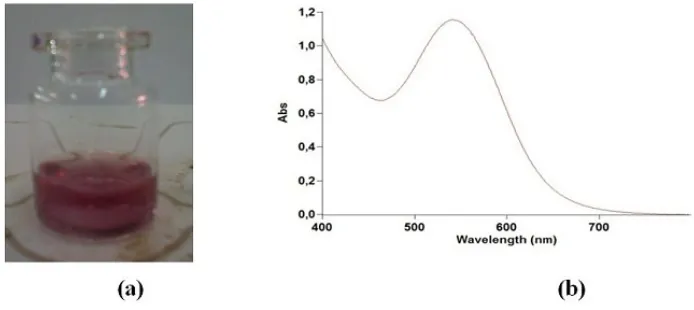
Figure. 1. (a) Photographs and (b) UV-Visible spectrum of M. malabathricum fruit extract
Fig. 2a. shows a photograph of the reaction product of silver nitrate and the M. malabathricum fruits extract. The color changed from the original color, as shown in Fig. 1, to that shown in Fig. 2a, indicating that a reaction had occurred in the reaction vessel. It has long been known that plant extracts are able to reduce metal ions, yielding nanoparticles [21]. The pH of the reaction mixture was 4, as determined using the universal indicator. This result indicated the formation of acid from the nitrate and hydrogen ions present in the reaction mixture. The UV-Vis absorption spectrum of the reaction mixture is shown in Fig. 2b. The presence of silver was indicated by the clear peaks at 426–427 nm, which correspond to the surface plasmon resonance (SPR) of silver nanoparticles. The progressive formation of silver nanoparticles with reaction time was monitored by measuring the continuing increase of their absorption intensity. Two secondary metabolite compounds of this plant are predicted to be able to reduce metal ions to metal nanoparticles [21].
(a) (b)
Figure 3. Particle size distribution of silver nanoparticles in M. malabathricum fruit extract
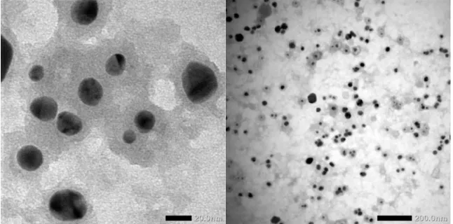
The TEM image in Fig. 4. illustrates that the silver nanoparticles were spherical in shape. This result clearly suggested that some of the compounds present in the M. malabathricum fruits extract could be used as reducing agents for silver ions as well as capping agents and stabilizers for the formed silver nanoparticles.
Figure 4. TEM pattern of silver nanoparticles in M. malabathricum fruit extract
Figure 5. Photographs of diluted silver nanoparticles in the presence of various concentrations of mercury (II) nitrate solutions
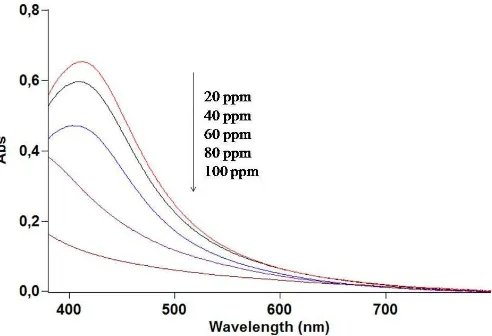
Fig. 5 shows the decreasing color intensities of the silver nanoparticles upon the addition of increasing concentrations of mercury (II) nitrate. Eventually, when 100 ppm of mercury (II) nitrate was added to the silver nanoparticles, total decolorization occurred. To supplement these visual observations, all the homogeneous mixtures were subjected to spectrophotometry analysis, and the results are shown in Fig. 6. The absorbances of the silver nanoparticles decreased as the concentration of mercury (II) nitrate solutions increased. These results show that the SPR of the silver nanoparticles at 427 nm was suppressed by the presence of mercury (II) nitrate. This clearly indicates an interaction between the two species. The color-change phenomenon suggests that the silver nanoparticles could be used as a visual sensor for high concentrations of mercury (II) nitrate.
Figure 6. UV-Visible spectra of solutions of silver nanoparticles (obtained from M. malabathricum fruits extract) in the presence of various concentrations of mercury (II) nitrate solutions
opportunity as Research Assistant at the Department of Chemistry.
REFERENCES
[1] Thakkar, K. N., Mhatre, S. S., Parikh, R.Y., Nanomed Nanotech Biol Med, 2009, 1–6. [2] Bryaskova , R., Pencheva, D., Nikolov, S., Kantardijev, T., J Chem Biol.,2011, 4, 185–
191.
[3] Suriati, G., Mariatti, M., Azizan, A., Intl. J. Automotive and Mech. Eng. (IJAME). 2014, 10, 1920–1927.
[4] Kumar, B., Smita, K., Cumbal, L., Debut, A., Pathak, R. N., Bioinorg Chem Appl., 2014, 2014, 1–8. Physicochem Eng Asp., 2014, 444, 180–188.
[8] Philip, D., Physica E., 2010, 42, 1417–1424.
[14] Farhadi, K., Farough, M., Molaei, R., Hajizadeh, S., Rafipour, A., Sensor Actuat B-Chem, 2011, 1–6.
[15] Chen, L., Fu, X., Lu, W., Chen, L., ACS Appl. Mater. Interfaces, 2013, 5, 284−290.
[16] Annadhasan, M., Muthukumarasamyvel, T., Babu, V.R.S., Rajendiran, N., ACS Sustainable Chem. Eng. 2 (4), 887–896.
[17] Some parts of this paper was presented (oral presentation only) in Green Development International Conference organized by University of Jambi, Indonesia. October 2016. [18] Joffry, M S. M., Yob, N. J., Rofiee, M. S., Affandi, M. M. R. M. M., Suhaili, Z.,
Othman, F., Akim, A. Md., Desa, M. N. M., Zakaria, Z. A., Evid. Based Complement. Alternat. Med., 2012, 2012, 1–48.
[20] Yudha S. S., Suharto, T.E., Angasa, E., Nishina, Y., Mardlia, Z.A., Sipriyadi, Oriental J. chem. 2017, 33(2), 745–751.
[21] Makarov, V. V., Love, A. J., Sinitsyna, O. V., Makarova, S. S., Yaminska, I. V., Taliansky, M. E., Kalinina, N. O., Acta Naturae., 2014, 6, 35 – 44.