R
ESEARCHA
RTICLECurcumin attenuates diabetic nephropathy by inhibiting
PKC-
a
and PKC-
b
1
activity in streptozotocin-induced type
I diabetic rats
Vivian Soetikno
1,2, Kenichi Watanabe
1, Flori R. Sari
1, Meilei Harima
1,
Rajarajan A. Thandavarayan
1, Punniyakoti T. Veeraveedu
1, Wawaimuli Arozal
1,
Vijayakumar Sukumaran
1, Arun Prasath Lakshmanan
1, Somasundaram Arumugam
1and
Kenji Suzuki
31Department of Clinical Pharmacology, Faculty of Pharmaceutical Sciences, Niigata University of Pharmacy and
Applied Life Sciences, Niigata City, Japan
2Department of Pharmacology, Faculty of Medicine, University of Indonesia, Jakarta, Indonesia
3Department of Gastroenterology and Hepatology, Niigata University Graduate School of Medical and Dental
Sciences, Niigata, Japan
Received: February 7, 2011 Revised: April 26, 2011 Accepted: June 20, 2011
Scope: We hypothesized that curcumin, a potent anti-oxidant, might be beneficial in ameli-orating the development of diabetic nephropathy through inhibition of PKC-a and PKC-b1 activity-ERK1/2 pathway.
Methods and results: Diabetes was induced by a single intraperitoneal injection of streptozo-tocin (STZ) (55 mg/kg) in rats. Three weeks after STZ injection, rats were divided into three groups, namely, normal, diabetic and diabetic treated with curcumin at 100 mg/kg/day, p.o., for 8 wk. At 11 wk after STZ injection, diabetic rats exhibited renal dysfunction, as evidenced by reduced creatinine clearance, increased blood urea nitrogen (BUN) and proteinuria, marked increases in lipid peroxidation, NOX4 and p67phox and decrease in anti-oxidant enzyme. All of these abnormalities were significantly reversed by curcumin. Furthermore, the high-glucose-induced PKC-aand PKC-b1 activities and phosphorylated ERK1/2 was significantly diminished by curcumin. Curcumin also attenuated the expression of TGF-b1, CTGF, osteopontin, p300 and ECM proteins such as fibronectin and type IV collagen. The high-glucose-induced expression of VEGF and its receptor VEGF receptor II (flk-1) was also ameliorated by curcumin.
Conclusion: These results prove that curcumin produces dual blockade of both PKC-aand PKC-b1 activities, which suggests that curcumin is a potential adjuvant therapy for the prevention and treatment of diabetic nephropathy.
Keywords:
Curcumin / Diabetic nephropathy / Mitogen-activated protein kinase / Oxidative stress / Protein kinase C
1
Introduction
Diabetic nephropathy (DN), one of the most serious complications of diabetes and the most common cause of end-stage renal failure, is characterized by glomerular hyperfiltration, glomerular and tubular epithelial
hyper-trophy, increased urinary albumin excretion, increased basement membrane thickness and mesangial expansion with the accumulation of extracellular matrix proteins (ECM) [1, 2]. The involvement of various growth factors such as transforming growth factor (TGF)-b1 and connective growth factor (CTGF) in the development of DN has also
Correspondence: Professor Kenichi Watanabe, Department of Clinical Pharmacology, Faculty of Pharmaceutical Sciences, Niigata University of Pharmacy and Applied Life Sciences, 265-1 Higashijima Akiha-ku, Niigata City 956-8603, Japan
E-mail:[email protected]
Fax:181-250-25-5021
Abbreviations: BUN,blood urea nitrogen;CCr,creatinine clearance;
CTGF,connective tissue growth factor;DN,diabetic nephropathy;
ECM,extracellular matrix;ERK,extracellular signal-regulated protein kinase; GPx, glutathione peroxidase; MAPK, mitogen-activated protein kinase;MDA,malondialdehyde;PKC,protein kinase C;ROS,
been reported [3, 4]. In addition to ECM expansion, the integrity of the glomerular capillary network is also recog-nized as a key determinant of renal pathology in both experimental and human settings [5, 6]. The development of this network is critically dependent on vascular endothelial growth factor (VEGF), a growth factor that not only stimu-lates endothelial cell proliferation and permeability but also has a role in the maintenance of endothelial cell integrity in the mature animal [7].
Protein kinase C (PKC) is a Ca21- and
phospholipid-dependent enzyme that phosphorylates serine and threonine residues in a wide variety of cellular proteins [8, 9]. In glomerular cells of diabetic animals or mesangial cells exposed to high glucose, de novo synthesis of DAG is postulated to activate PKC, causing cytosol-to-membrane translocation of selective PKC isoforms [10, 11]. Furthermore, activated PKC triggers the mitogen-activated protein kinase (MAPK) cascade [12, 13]. Previous studies have reported that PKC-a and -b1
isoforms were increased in vivo in membranous fractions of diabetic rat glomeruli and in vitro in mesangial cells exposed to elevated glucose levels [14], whereas PKC-b2was reported to be preferentially activated in the aorta and heart of diabetic rats [15]. Studies have also reported that deletion of the PKC-bgene or treatment with LY333531 protects against diabetes by causing increases in renal hypertrophy, glomerular hyperfil-tration, ECM production, and reactive oxygen species (ROS) whereas deletion of the PKC-areduces proteinuria [14, 16, 17]. These data suggest that two physiologically important features of DN, renal hypertrophy and proteinuria, are regulated through different PKC-signaling events.
Curcumin, a powerful anti-oxidant, is a component of turmeric found in theCurcuma longaplant and has been used for centuries in treating inflammatory ailments and condi-tions. Accumulating evidence suggests that curcumin has a diverse range of molecular targets. Among its molecular targets are transcription factors, transcriptional coactivator (e.g. p300), growth factors and their receptors, cytokines, enzymes and genes regulating cell proliferation and apopto-sis. Other effects of curcumin are complete inhibition of the activity of several protein kinases including phosphorylase kinase, PKC and protamine kinase [18, 19]. However, little is known about whether curcumin inhibits only the activity of PKC-a, that of PKC-b1, or both. Accordingly, we sought to
examine the effects of curcumin on ameliorating ECM accumulation in streptozotocin (STZ)-induced diabetic rats through PKC-ERK1/2 pathway and its downstream signaling events, including generation of ROS, induction of VEGF and accumulation of basement membrane proteins.
2
Materials and methods
2.1 Drug and chemicals
Unless otherwise stated, all reagents were of analytical grade and were purchased from Sigma-Aldrich (Tokyo, Japan).
2.2 Experimental animals
Male Sprague–Dawley rats (8 wk of age) weighing 250–280 g were obtained from Charles River Japan (Kanagawa, Japan). Diabetes was induced by a single intraperitoneal injection of STZ at a dose of 55 mg/kg body weight diluted in 20 mM sodium citrate saline buffer, pH 4.5, and injected within 5 min of preparation (n510). Age-matched non-diabetic control rats (group N;n55) were each injected with an equal volume of citrate buffer. Rats were maintained with free access to water and chow throughout the period of study, and the animals were treated in accordance with the guidelines for animal experimentation of our institute. Each week, rats were weighed and their blood glucose levels were measured using Medi-safe chips (Terumo, Tokyo, Japan) and only STZ-treated animals with blood glucose Z300 mg/dL were considered diabetic. Three wks after the induction of diabetes, rats were divided randomly into two groups, namely, diabetic (group D;n55) and diabetic/treated with curcumin at a dose of 100 mg/kg body weight/day (group Cur; n55) [20]. Curcumin was dissolved in 1% gum Arabic. Each animal in the group D received vehicle 1% gum Arabic. Drug and vehicle were administered daily by oral gavage for 8 wk. Before sacrificed, individual rats were placed in metabolic cages to obtain 24 h urine collections for the measurement of urine creatinine and protein concentrations determined by the Jaffe and Bradford method, respectively.
2.3 Estimation of biochemical parameters
Blood samples were collected via heart puncture in each animal at the time of death into EDTA vacutainer tubes. EDTA-blood was centrifuged at 3000g, 41C, for 15 min for
separation of plasma. Plasma was stored at 801C until
assays were performed. The plasma was used for the estimation of glucose, blood urea nitrogen (BUN) and creatinine.
2.4 Measurement of lipid peroxidation
Lipid peroxidation was assessed by measuring the thio-barbituric acid (TBA) reactivity of malondialdehyde (MDA), an end product of fatty acid peroxidation. For this purpose, kidney tissue was rinsed, weighed, resuspended at 50 mg/mL in normal saline, homogenized and analyzed using thiobarbituric acid-reacting substances (TBARS) assay kit (OXItek, ZeptoMetrix, NY, USA).
2.5 Measurement of total glutathione peroxidase (GPx) activity
centrifuged for 8000 rpm, 41C, for 15 min in accordance
with the total GPx assay kit instructions (OXItek, Zepto-Metrix. GPx activity in kidney tissue was measured using a kinetic ultraviolet–visible spectrophotometer (Ultraspec 3100, Amersham Biosciences, Cambridge, UK). The oxida-tion of NADPH to NADP1was measured by the decrease in
absorbance at 340 nm.
2.6 Histopathological analysis
The kidney was decapsulated. Half of the kidney was immediately snap-frozen in liquid nitrogen for subsequent protein extraction and enzymatic assays. The remaining excised kidney was cut into about 2-mm-thick transverse slices and fixed in 10% formalin. After being embedded in paraffin, several transverse sections were obtained from the kidney and stained with hematoxylin and eosin and periodic acid-Schiff (PAS) for histological evaluation, and also stained with Azan-Mallory to demonstrate fibrosis in kidney tissues. The frequency and the severity of lesions in kidney were assessed semi-quantitatively by light microscopy using the following scores: 0, normal; 1, mild; 2, moderate; and 3, severe. The criteria for kidney lesions were degree of glomerular thickening, interstitial fibrosis, arteriolopahty, hyaline cast and tubular degenera-tion [21].
2.7 Immunohistochemistry for type IV collagen, VEGFR-II (flk-1) and fibronectin
Formalin-fixed, paraffin-embedded kidney tissue sections were used for immunohistochemical staining. After depar-affinization and hydration, the slides were washed in Tris-buffered saline (TBS; 10 mM/L Tris-HCl, 0.85% NaCl, pH 7.2). Endogenous peroxidase activity was quenched by incubating the slides in methanol and 0.3% H2O2 in
methanol. After overnight incubation with the primary antibody, rabbit polyclonal anti-collagen IV (Abcam 6586, Cambridge, UK), mouse monoclonal anti-flk-1 (sc-6251, Santa Cruz Biotechnology, Santa Cruz, CA, USA) and mouse monoclonal anti-fibronectin (sc-8422, Santa Cruz), diluted 1:50, at 41C, the slides were washed in TBS and then HRP-conjugated rabbit anti-goat secondary antibody or HRP-conjugated mouse anti-goat secondary antibody was added and incubated at room temperature for 45 min. The slides were washed in TBS and incubated with diamino-benzidine tetrahydrochloride as the substrate, and then counterstained with hematoxylin. A negative control without primary antibody was included in the experiment to verify the antibody specificity. Semi-quantitative analysis of type IV collagen, flk-1 and fibronectin was carried out by counting the numbers of glomeruli with strong, moderate and weak expression. A total 40 glomeruli/animal were counted [17].
2.8 Protein analyses by Western blotting
The frozen colonic tissues were weighed and homogenized in an ice-cold buffer (50 mM Tris-HCl, pH 7.4, 200 mM NaCl, 20 mM NaF, 1 mM Na3VO4, 1 mM 2-mercaptoethanol,
0.01 mg/mL leupeptin, 0.01 mg/mL aprotinin). Homogenates were then centrifuged (3000g, 10 min, 41C) and the
super-natants were collected and stored at 801C. The total protein
concentration in samples was measured by the bicinchoninic acid method [22]. Aliquots of supernatants containing equal amounts of protein (70mg) were evaluated to determine p67phox (sc-7663; Santa Cruz), Nox4 (ab60940; Abcam), osteopontin (sc-21742; Santa Cruz) and TGF-b1 (Promega G-1221). The expression of the above antibodies was normalized by b-actin protein expression in the same sample. The expression of phospho-ERK1/2 (Cell Signaling-9106) was normalized by the basal level of ERK1/2 (Cell Signaling-9102). All primary antibodies were used at a dilution of 1:1000 and secondary antibodies were used at a dilution of 1:5000.
2.9 PKC-aandb1activity
Kidney cortex was homogenized in ice-cold buffer A (20 mmol/L Tris-HCl, pH 7.5; 2 mmol/L EDTA; 10 mmol/L EGTA; and 0.25 mol/L sucrose containing complete protease and phosphatase inhibitors). The homogenates were centri-fuged at 1000gfor 10 min. These supernatants were then
centrifuged at 10 000gat 41C for 20 min. The supernatant
was used as a total cell lysate. The supernatants were then ultracentrifuged at 100 000gfor 1 h at 41C. This supernatant
was retained as the cytosolic fraction, and the pellet was resuspended in buffer B (buffer A with 1% Triton X-100) and ultracentrifuged at 100 000gfor 1 h at 41C. This supernatant
was retained as the membranous fraction [23]. Western blot analysis was then performed on total cell lysate, cytosol and membrane fraction using PKC-a(sc-208; Santa Cruz) andb1 (sc-209; Santa Cruz) antibodies and activation was inferred from the ratio of membrane–to-cytosol fractions [24].
2.10 Quantitative RT-PCR to detect p300
Quantitative real-time polymerase chain reaction was performed, as previously described, with the abundance of transcript expressed relative to that of GAPDH mRNA [25]. Nucleotide sequences of primers and probes were p300 (forward) GGGACTAACCAATGGTGGTG, (reverse) ATTGG GAGAAGTCAAGCCTG; GAPDH (forward) GCTCATTTCCT GGTATGACAACG, (reverse) AGGGGTCTACATGGCAACTG.
2.11 Statistical analysis
methods for post-hoc analysis and two-tailed t-test when appropriate. A value ofpo0.05 was considered statistically
significant. For statistical analysis, GraphPad Prism 5 soft-ware (San Diego, CA, USA) was used.
3
Results
3.1 Effect of curcumin treatment on body weight, kidney weight/body weight ratio, blood glucose, creatinine clearance (Ccr), BUN and protein urine
At the end of the 11th wk, diabetic rats exhibited significantly increased plasma glucose levels and decreased body weight compared with normal rats. Chronic treatment with curcumin in diabetic rats from 3 to 11 wk altered plasma glucose levels significantly compared with those of untreated rats. Treatment with curcumin also prevented body weight loss in diabetic rats, but this effect was not significant compared with that of untreated diabetic rats. Diabetic rats had increased kidney weight/body weight ratio, a marker for the development of DN, and this ratio was significantly decreased by treatment with curcumin (Table 1). Diabetic rats also exhibited increased plasma creatinine, increased BUN, increased urinary protein excretion and decreased CCr and curcumin treatment signifi-cantly reduced plasma creatinine, reduced BUN, reduced urinary protein excretion and increased CCr (Table 1).
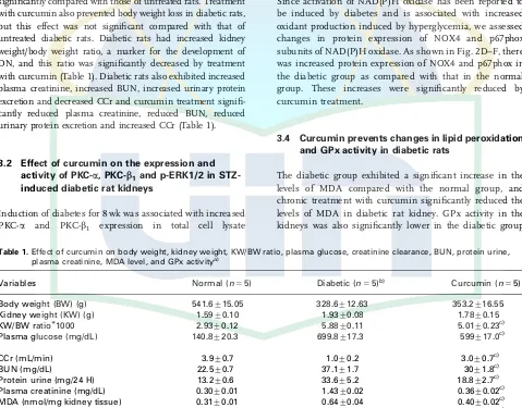
3.2 Effect of curcumin on the expression and activity of PKC-a, PKC-b1and p-ERK1/2 in STZ-induced diabetic rat kidneys
Induction of diabetes for 8 wk was associated with increased PKC-a and PKC-b1 expression in total cell lysate
(Fig. 1A–C), which was significantly reduced by curcumin treatment. In Fig. 1D and E, evidence is shown that diabetic animals demonstrated PKC activation, with a significant increase in the ratio of membrane to cytosolic PKC-aand-b1
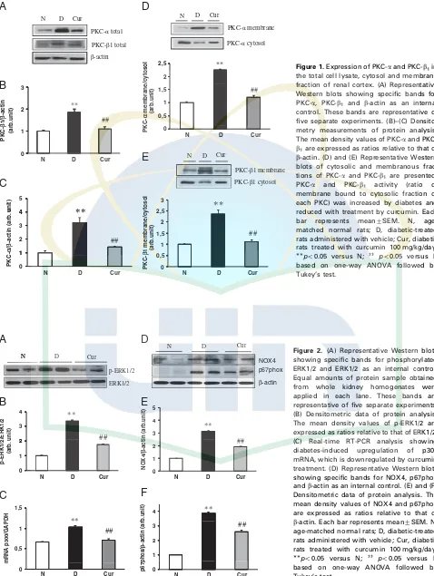
which was reduced by curcumin. Moreover, Western blot-ting analysis using anti-phospho ERK1/2 antibody, which recognizes phosphorylated threonine 202 and tyrosine 204 of p44/p42 ERK, detected enhanced phosphorylation of p44/ p42 ERK in the diabetic group (Fig. 2A and B). Curcumin treatment significantly inhibited the phosphorylation of ERK in diabetes.
3.3 Curcumin reduced protein expression of NAD(P)H oxidase subunits in STZ-induced diabetic rat kidneys
Since activation of NAD(P)H oxidase has been reported to be induced by diabetes and is associated with increased oxidant production induced by hyperglycemia, we assessed changes in protein expression of NOX4 and p67phox subunits of NAD(P)H oxidase. As shown in Fig. 2D–F, there was increased protein expression of NOX4 and p67phox in the diabetic group as compared with that in the normal group. These increases were significantly reduced by curcumin treatment.
3.4 Curcumin prevents changes in lipid peroxidation and GPx activity in diabetic rats
The diabetic group exhibited a significant increase in the levels of MDA compared with the normal group, and chronic treatment with curcumin significantly reduced the levels of MDA in diabetic rat kidney. GPx activity in the kidneys was also significantly lower in the diabetic group
Table 1.Effect of curcumin on body weight, kidney weight, KW/BW ratio, plasma glucose, creatinine clearance, BUN, protein urine, plasma creatinine, MDA level, and GPx activitya)
Variables Normal (n55) Diabetic (n55)b) Curcumin (n
55)
Body weight (BW) (g) 541.6715.05 328.6712.63 353.2716.55 Kidney weight (KW) (g) 1.5970.10 1.9370.08 1.7870.15 KW/BW ratio1000 2.9370.12 5.8870.11 5.0170.23c)
Plasma glucose (mg/dL) 140.8720.3 699.8717.3 599717.0c)
CCr (mL/min) 3.970.7 1.070.2 3.070.7c)
BUN (mg/dL) 22.570.7 37.171.7 3071.8c)
Protein urine (mg/24 H) 13.270.6 33.675.2 18.872.7c)
Plasma creatinine (mg/dL) 0.3070.01 1.4370.02 0.3670.02c)
MDA (nmol/mg kidney tissue) 0.3170.01 0.6470.04 0.4070.02c)
GPx (U/mg protein kidney tissue) 0.1770.03 0.0470.01 0.1370.01c)
BUN: blood urea nitrogen, CCr: creatinine clearance, GPx: glutathione peroxidase, KW/BW: kidney weight/body weight, MDA: malondialdehyde.
a) Values are expressed as mean7SEM
b)po0.05 versus.to normal group based on two-tailedt-test.
N D Cur
PKCαcytosol PKC-αmembrane Cur
D N
D
A
PKC-β1 total PKC-αtotal
β-actin -2 2,5 ** 3 ** 0,5 1 1,5 ##
B
Cur D N 1 2 PKC-β 1/ β -actin (arb.unit) ## 0N D Cur
(arb.unit)
PKC-α
membrane/cytosol
PKC-β1 cytosol PKC-β1 membrane
0
N D Cur
2,5 3
**
4 5**
C
E
1 1,5 2 ## 1 2 3 4 ##**
0 0,5N D Cur
(arb.unit) PKC-β 1 membrane/cytosol 0 1
N D Cur
un it ) C-α / β -actin (arb. u PK
Figure 1.Expression of PKC-aand PKC-b1in
the total cell lysate, cytosol and membrane fraction of renal cortex. (A) Representative Western blots showing specific bands for PKC-a, PKC-b1 and b-actin as an internal
control. These bands are representative of five separate experiments. (B)–(C) Densito-metry measurements of protein analysis. The mean density values of PKC-aand PKC-b1are expressed as ratios relative to that of
b-actin. (D) and (E) Representative Western blots of cytosolic and membranous frac-tions of PKC-a and PKC-b1 are presented.
PKC-a and PKC-b1 activity (ratio of
membrane bound to cytosolic fraction of each PKC) was increased by diabetes and reduced with treatment by curcumin. Each bar represents mean7SEM. N, age-matched normal rats; D, diabetic-treated rats administered with vehicle; Cur, diabetic rats treated with curcumin 100 mg/kg/day. po0.05 versus N; ]] po0.05 versus D
based on one-way ANOVA followed by Tukey’s test.
N D Cur
NOX4
N D Cur
p-ERK1/2 ERK1/2 N p67phox β-actin
A
D
2 3 4 ** ## 3 4 5 ## **B
E
0 1 2N D Cur
p-ERK1/2 /ERK1/2 (arb. unit) 0 1 2 NOX4/ β -actin (arb.unit)
N D Cur
1,5 ** 4 **
C
F
0,5 1 ## 1 2 3 ## 0N D Cur
m R N A p 3 0 0 /G A P D H 0
N D Cur
p67phox/
β
-actin (arb.unit)
Figure 2. (A) Representative Western blots showing specific bands for phosphorylated ERK1/2 and ERK1/2 as an internal control. Equal amounts of protein sample obtained from whole kidney homogenates were applied in each lane. These bands are representative of five separate experiments. (B) Densitometric data of protein analysis. The mean density values of p-ERK1/2 are expressed as ratios relative to that of ERK1/2. (C) Real-time RT-PCR analysis showing diabetes-induced upregulation of p300 mRNA, which is downregulated by curcumin treatment. (D) Representative Western blots showing specific bands for NOX4, p67phox andb-actin as an internal control. (E) and (F) Densitometric data of protein analysis. The mean density values of NOX4 and p67phox are expressed as ratios relative to that of b-actin. Each bar represents mean7SEM. N, age-matched normal rats; D, diabetic-treated rats administered with vehicle; Cur, diabetic rats treated with curcumin 100 mg/kg/day. po0.05 versus N; ]] po0.05 versus D
than in the normal group. However, chronic treatment with curcumin prevented this decrease in GPx activity in the diabetic group (Table 1).
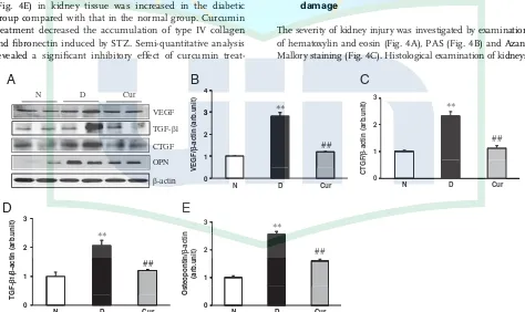
3.5 Effect of curcumin on extracellular matrix protein expression
Increases in the expressions of fibrotic factors, such as
TGF-b1, CTGF and osteopontin, and extracellular matrix proteins, such as type IV collagen and fibronectin, are believed to be partly responsible for the glomerular enlar-gements and fibrosis [25]. Western blotting analysis demonstrated significant increases of TGF-b1 and CTGF
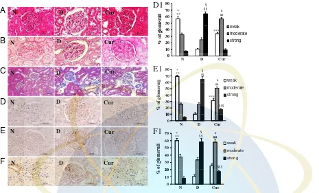
expression in the diabetic group compared with those in the normal group, which were significantly attenuated by treatment with curcumin (Fig. 3A, C, D). Moreover, protein expression of osteopontin was also significantly increased in the diabetic group compared with that in the normal group, which was significantly reduced by curcumin treat-ment (Fig. 3A and E). To further evaluate the effect of curcumin treatment in the STZ-induced DN model, the expressions of type IV collagen and fibronectin were exam-ined by immunohistochemical staining. The immunor-eactivity of type IV collagen (Fig. 4D) and fibronectin (Fig. 4E) in kidney tissue was increased in the diabetic group compared with that in the normal group. Curcumin treatment decreased the accumulation of type IV collagen and fibronectin induced by STZ. Semi-quantitative analysis revealed a significant inhibitory effect of curcumin
treat-ment on kidney tissue accumulation of type IV collagen and fibronectin induced by STZ compared with that of vehicle (Fig. 4D1 and E1).
3.6 Effect of curcumin treatment on pro-angiogenic VEGF and VEGF-RII (flk-1)
We next examined the expression of pro-angiogenic factor VEGF and corresponding receptors flk-1 by Western blot analysis and immunohistochemistry, respectively. The levels of VEGF (Fig. 3A and B) and flk-1 (Fig. 4F, 4F1) were significantly increased in the diabetic group, which were significantly suppressed by treatment with curcumin.
3.7 p300 level assessed by RT-PCR
Rats with DN had upregulated expression of p300 mRNA in comparison with group N, and the treatment with curcumin significantly reversed the renal mRNA levels of p300 in rats with DN (Fig. 2C).
3.8 Effect of curcumin against renal histological damage
The severity of kidney injury was investigated by examination of hematoxylin and eosin (Fig. 4A), PAS (Fig. 4B) and Azan-Mallory staining (Fig. 4C). Histological examination of kidneys
VEGF TGF-β1 N D Cur
A
CTGF OPN
β-actin
3
**
3 4
**
B
D
1 2
##
1 2 3
VEGF/
β
-actin (arb.unit) ##
0
N D Cur
TGF-β
1/
β
-actin (arb.unit)
0
N D Cur
3
**
3
**
C
E
1
2 ##
1 2
##
0
N D Cur
(arb.unit)
Osteopontin/
β
-actin
0
N D Cur
CTGF/
β
-actin (arb.unit)
Figure 3.Renal expression of VEGF, TGF-b1, CTGF and osteopontin. (A) Representative Western blots showing specific bands for VEGF,
TGF-b1, CTGF, osteopontin andb-actin as an internal control. Equal amounts of protein sample obtained from whole kidney homogenate
were applied in each lane. These bands are representative of five separate experiments. (B) and (E) Densitometric data of protein analysis. The mean density values of VEGF, TGF-b1, CTGF and osteopontin are expressed as ratios relative to that ofb-actin. Each bar represents
from the normal group showed normal glomerulus and tubules. The kidneys of the diabetic group showed marked histological changes in the cortex and outer medulla as hyaline casts, glomerular thickening and moderate interstitial fibrosis and arteriolopathy. Renal changes in diabetic group were improved in the group treated with curcumin (Table 2).
4
Discussion
We here provide evidence that protection against the development of DN by curcumin treatment involves chan-ges in the PKC-aand PKC-b1activities as well as expression
of phophorylated ERK1/2. Moreover, curcumin treatment
N D Cur
D1
50 60 70 80
* **
$ $$
## #
N D Cur
A
0 10 20 30 40
% of glomeruli
weak moderate strong ***
N D Cur
B
E1
0
N D Cur
60 70 80 *
** $$ $
#
N D Cur
C
D
10 20 30 40 50
% of glom
eru
li weak
moderate strong ***
##
$$$
N D Cur
E
F1
0 10
N D Cur
70
*** $$ $ #
##
N D Cur
F
20 30 40 50 60
weak moderate ##
$$$
0 10 20
N D Cur
% of glomeruli
strong
Figure 4.(A) Hematoxylin and eosin staining of the cross-sectional tissue slices of kidney depicting glomerular thickening, interstitial fibrosis, arteriolopathy, hyaline cast and tubular degeneration (x400). (B) Periodic acid-Schiff staining of kidney depicting glomerular lesions (x400). (C) Azan-Mallory staining for fibrosis of the cross-sectional tissue slices of kidney. Fibrosis is indicated by the blue area (x200). Immunohistochemistry of type IV collagen (D), fibronectin (E) and flk-1 (F) including semiquantitative analysis of type IV collagen (D1), fibronectin (E1) and flk-1 (F1) in normal, diabetic and curcumin group. The number of glomeruli with a strong expression of type IV collagen, fibronectin and flk-1 was significantly higher in the diabetic group than in the normal group ($p
o0.01) and the number
of glomeruli with a moderate expression of type IV collagen, fibronectin and flk-1 was significantly higher in the curcumin group than in the diabetic group (]
po0.05). The values are mean7SEM. po0.01 N versus D; po0.01 N versus Cur;po0.01 Cur versus D;
]
po0.05 Cur versus D;]]po0.05 Cur versus N;$po0.01 D versus N;$$po0.01 D versus Cur;$$$po0.05 Curversus N based on two-tailed t-test.
Table 2.Effect of curcumin on histopathological changes in kidney tissues after 8 wk of treatment in streptozotocin-induced diabetic ratsa)
Histopathological finding Normal (n55) Diabetic (n55)b) Curcumin (n
55)c)
Glomerular thickening 0.070.0 2.6770.07 1.6470.06 Interstitial fibrosis 0.070.0 2.5770.06 1.4670.04
Arteriolopathy 0.070.0 2.3470.13 1.4670.12
Hyaline cast 0.070.0 2.3670.1 1.3670.02
Tubular degeneration 0.070.0 2.4670.06 1.4770.13
a) Values are expressed as mean7SEM
b)po0.05 versus to normal group based on two-tailedt-test.
also decreases the expression of NAD(P)H oxidase subunits, NOX4 and p67phox, which is an essential mechanism responsible for increased ROS production. In this study, we also showed that STZ administration produced significant increases in blood glucose level and in protein urine, along with reduction of body weight, typical characteristics of diabetes mellitus. With the onset of diabetes mellitus, there is a subsequent decrease in CCr and increase in BUN, which indicate progressive renal damage. Curcumin treatment prevented the development of DN by significantly lowering BUN and protein urine and increasing CCr. Furthermore, curcumin also prevented the rise in blood glucose level and maintained the body weight of diabetic animals throughout the study period.
Previous studies have demonstrated increased membrane recovery of PKC-a and -b in the cytosolic and membrane cellular fractions of glomeruli isolated from the STZ-diabetic rat, suggesting an activation/translocation pattern of these PKC isozymes in the diabetic milieu [16, 26–29]. Meier et al., reported that diabetic PKC-b /
mice demonstrate reduced renal hypertrophy, as well as diminished high-glucose-induced expression of ECM proteins and of the profibrotic cytokines TGF-b1and CTGF
[30], which were previously demonstrated to be unaltered in diabetic PKC-a / mice [17]. The above data suggest that activation of the PKC-b isoform signaling pathway in the diabetic state directly contributes in the regulation of
TGF-b1in experimental DN, whereas PKC-aisoform signaling is
not involved. In this view, curcumin is a well-known anti-oxidant and anti-inflammatory agent that can inhibit 12-O -tetradecanoyl phorbol-13-acetate (TPA)-induced PKC activity in NIH 3T3 cells as well as ROS generation in phorbol-12 myristate-13 acetate (PMA)-treated lymphocytes through inhibition of PKC activity [19, 31]. Mahmmoud reported that curcumin competes with Ca21for the regulatory domain of
PKC, producing inhibition of PKC activity at low Ca21
concentrations [32]. Consistent with other reports, we found that there was an increase in activity of PKC-aand PKC-b1,
which is shown by their increased expression in membrane fractions as well as increased phosphorylated ERK1/2 in diabetic kidney. Interestingly, curcumin treatment could prevent this increase in PKC-aand PKC-b1activity as well as phosphorylated ERK1/2 expression.
VEGF is an important mediator in maintaining normal kidney functions. In the diabetic kidney, the expression level of this normally expressed VEGF protein as well as VEGF receptors can be markedly increased from the early stages of DN and is associated with proteinuria, in both diabetic animal models as well as patients with diabetes [33–36]. A previous study also reported that the translocation of PKC-a
to renal cortical membranes by diabetes is associated with NADPH-dependent superoxide production and elevated renal serum and urinary VEGF [23]. In the present study, we observed that diabetic rats treated with curcumin showed decreased proteinuria compared with diabetic rats treated with vehicle. Furthermore, we also showed that STZ
administration induced increased expression of VEGF and renal positive immunostaining of its receptor, flk-1, which was reversed by curcumin treatment. Additionally, curcu-min treatment decreased the expression of PKC-a in the membrane fraction of diabetic kidney. Therefore, it is reasonable to speculate that curcumin treatment amelio-rates proteinuria as well as increases VEGF and its receptor by inhibiting the activity of PKC-a.
Numerous reports have demonstrated that high-glucose concentration-induced PKC and MAPK activation causes increased production of ROS in the diabetic kidney, which may lead to increased cytokine and growth factor production [37, 38]. Very recent study demonstrated that p300, a tran-scriptional coactivator with histone acetyl transferase activ-ity, regulates glucose-induced activation of transcription factors and subsequent upregulation of vasoactive factors and ECM proteins, such as fibronectin and collagen, in human umbilical vein endothelial cells. This study also reported that glucose-induced p300 upregulation was blocked after exposure of the cells to PKC as well as MAPK blockers with further prevention of glucose-induced upre-gulation of ECM proteins [39]. Therefore, the upstream mechanism of glucose-induced p300 possibly involves acti-vation of MAPK and PKC pathways [40]. Curcumin is a known p300 inhibitor and has been demonstrated to reduce p300 acetylation by binding to p300 and causing its degra-dation [41]. Previous study also showed that curcumin attenuates the upregulation of ECM proteins in the kidneys by inhibiting oxidative stress and p300 as well as nuclear factor-kB [42]. In agreement with previous studies, the findings from this study demonstrate that STZ administra-tion induces glomerular enlargement and ECM accumula-tion, which was confirmed by upregulation of TGF-b1,
CTGF and osteopontin in kidney of diabetic rats and increased immunostaining of fibronectin and type IV collagen under diabetic conditions. Interestingly, curcumin treatment decreased the overexpression of fibrotic cytokine and ECM molecules and this effect might be mediated by inhibition on PKC-b activity, which subsequently down-regulated the expression of mRNA p300 (Fig. 2C).
which is an anti-oxidant defense system component. All such abnormalities were completely normalized by curcu-min treatment, which has been shown to prevent oxidative stress by inhibition of PKC activity. In agreement, others have shown that curcumin can prevent diabetes-induced decrease in the antioxidant capacity in the retina and kidney [21, 49].
The concentration of curcumin used in our study was 100 mg/kg body weight. A similar dose of curcumin was shown to be effective in preventing nephropathy and decreasing cellular oxidative stress, as well as reducing the blood levels of proinflammatory cytokines in animal studies [20]. This dosage is equivalent to 6 g per adult, assuming 60 kg body weight per adult, and has been used in humans [50, 51].
In conclusion, our results show that curcumin protects against the development of DN, which involves dual block-ade of activities of both classical PKC isoforms, PKC-aand PKC-b1, as well as the downstream pathway, ERK1/2.
Moreover, the findings of this study also suggest that the principal mechanism involved in the anti-fibrotic effect of curcumin is its strong anti-oxidant property. Owing to its extremely good safety profile and long history of safe use, curcumin may find clinical application in preventing complications in diabetic patients.
This research was supported by a Yujin Memorial Grant, Ministry of Education, Culture, Sports and Technology of Japan, and by a grant from the Promotion and Mutual Aid Corpora-tion for Private Schools, Japan. The authors thank Sayaka Mito and Yoshiyasu Kobayashi for their assistance in this research work.
The authors have declared no conflict of interest.
5
References
[1] Zelmanovitz, T., Gerchman, F., Balthazar, A. P. S., Thoma-zelli, F. C. S. et al., Diabetic nephropathy.Diabetol. Metab. Syndr.2009,21, 1–10.
[2] Kanwar, Y. S., Wada, J., Sun, L., Xie, P. et al., Diabetic nephropathy: Mechanisms of renal disease progression.
Exp. Biol. Med.2008,233, 4–11.
[3] Sharma, K., Ziyadeh, F. N., Hyperglycemia and diabetic kidney disease. The case for transforming growth factor-beta as a key mediator.Diabetes1995,44, 1139–1146. [4] Riser, B. L., Denichilo, M., Cortes, P., Baker, C., Regulation of
connective tissue growth factor activity in cultured rat mesangial cells and its expression in experimental diabetic glomerulosclerosis.J. Am. Soc. Nephrol.2000,11, 25–38.
[5] Remuzzi, G., Benigni, A., Remuzzi, A., Mechanisms of progression and regression of renal lesions of chronic nephropathies and diabetes. J. Clin. Invest. 2006, 116, 288–296.
[6] Mauer, S. M., Steffes, M. W., Ellis, E. N., Sutherland, D. E., Structural–functional relationships in diabetic nephropathy.
J. Clin. Invest.1984,74, 1143–1155.
[7] Ferrara, N., Vascular endothelial growth factor: basic science and clinical progress. Endocr. Rev. 2004, 25, 581–611.
[8] Meier, M., King, G. L., Protein kinase C activation and its pharmacological inhibition in vascular disease.Vasc. Med.
2000,5, 173–185.
[9] Dempsey, E. C., Newton, A. C., Mochly-Rosen, D., Fields, A. P., Protein kinase C isozymes and the regulation of diverse cell responses. Am. J. Physiol. Lung. Cell. Mol. Physiol.2000,279, L429–L438.
[10] Kikkawa, R., Haneda, M., Uzu, T., Koya, D. et al., Translo-cation of protein kinase C alpha and zeta in rat glomerular mesangial cells cultured under high glucose conditions.
Diabetologia1994,37, 838–841.
[11] Ayo, S. H., Radnik, R., Garoni, J. A., Troyer, D. A. et al., High glucose increases diacylglycerol mass and activates protein kinase C in mesangial cell cultures.Am. J. Physiol.1991,
261, F571–F577.
[12] Seger, R., Krebs, E. G., The MAPK signaling cascade.FASEB J.1995,9, 726–735.
[13] Haneda, M., Koya, D., Isono, M., Kikkawa, R., Overview of glucose signaling in mesangial cells in diabetic nephro-pathy.J. Am. Soc. Nephrol.2003,14, 1374–1382.
[14] Koya, D., Jirousek, M. R., Lin, Y. W., Ishii, H., Characteriza-tion of protein kinase C beta isoform activaCharacteriza-tion on the gene expression of transforming growth factor-beta, extracellular matrix components, and prostanoids in the glomeruli of diabetic rats.J. Clin. Invest.1997,100, 115–126.
[15] Inoguchi, T., Battan, R., Handler, E., Sportsman, J. R. et al., Preferential elevation of protein kinase C isoform beta II and diacylglycerol levels in the aorta and heart of diabetic rats: differential reversibility to glycemic control by islet cell transplantation. Proc. Natl. Acad. Sci. USA 1992, 89, 11059–11063.
[16] Koya, D., Haneda, M., Nakagawa, H., Isshiki, K. et al., Amelioration of accelerated diabetic mesangial expansion by treatment with a PKC beta inhibitor in diabetic db/db mice, a rodent model for type 2 diabetes.FASEB J.2000,14, 439–447.
[17] Menne, J., Park, J. K., Boehne, M., Elger, M. et al., Dimin-ished loss of proteoglycans and lack of albuminuria in protein kinase C-alpha-deficient diabetic mice. Diabetes
2004,53, 2101–2109.
[18] Lin, J. K., Molecular targets of curcumin.Adv. Exp. Med. Biol.2007,595, 227–243.
[19] Liu, J. Y., Lin, S. J., Lin, J. K., Inhibitory effects of curcumin on protein kinase C activity induced by TPA in NIH3T3 cells.
Carcinogenesis1993,14, 857–861.
[20] Jain, S. K., Rains, J., Croad, J., Larson, B. et al., Curcumin supplementation lowers TNF-a, IL-6, IL-8, MCP-1 secretion in high glucose-treated cultured monocytes and blood levels of TNF-a, IL-6, MCP-1, glucose, and glycosylated hemoglobin in diabetic rats.Antioxid. Redox. Signal.2009,
[21] Sharma, S., Kulkarni, S. K., Chopra, K., Curcumin, the active principle of turmeric (Curcuma longa), ameliorates diabetic nephropathy in rats.Clin. Exp. Pharmacol. Physiol.2006,33, 940–945.
[22] Smith, P. K., Krohn, R. I., Hermanson, G. T., Mallia, A. K. et al., Measurement of protein using bicinchoninic acid.
Anal. Biochem.1985,150, 76–85.
[23] Thallas-Bonke, V., Thorpe, S. R., Coughlan, M. T., Fukami, K. et al., Inhibition of NADPH oxidase prevents advanced glycation end product-mediated damage in diabetic nephropathy through a protein kinase C-alpha-dependent pathway.Diabetes2008,57, 460–469.
[24] Connelly, K. A., Kelly, D. J., Zhang, Y., Prior, D. L. et al., Inhibition of protein kinase C-beta by ruboxistaurin preserves cardiac function and reduces extracellular matrix production in diabetic cardiomyopathy. Circ. Heart Fail.
2009,2, 129–137.
[25] Wolf, G., Chen, S., Ziyadeh, F. N., From the periphery of the glomerular capillary wall toward the center of disease: podocyte injury comes of age in diabetic nephropathy.
Diabetes2005,54, 1626–1634.
[26] Babazono, T., Kapor-Drezgic, J., Dlugosz, J. A., Whiteside, C., Altered expression and subcellular localization of diacylglycerol-sensitive protein kinase C isoforms in diabetic rat glomerular cells.Diabetes1998,47, 668–676. [27] Haller, H., Baur, E., Quass, P., Behrend, M. et al., High
glucose concentrations and protein kinase C isoforms in vascular smooth muscle cells. Kidney Int. 1995, 47, 1057–1067.
[28] Whiteside, C. I., Dlugosz, J. A., Mesangial cell protein kinase C isozyme activation in the diabetic milieu.Am. J. Physiol. Renal Physiol.2002,282, F975–F980.
[29] Ohshiro, Y., Ma, R. C., Yasuda, Y., Hiraoka-Yamamoto, J. et al., Reduction of diabetes-induced oxidative stress, fibrotic cytokine expression, and renal dysfunction in protein kinase C beta-null mice. Diabetes 2006, 55, 3112–3120.
[30] Meier, M., Park, J. K., Overheu, D., Kirsch, T. et al., Deletion of protein kinase C-beta isoform in vivo reduces renal hypertrophy but not albuminuria in the streptozotocin-induced diabetic mouse model. Diabetes 2007, 56, 346–354.
[31] Balasubramanyam, M., Koteswari, A. A., Kumar, R. S., Monickaraj, S. F. et al., Curcumin-induced inhibition of cellular reactive oxygen species generation: novel thera-peutic implications.J. Biosci.2003,28, 715–721.
[32] Mahmmoud, Y. A., Modulation of protein kinase C by curcumin; inhibition and activation switched by calcium ions.Br. J. Pharmacol.2007,150, 200–208.
[33] Cooper, M. E., Vranes, D., Youssef, S., Stacker, S. A. et al., Increased renal expression of vascular endothelial growth factor (VEGF) and its receptor VEGFR-2 in experimental diabetes.Diabetes1999,48, 2229–2239.
[34] Liu, E., Morimoto, M., Kitajima, S., Koike, T. et al., Increased expression of vascular endothelial growth factor in kidney leads to progressive impairment of glomerular functions.J. Am. Soc. Nephrol.2007,18, 2094–2104.
[35] Kim, N. H., Oh, J. H., Seo, J. A., Lee, K. W. et al., Vascular endothelial growth factor (VEGF) and soluble VEGF recep-tor FLT-1 in diabetic nephropathy. Kidney Int. 2005, 67, 167–177.
[36] de Vriese, A. S., Tilton, R. G., Elger, M., Stephan, C. C. et al., Antibodies against vascular endothelial growth factor improve early renal dysfunction in experimental diabetes.
J. Am. Soc. Nephrol.2001,12, 993–1000.
[37] Haneda, M., Araki, S., Togawa, M., Sugimoto, T. et al., Mitogen-activated protein kinase cascade is activated in glomeruli of diabetic rats and glomerular mesangial cells cultured under high glucose conditions.Diabetes1997,46, 847–853.
[38] Toyoda, M., Suzuki, D., Honma, M., Uehara, G. et al., High expression of PKC-MAPK pathway mRNAs correlates with glomerular lesions in human diabetic nephropathy.Kidney Int.2004,66, 1107–1114.
[39] Chen, S., Feng, B., George, B., Chakrabarti, R. et al., Tran-scriptional coactivator p300 regulates glucose-induced gene expression in endothelial cells.Am. J. Physiol. Endo-crinol. Metab.2010,298, E127–E137.
[40] Kaur, H., Chen, S., Xin, X., Chiu, J. et al., Diabetes-induced extracellular matrix protein expression is mediated by transcription coactivator p300. Diabetes 2006, 55, 3104–3111.
[41] Morimoto, T., Sunagawa, Y., Kawamura, T., Takaya, T. et al., The dietary compound curcumin inhibits p300 histone acetyltransferase activity and prevents heart failure in rats.
J. Clin. Invest.2008,118, 868–878.
[42] Chiu, J., Khan, Z. A., Farhangkhoee, H., Chakrabarti, S., Curcumin prevents diabetes-associated abnormalities in the kidneys by inhibiting p300 and nuclear factor-kappaB.
Nutrition2009,25, 964–972.
[43] Lee, H. B., Yu, M. R., Yang, Y., Jiang, Z. et al., Reactive oxygen species-regulated signaling pathways in diabetic nephropathy. J. Am. Soc. Nephrol. 2003, 14, S241–S245.
[44] Etoh, T., Inoguchi, T., Kakimoto, M., Sonoda, N. et al., Increased expression of NAD(P)H oxidase subunits, NOX4 and p22phox, in the kidney of streptozotocin-induced diabetic rats and its reversibility by interventive insulin treatment.Diabetologia2003,46, 1428–1437.
[45] Satoh, M., Fujimoto, S., Haruna, Y., Arakawa, S. et al., NAD(P)H oxidase and uncoupled nitric oxide synthase are major sources of glomerular superoxide in rats with experimental diabetic nephropathy.Am. J. Physiol. Renal Physiol.2005,288, F1144–F1152.
[46] Inoguchi, T., Sonta, T., Tsubouchi, H., Etoh, T. et al., Protein kinase C-dependent increase in reactive oxygen species (ROS) production in vascular tissues of diabetes: role of vascular NAD(P)H oxidase.J. Am. Soc. Nephrol.2003,14, S227–S232.
[48] Gorin, Y., Block, K., Hernandez, J., Bhandari, B. et al., Nox4 NAD(P)H oxidase mediates hypertrophy and fibronectin expression in the diabetic kidney.J. Biol. Chem.2005,280, 16–26.
[49] Kowluru, R. A., Kanwar, M., Effects of curcumin on retinal oxidative stress and inflammation in diabetes.Nutr. Metab. (Lond)2007,16, 4–8.
[50] Anand, P., Kunnumakkara, A. B., Newman, R. A., Aggarwal, B. B., Bioavailability of curcumin: problems and promises.
Mol. Pharm.2007,4, 807–818.