Published Ahead of Print 31 October 2012.
10.1128/CVI.00536-12.
2013, 20(1):52. DOI:
Clin. Vaccine Immunol.
Bauman, Thomas R. Kozel and Kimberly E. Hanson
Sean
Gates-Hollingsworth, Brandon Neary, Adam P. Barker,
Jessica Hansen, E. Susan Slechta, Marcellene A.
Serum and Cerebrospinal Fluid
Detection of Cryptococcal Antigen in
Enzyme-Linked Immunoassays for
Immuno-Mycologics Lateral Flow and
Large-Scale Evaluation of the
http://cvi.asm.org/content/20/1/52
Updated information and services can be found at:
These include:
REFERENCES
http://cvi.asm.org/content/20/1/52#ref-list-1
at:
This article cites 20 articles, 11 of which can be accessed free
CONTENT ALERTS
more»
articles cite this article),
Receive: RSS Feeds, eTOCs, free email alerts (when new
http://journals.asm.org/site/misc/reprints.xhtml Information about commercial reprint orders:
http://journals.asm.org/site/subscriptions/ To subscribe to to another ASM Journal go to:
on January 25, 2013 by CDC Public Health Library & Information Center
http://cvi.asm.org/
Enzyme-Linked Immunoassays for Detection of Cryptococcal Antigen
in Serum and Cerebrospinal Fluid
Jessica Hansen,aE. Susan Slechta,aMarcellene A. Gates-Hollingsworth,bBrandon Neary,cAdam P. Barker,dSean Bauman,c
Thomas R. Kozel,bKimberly E. Hansond
ARUP Laboratories, Salt Lake City, Utah, USAa; Department of Microbiology and Immunology, University of Nevada School of Medicine, Reno, Nevada, USAb; Immuno-Mycologics Inc., Norman, Oklahoma, USAc; Department of Pathology, University of Utah School of Medicine, Salt Lake City, Utah, USAd
Cryptococcosis is a systemic infection caused by the pathogenic yeasts
Cryptococcus neoformans
and
C. gattii. Detection of
cryp-tococcal capsular antigen (CrAg) in serum and cerebrospinal fluid (CSF) plays an important diagnostic role. We prospectively
compared the new Immuno-Mycologics Inc. (IMMY) lateral flow assay (LFA) and enzyme immunoassay (EIA) to our current
CrAg test (Premier EIA; Meridian Bioscience Inc.). Discordant samples were retested with the latex-Cryptococcus
antigen test
(IMMY) and using serotype-specific monoclonal antibodies (MAbs). A total of 589 serum and 411 CSF specimens were tested in
parallel. Qualitative agreement across assays was 97.7%. In all, 56 (41 serum and 15 CSF) samples were positive and 921 (527
se-rum and 394 CSF) samples were negative by all three assays. The 23 discrepant specimens were all Meridian EIA negative. Of 23
discordant specimens, 20 (87.0%) were positive by both the IMMY LFA and EIA, 2 were LFA positive only, and 1 was EIA positive
only. Eleven discrepant specimens had adequate volume for latex agglutination (LA) testing; 8 were LA positive, and 3 were LA
negative. LA-negative samples (2 CSF samples and 1 serum) had low IMMY LFA/EIA titers (
<
1:10). Serotype-specific MAb
anal-ysis of the LA-positive samples suggested that these specimens contained CrAg epitopes similar to those of serotype C strains. In
conclusion, the IMMY assays showed excellent overall concordance with the Meridian EIA. Assay performance differences were
related to issues of analytic sensitivity and possible serotype bias. Incomplete access to patient-level data combined with low
specimen volumes limited our ability to fully resolve discrepant results.
C
ryptococcus
spp. are encapsulated, yeast-like fungi that exist as
saprobes in nature. Cryptococcosis, an invasive disease caused
primarily by the pathogenic species
Cryptococcus neoformans
and
C. gattii
, is one of the most important opportunistic infections
affecting immunocompromised patients worldwide.
Immunoas-says for the detection of cryptococcal capsular polysaccharide
an-tigen (CrAg) in serum and cerebrospinal fluid (CSF) have played
an integral role in the diagnosis of invasive disease since the first
description of a latex agglutination assay nearly 50 years ago (
1
).
A variety of different immunoassays are cleared by the U.S.
Food and Drug Administration (FDA) for the diagnosis of
cryp-tococcosis. These assays include latex agglutination (LA)-based
tests, antigen capture sandwich enzyme immunoassays (EIAs),
and a lateral flow immunochromatographic assay (LFA) (
2
–
4
).
The antigen target for all tests is glucuronoxylomannan (GXM),
the primary polysaccharide component of the cryptococcal
cap-sule. GXM occurs as four major serotypes—A, B, C, and D—and
a hybrid serotype, AD (
5
,
6
). Serotypes A and D make up the large
majority of
C. neoformans
clinical isolates. Serotype B and C
iso-lates are classified as
C. gattii
based on biochemical and molecular
genetic features that differentiate them from serotype A and D
isolates (
7
).
The sensitivities of four commercially available CrAg
immu-noassays were recently evaluated using purified GXM isolated
from serotype A, B, C, and D strains (
8
). Several of the assays
tested, including the kit currently used in our laboratory, showed
reduced sensitivity for serotype C GXM (
8
). The purpose of this
study was to evaluate a new CrAg LFA and EIA
(Immuno-Myco-logics Inc. [IMMY], Norman, OK) in comparison to our current
EIA (Meridian Bioscience Inc., Cincinnati, OH). We also sought
to determine whether serotype bias influences assay test
perfor-mance by using a large number of serum and CSF specimens and
anti-GXM monoclonal antibodies (MAbs) with differing
reactiv-ities toward each of the major cryptococcal serotypes.
(This study was presented in part at the 112th General Meeting
of the American Society for Microbiology, San Francisco, CA.)
MATERIALS AND METHODS
Serum and CSF specimens submitted to ARUP Laboratories for CrAg testing between May and November 2011 were included in the analysis. Specimens with sufficient volume were tested in parallel using the IMMY CrAg LFA, IMMY Alpha EIA, and Meridian Premier EIA per the manu-facturers’ instructions. Laboratory records were reviewed to confirm the specimen type and the geographic location (state) of the patient. Qualita-tive test results and endpoint titers (!1 dilution) were compared with the percent agreement and the kappa statistic. Measures of agreement by the kappa statistic were categorized as near perfect (0.8 to 0.99), substantial (0.61 to 0.8), moderate (0.41 to 0.6), fair (0.21 to 0.4), slight (0.01 to 0.2), or poor (0). Differences in proportions were assessed with the Fisher exact test or the chi-square test. Statistical analyses were performed using Ana-lyze-it software, version 2.26 (Leeds, United Kingdom). Indeterminate
Received14 September 2012Returned for modification23 October 2012
Accepted25 October 2012
Published ahead of print31 October 2012
Address correspondence to Kimberly E. Hanson, [email protected].
J.H. and E.S.S. contributed equally to this article.
Copyright © 2013, American Society for Microbiology. All Rights Reserved.
doi:10.1128/CVI.00536-12
on January 25, 2013 by CDC Public Health Library & Information Center
http://cvi.asm.org/
CrAg results were considered to be negative for the comparison studies. The study was approved by the University of Utah Institutional Review Board (IRB).
IMMY LFA.The IMMY LFA is a dipstick sandwich immunochro-matographic assay that utilizes specimen wicking to capture gold-conju-gated, anti-CryptococcusMAbs deposited on the test membrane. An opti-mized mixture of two anti-GXM MAbs, F12D2 and 339, is used to capture and then detect CrAg (3). No reagent preparation is required. Test results are read after 10 min, as the presence or absence of a positive-control line with or without a visible specimen test line. In our study, LFA titers were then determined by diluting patient samples in diluent and assessing the reactivity of the control and specimen lines. A single technologist per-formed and interpreted all of the LFA results for this study. An evaluation of clinical test characteristics was previously reported, using serum and urine specimens collected from HIV-positive patients (3,9).
IMMY EIA.The Alpha EIA is a direct, microplate-based, immunoen-zymatic sandwich assay. The test protocol includes wash buffer and dilu-ent preparation, 3 incubation steps (two for 30 min and one for 10 min), and 6 washes. The same mixtures of capture and detection MAbs that are utilized in the LFA strips are combined in this EIA format. In the EIA, however, the detection MAbs are conjugated to horseradish peroxidase. Qualitative test results were determined using a spectrophotometer set at wavelengths of 450 nm and 630 nm. A positive result was defined as an optical density at 450/630 nm (OD450/630) of"0.265, and a negative result was defined as an OD450/630of!0.265. Positive specimens were serially diluted and the EIA titer calculated using an equation based on the OD of the blank and a multiplication factor.
Meridian EIA.The Meridian Premier EIA utilizes an anti- Cryptococ-cuspolyclonal capture antibody adsorbed to microwell plates in combi-nation with a MAb-peroxidase conjugate. Similar to the IMMY EIA, per-formance of the Meridian assay requires preparation of a wash buffer, 3 incubation steps (10 min each), and 8 washes. Qualitative results are also read on a dual-wavelength plate reader set at 450/630 nm. The following test cutoffs were applied per the instructions in the package insert: nega-tive result, OD450/630 of #0.070; indeterminate result, OD450/630 of
"0.070 to#0.100; and positive result, OD450/630of"0.100. Positive spec-imens were serially diluted and the EIA titer calculated using an equation based on the OD of the blank and a multiplication factor.
Discrepancy testing.Specimens displaying discordant results were reanalyzed with the latex-Cryptococcusantigen test (IMMY), a polyclonal LA test shown to detect serotypes A, B, C, and D (8). The manufacturer’s instructions were modified slightly to accommodate repeat testing of low-volume specimens. Briefly, the specimen low-volume was reduced from 300$l to 150$l, and the serum pronase treatment was also cut by half.
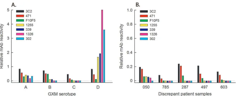
In an attempt to determine the GXM epitopes present in discordant specimens, samples with adequate volume were reanalyzed using a panel
of MAbs known to have different serotype recognition patterns (Fig. 1A). The antibody panel included three MAbs that recognize serotypes A, B, C, and D (F12D2, 3C2, and 471), one that detects serotypes A and B (F10F5), two that react with serotypes A, B, and D (1255 and 339), and two directed against serotypes A and D (1326 and 302) (10–15). GXM EIAs were per-formed as previously described (8), with the following modifications. Plates were coated overnight with a 1:1 combination of capture MAbs F12D2 and 339. Discrepant specimens and purified GXM were serially diluted across plates in phosphate-buffered saline (PBS) containing 0.05% Tween. Bound polysaccharide was detected by incubation with each of eight horseradish peroxidase-conjugated MAbs followed by a per-oxidase substrate. Assays were completed by adding stop solution, and results were read at 450 nm. The log OD was plotted against the log titer or log concentration (ng/ml) and fit to a linear regression with correction for background, and the titer/concentration that produced an OD of 0.5 was calculated from the regression and taken as the endpoint. F12D2, a MAb that has strong reactivity with all four serotypes, was used to normalize endpoints to account for differing GXM concentrations in clinical speci-mens. Normalized values were calculated as the quotient of each MAb endpoint (ng/ml) divided by the F12D2 endpoint (ng/ml) and are re-ported as “relative MAb reactivities” for discrepant samples and GXMs.
RESULTS
Over the 5-month study period, 1,000 specimens (589 serum and
411 CSF specimens) were tested in parallel. The LFA required the
fewest steps and the least hands-on time to perform, but the
in-terpretation of results was more subjective than that of the
spec-trophotometric methods.
Assay comparisons.
Summaries of the test results are
pre-sented in
Tables 1
and
2
. In all, 56 (41 serum and 15 CSF)
[image:3.585.101.494.64.220.2]speci-FIG 1MAb binding patterns for purified GXM (A) and discordant patient specimens (B). Refer to Materials and Methods for a detailed description of the anti-GXM EIA. MAb, monoclonal antibody; GXM, glucuronoxylomannan.
TABLE 1Qualitative test results
IMMY test result
No. of samples with Meridian EIA result
CSF Serum
Positive Negative Positive Negative
IMMY LFA results
Positive 15 1 41 21
Negative 0 395 0 527
IMMY EIA results
Positive 15 2 41 19
Negative 0 394 0 529
Evaluation of New Cryptococcal Antigen LFA and EIA
January 2013 Volume 20 Number 1 cvi.asm.org 53
on January 25, 2013 by CDC Public Health Library & Information Center
http://cvi.asm.org/
[image:3.585.298.544.606.723.2]mens were positive and 921 (527 serum and 394 CSF) specimens
were negative by all three methods. Qualitative agreement across
assays was 97.7% (kappa value
%
0.82; 95% confidence interval
[95% CI]
%
0.75 to 0.89). Concordance was highest for CSF
com-pared to serum specimens (99.5% versus 96.4% agreement,
re-spectively [
P
%
0.0013]). Similarly, agreement was significantly
greater for CrAg-negative than CrAg-positive specimens (97.9%
versus 73.7% agreement, respectively [
P
#
0.0001]). Endpoint
titers showed little to no correlation between assays, and on
aver-age, the IMMY tests produced severalfold higher titers than the
Meridian test (data not shown).
Discrepant specimens.
Twenty-three specimens (2.3%)
yielded discordant results across the assay comparisons, and all of
these were Meridian EIA negative (
Table 3
). Of the 23 discrepant
specimens, 20 (87.0%) were positive by both the IMMY LFA and
EIA, 2 (8.7%) were LFA positive only, and 1 (4.3%) was EIA
pos-itive only. Eleven samples had an adequate volume for LA testing;
8/11 samples (72.7%) were LA positive, and 3/11 samples (27.3%)
were LA negative. LA-negative samples (2 CSF samples and 1
se-rum) displayed low titers by IMMY EIA/LFA (
!
1:10).
The same 11 specimens analyzed by LA assay were retested
using a panel of anti-GXM MAbs with distinct recognition
pat-terns for the four major GXM serotypes (
Table 3
). Low-titer
spec-imens could not be evaluated using the GXM EIA (6/11 samples
[54.5%]). The pattern of MAb recognition for 4 of 5 higher-titer
specimens (i.e., discrepant patient samples 785, 287, 497, and 603)
was consistent with the expression of serotype C epitopes (
Fig.
1B
). Specimens containing serotype C-like GXM were sent to
ARUP Laboratories from referring hospitals in Louisiana,
Califor-nia, Washington, and Missouri.
DISCUSSION
Immunoassays designed to detect
Cryptococcus
capsular antigens
are important tools for the diagnosis of cryptococcosis. We
com-pared three commercially available CrAg assays by using a large
number of serum and CSF samples that were collected as a part of
routine patient care. The new IMMY LFA and EIA showed almost
perfect overall agreement (kappa statistic of 0.82) with our current
test, the Meridian Premier EIA. Qualitative agreement was
signif-icantly greater for CrAg-negative than CrAg-positive specimens,
likely due to sensitivity differences between the IMMY and
Merid-ian assays. In addition, interassay agreement was higher for CSF
than for serum specimens. This may have been a result of higher
antigen concentrations present in the CSF of patients with
cryp-tococcal meningitis. The highest percent agreement (99.7%) was
observed between the two IMMY tests, which reflects the use of
the same MAbs for CrAg detection in both platforms.
TABLE 2Qualitative assay agreement
Comparison and agreement % Agreement Kappa value (95% CI)
Meridian EIA vs IMMY LFA 97.8 0.82 (0.75–0.9)
Positive agreement 71.8
Negative agreement 97.7
Meridian EIA vs IMMY EIA 98.0 0.84 (0.77–0.91)
Positive agreement 73.7
Negative agreement 97.9
IMMY LFA vs IMMY EIA 99.7 0.98 (0.96–1.00)
Positive agreement 97.4
Negative agreement 99.9
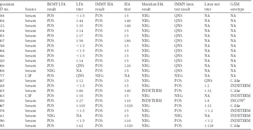
TABLE 3Discrepant specimen resultsa
Specimen
ID no. Source
IMMY LFA result
LFA titer
IMMY EIA result
EIA titer
Meridian EIA result
IMMY latex test result
Latex test titer
GXM serotype
058 Serum POS #1:5 POS 1:5 NEG QNS NA NA
804 Serum POS 1:44 POS 1:40 NEG QNS NA NA
122 Serum POS 1:35 POS 1:40 NEG QNS NA NA
104 Serum POS 1:14 POS 1:5 NEG QNS NA NA
953 Serum POS 1:17 POS 1:5 NEG QNS NA NA
207 Serum POS 1:59 POS 1:80 NEG QNS NA NA
749 Serum POS #1:5 POS 1:5 NEG QNS NA NA
894 Serum POS #1:5 POS 1:5 NEG QNS NA NA
046 Serum POS #1:5 POS 1:5 NEG QNS NA NA
520 Serum POS 1:14 POS 1:5 NEG QNS NA NA
806 Serum POS QNS POS 1:20 NEG QNS NA NA
309 Serum NEG NA POS 1:5 NEG QNS NA NA
777 CSF POS QNS NEG NA NEG NEG NA NA
287 Serum POS 1:12 POS 1:5 NEG POS QNS C-like
028 Serum POS #1:5 POS 1:5 NEG POS 1:2 INDETERM
603 Serum POS 1:86 POS 1:40 INDETERM POS 1:32 C-like
963 CSF POS 1:10 POS 1:5 NEG NEG NA INDETERM
050 Serum POS 1:27 POS 1:10 INDETERM POS 1:8 INCONb
497 Serum POS 1:105 POS 1:320 NEG POS 1:32 C-like
378 Serum POS #1:5 POS 1:5 NEG POS #1.2 INDETERM
161 Serum NEG NA POS 1:5 NEG NEG NA INDETERM
780 Serum POS #1:5 POS 1:10 NEG POS #1:2 INDETERM
785 Serum POS 1:62 POS 1:320 NEG POS 1:128 C-like
aAbbreviations: POS, positive; NEG, negative; NA, not applicable; QNS, quantity of the specimen was not sufficient for repeat testing; INDETERM, indeterminate; INCON,
inconclusive; GXM, glucuronoxylomannan.
bMAb reactivity was not typical of patterns observed with GXM from the four major serotypes.
on January 25, 2013 by CDC Public Health Library & Information Center
http://cvi.asm.org/
The IMMY assays had the highest sensitivities, with positivity
rates of 7.8% and 7.6% for the IMMY LFA and IMMY EIA,
re-spectively, compared to the Meridian EIA positivity rate of 5.6%.
Analysis of discrepant specimens by use of a separate latex
agglu-tination kit suggested that the majority of IMMY-positive but
Me-ridian-negative specimens with adequate volume for retesting did
contain CrAg. The IMMY LFA- and/or EIA-positive specimens
displayed low endpoint titers. The sensitivity of the LA method
may have been influenced by the sample volume used to
accom-modate retesting of low-volume specimens (150
$l versus the 300
$l recommended in the manufacturer’s instructions). An
alterna-tive explanation is that the low-titer, IMMY LFA- and/or
EIA-positive results represent false-EIA-positive test results. Incomplete
ac-cess to patient-level data limited our ability to fully resolve
discrepancies. However, in a recent study comparing the IMMY
LFA to the Meridian cryptococcal antigen latex system (CALAS), 2
of 76 specimens (2.6%) produced discordant results; both of these
were IMMY-positive but Meridian-negative samples that came
from patients with a previous documented history of
cryptococ-cosis (
16
).
Discrepant specimen analysis using anti-GXM MAbs with
differing serotype reactivity patterns was limited by the small
number of specimens with adequate volume for retesting and
by an overrepresentation of low-titer samples. However, most
discordant specimens with high enough titers for GXM EIA
analysis (4/5 samples [80%]) were shown to contain CrAg
epitopes consistent with serotype C. This observation is in line
with previous reports that the Meridian assays (both the EIA
and CALAS kits) have reduced sensitivity for detection of
pu-rified GXM of serotype C (
8
). Furthermore, the IMMY assays
were constructed using a combination of MAbs that recognize
all 4 major serotypes (
3
,
8
,
11
).
Serotype C (i.e.,
C. gattii
molecular type VGIV) has emerged as
a relatively common cause of cryptococcosis in HIV-positive
pa-tients from sub-Saharan Africa (
17
,
18
) and has also been reported
in India and South America (
19
–
21
). Patient race, HIV status, and
travel histories were not available to determine whether the
spec-imens with serotype C-like reactivity patterns in this study came
from patients who had either lived in or traveled to geographic
regions known to harbor serotype C
Cryptococcus
. ARUP
Labora-tories is a large national reference laboratory, and it is conceivable
that serotype C patients were originally from one of these areas.
Alternatively, these results may be the result of a heretofore
un-recognized complexity in expression of CrAg epitopes.
In conclusion, the IMMY assays showed excellent overall
con-cordance with the Meridian EIA. Assay performance differences
appear to be related to issues of analytic sensitivity and serotype
bias. Incomplete access to patient-level data, as well as low
speci-men volumes, which precluded our ability to perform repeat
test-ing, affected our ability to fully resolve discrepant results.
ACKNOWLEDGMENTS
The IMMY reagents were donated by the manufacturer.
J.H. performed and interpreted all of the LFA results for this study.
REFERENCES
1.Bloomfield N, Gordon MA, Elmendorf DF, Jr.1963. Detection of Cryp-tococcus neoformans antigen in body fluids by latex particle agglutina-tion. Proc. Soc. Exp. Biol. Med.114:64 – 67.
2.Babady NE, Bestrom JE, Jespersen DJ, Jones MF, Beito EM, Binnicker MJ, Wengenack NL.2009. Evaluation of three commercial latex aggluti-nation kits and a commercial enzyme immunoassay for the detection of cryptococcal antigen. Med. Mycol.47:336 –338.
3.Jarvis JN, Percival A, Bauman S, Pelfrey J, Meintjes G, Williams GN, Longley N, Harrison TS, Kozel TR.2011. Evaluation of a novel point-of-care cryptococcal antigen test on serum, plasma, and urine from pa-tients with HIV-associated cryptococcal meningitis. Clin. Infect. Dis.53: 1019 –1023.
4.Tanner DC, Weinstein MP, Fedorciw B, Joho KL, Thorpe JJ, Reller L.
1994. Comparison of commercial kits for detection of cryptococcal anti-gen. J. Clin. Microbiol.32:1680 –1684.
5.Ikeda R, Nishikawa A, Shinoda T, Fukazawa Y.1985. Chemical charac-terization of capsular polysaccharide from Cryptococcus neoformans se-rotype A-D. Microbiol. Immunol.29:981–991.
6.Wilson DE, Bennett JE, Bailey JW.1968. Serologic grouping of Crypto-coccus neoformans. Proc. Soc. Exp. Biol. Med.127:820 – 823.
7.Byrnes EJ, 3rd, Bartlett KH, Perfect JR, Heitman J.2011. Cryptococcus gattii: an emerging fungal pathogen infecting humans and animals. Mi-crobes Infect.13:895–907.
8.Percival A, Thorkildson P, Kozel TR. 2011. Monoclonal antibodies specific for immunorecessive epitopes of glucuronoxylomannan, the ma-jor capsular polysaccharide of Cryptococcus neoformans, reduce serotype bias in an immunoassay for cryptococcal antigen. Clin. Vaccine Immunol.
18:1292–1296.
9.Lindsley MD, Mekha N, Baggett HC, Surinthong Y, Autthateinchai R, Sawatwong P, Harris JR, Park BJ, Chiller T, Balajee SA, Poonwan N.
2011. Evaluation of a newly developed lateral flow immunoassay for the diagnosis of cryptococcosis. Clin. Infect. Dis.53:321–325.
10. Belay T, Cherniak R, Kozel TR, Casadevall A.1997. Reactivity patterns and epitope specificities of anti-Cryptococcus neoformans monoclonal antibodies by enzyme-linked immunosorbent assay and dot enzyme assay. Infect. Immun.65:718 –728.
11. Brandt S, Thorkildson P, Kozel TR.2003. Monoclonal antibodies reac-tive with immunorecessive epitopes of glucuronoxylomannan, the major capsular polysaccharide of Cryptococcus neoformans. Clin. Diagn. Lab. Immunol.10:903–909.
12. Eckert TF, Kozel TR.1987. Production and characterization of mono-clonal antibodies specific for Cryptococcus neoformans capsular polysac-charide. Infect. Immun.55:1895–1899.
13. Gates-Hollingsworth MA, Kozel TR.2009. Phenotypic heterogeneity in expression of epitopes in the Cryptococcus neoformans capsule. Mol. Mi-crobiol.74:126 –138.
14. Netski D, Kozel TR.2002. Fc-dependent and Fc-independent opsoniza-tion of Cryptococcus neoformans by anticapsular monoclonal antibodies: importance of epitope specificity. Infect. Immun.70:2812–2819. 15. Spiropulu C, Eppard RA, Otteson E, Kozel TR.1989. Antigenic variation
within serotypes of Cryptococcus neoformans detected by monoclonal antibodies specific for the capsular polysaccharide. Infect. Immun.57: 3240 –3242.
16. Clarke S, Gibb R.2012. Evaluation of the IMMY CrAg lateral flow assay for detection of cryptococcal antigen [abstract]. Aust. Soc. Microbiol. 2012 Annu. Sci. Meet., Brisbane, Australia.
17. Litvintseva AP, Thakur R, Reller LB, Mitchell TG.2005. Prevalence of clinical isolates of Cryptococcus gattii serotype C among patients with AIDS in sub-Saharan Africa. J. Infect. Dis.192:888 – 892.
18. Karstaedt AS, Crewe-Brown HH, Dromer F.2002. Cryptococcal men-ingitis caused by Cryptococcus neoformans var. gattii, serotype C, in AIDS patients in Soweto, South Africa. Med. Mycol.40:7–11.
19. Cogliati M, Chandrashekar N, Esposto MC, Chandramuki A, Petrini B, Viviani MA.2012. Cryptococcus gattii serotype-C strains isolated in Ban-galore, Karnataka, India. Mycoses55:262–268.
20. Perez C, Dolande M, Moya M, Rosello A, de Capriles CR, Landaeta ME, Mata-Essayag S.2008. Cryptococcus neoformans, Cryptococcus gattii: serotypes in Venezuela. Mycopathologia166:149 –153.
21. Trilles L, Lazera Mdos S, Wanke B, Oliveira RV, Barbosa GG, Ni-shikawa MM, Morales BP, Meyer W. 2008. Regional pattern of the molecular types of Cryptococcus neoformans and Cryptococcus gattii in Brazil. Mem. Inst. Oswaldo Cruz103:455– 462.
Evaluation of New Cryptococcal Antigen LFA and EIA
January 2013 Volume 20 Number 1 cvi.asm.org 55
on January 25, 2013 by CDC Public Health Library & Information Center
http://cvi.asm.org/