* corresponding author: [email protected]
Clinical signs as diagnostic test to assess
hypoxemia in children with acute asthma
exacerbation
Latifah Hanum*, Roni Naning, Endy Paryanto Prawirohartono
Department of Pediatrics, Dr. Sardjito General Hospital/Faculty of Medicine, Universitas Gadjah Mada, Yogyakarta, Indonesia
ABSTRACT
Asthma attack can cause hypoxemia. One of the methods to detect hypoxemia is by using pulse oximetry. However, this tool is not always available in some health care centres. Therefore, a more rapid and simple diagnostic tool is needed as an alternative method to detect hypoxemia. This study aimed to assess signs and symptoms as diagnostic tools for hypoxemia in children with asthma. This was an analytical observational with cross-sectional design performed in Department of Pediatrics, Dr. Sardjito General Hospital/Faculty of Medicine, Universitas Gadjah Mada, Yogyakarta. The study was started in Februari 2010 until the sample size was sufficient. Seventy seven children with asthma between 0 – 18 years old who were presented to Emergency Department and Respiratory Outpatient Clinic were involvoled in this study. All subjects were examined for clinical signs and oxygen saturation as the gold standard. The prevalence of hypoxemia in children with asthma in this study was 18.2%. The best single clinical predictor of hypoxemia was tachycardia that yielded a sensitivity of 86% (95%CI: 67 – 100%) and specificity of 59% (95%CI: 49 – 71%), and nasal flaring yielded a sensitivity of 79% and specificity of 79%. The combination of 2 clinical signs namely chest wall retraction-nasal flaring increased a sensitivity of 79% and specificity of 71%, chest wall retraction-tachycardia increased a sensitivity of 86% and a specificity of 76%, chest wall retraction-tachypnoe increased a sensitivity of 86% and a specificity of 51%, tachycardia-tachypnoea increased a sensitivity of 79% and a specificity of 76%. The combination 3 clinical sign namely chest wall retraction-tachycardia-tachypnoea yielded a sensitivity of 79% and specificity of 79%. In conclusion, chest wall retraction and tachycardia have higher diagnostic score than other clinical signs to assess hypoxemia in children with asthma on acute exacerbation.
ABSTRAK
2 gejala klinik dapat meningkatkan sensitivitas dan spesifisitas yaitu retraksi dinding dada-pernafasan cuping hidung meningkatkan sensitivitas 79% dan spesifisitas 71%, retraksi dinding takikardia meningkatkan sensitivitas 86% dan spesifisitas 76%, retraksi dinding dada-takipnea meningkatkan sensitivitas 86% dan spesifisitas 51%, takikardia-dada-takipnea meningkatkan sensitivitas 79% dan spesifisitas 76%. Kombinasi 3 gejala klinik yaitu retraksi dinding dada-takikardia-takipnea meningkatkan sensitifitas 79% dan spesifisitas 79%. Dapat disimpulkan retraksi dinding dada dan takikardia mempunyai nilai diaknosis lebih tinggi dari pada gejala klinik lain untuk menilai hipoksia pada anak penderita asma saat eksaserbasi akut.
Keywords: asthma - hypoxemia - clinical signs - children - assessment
INTRODUCTION
Asthma is a common chronic respiratory disease in children characterized by episodes or attacks of impaired breathing. The asthma symptoms are caused by inflammation and narrowing of small airways and may include shortness of breath, coughing, wheezing, and chest pain.1The prevalence of asthma increases
gradually each year in both developed and developing countries. The current prevalence of asthma ranges from 2 – 30%.2,3The episode
of asthma attack ranges from mild to severe which may cause death. Asthma is one of the five major diseases that causes death. It is estimated that about 17.4% of death are caused by asthma.4
Due to better understanding of patho-physiology, the availability of effective therapy and extensive application of evidence-base treatment guidelines in the past decade has made the asthma mortality rate gradually in decline in several reports.5,6However, if asthma is not
prevented and treated properly, the prevalence of asthma may continue to increase and disturb the growth and development of children as well as their quality of life.
Hypoxemia is one of the imporant risk factors that may contribute to death of asthma patients. Hypoxemia is a condition in which
level of arterial oxygen saturation (SaO2) of hemoglobin. It is often used to detect the hypoxemia condition of asthma patients. However, this device is often not available in some health care centres especially in develop-ing countries. Therefore, a more rapid and simple diagnostic method to estimate SaO2 is urgently needed as an alternative method to detect hypoxemia in asthma patients. This study aimed to assess signs and symptoms as diagnostic tools for detecting hypoxemia (SpO2 of 90%) in children with asthma.
MATERIALS AND METHODS
Subjects
Health Research Ethics Committee, Faculty of Medicine, Universitas Gadjah Mada, Yogya-karta.
Protocols
The signs and symptoms of children who fulfilled the inclusion criteria were recorded using standadized forms. Children were diagnosed with asthma if they showed recurrent wheezing with persistent cough, particularlyat night, after physical activity, and also had a family history of asthma and allergy. Asthma on acute exacerbation was defined when the shortness of breath worsened. The children were then examined and counted for their clinical signs namely pulse rate, respiratory rate, tachypnoea, retraction, head nodding, nasal flaring, cyanosis and conciousness level by a physician.
Pulse rate was counted from the palpation of radial artery per minutes. Tachycardia was defined if pulse rate was >60 times/minutes in children aged <1 year, >120 times/ minutes in children aged 2 – 8 years, >100 times/minutes in children aged 8 – 18 years. Respiratory rate was counted per minute by observing the movement of abdominal wall and one breath was counted from the upward movement of abdomen. Tachypnoea was defined when the respiratory rate was >50 times/minute in children aged <1 year, >40 times/minute in children aged 1 – 5 years, >30 times/minute in children aged 6 – 18 years. Retraction was assessed by the presence of retraction on the chest wall (suprasternal and/or subcostal, intercostal). Head nodding was noted by observing movement of the head for every breath. Nasal flaring was assessed by observing movement of the nostrils for 30 seconds.
Cyanosis was observed by looking at the bluish color of the lips and tongue. Cyanosis was defined when the color was bluish, but, reddish if it was not cyanotic. Consciousness level was noted by the Glascow’s coma scale. Decreased consciousness was defined when the coma scale was below normal.
The children’s arterial oxygen saturation was then measured with a pulse oximeter placed on a toe or finger, while the patient breathed room air. Hypoxemia was defined as an arterial
oxygen saturation 90% recorded by the pulse
oximeter.
Stastitical analysis
Statistical analysis was performed using SPSS. Data were expressed as mean ± standard deviation (SD) for quantitative variables and as number and percentage for qualitative values. Clinical signs between hypoxemic and non-hypoxemic children with asthma were compared using the Chi square test, or by the Fisher’s exact test if the expected frequencies were less than 5. A p value of 0.05 was considered significant. The sensitivity and specificity of each clinical sign in its ability to predict hypoxemia was also calculated.
RESULTS
TABLE 1. Characteristics of subjects according to the percentage in the group of hypoxemia and hypoxemia
The hypoxemia was observed in 14 (18.2%) patients among 77 patients involved during the study. Mean of SpO2of all asthma patients is presented in FIGURE 1. Mean SpO2
Diagnostic test on each clinical signs of hypoxemia on asthma patients is presented in
FIGURE 1. Mean of SpO2of asthma patients in each group
of all patients, patients with hypoxemia and patents without hypoxemia were 94.3 ± 3.1%, 89.4 ± 0.9% and 95.4 ± 2.1%, respectively.
TABLE 2. The value of sensitivity and specificity of single clinical signs for for detection hypoxemia in children with acute asthma exacerbation
Note: Sen = sensitivity, Spe = specificity, CI = confidence interval
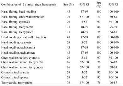
The combination of 2 clinical signs for detecting hypoxemia on asthma patients is presented in TABLE 3. This combination can increase both sensitivity and specificity for detecting hypoxemia on asthma patients. The combination of 2 clinical signs that did not show wide range of difference between sensitivity and
specificity were chest wall retraction and nasal flaring (79% for sensitivity and 71% for specificity), chest wall retraction and tachycardia (86% and 76%), chest wall retraction and tachypnoea (86% and 51%), tachycardia and tachypnoea (79% and 76%).
TABLE 3. The value of sensitivity and specificity of 2 clinical signs combination for detection hypoxemia in children with acute asthma exacerbation
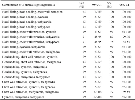
After the 3 clinical signs were combined, only one combination having good sensitivity and specificity was obtained (TABLE 4). This combination was chest wall retraction, tachycardia and tachypnoea with sensitivity of
79% and specificity of 79%. This means that the chest wall retraction, tachycardia and tachypnoea had 79% of positive results in detecting hypoxemia as well as determining the patients without hypoxemia.
TABLE 4. The value of sensitivity and specificity of 3 clinical signs combination for for detection hypoxemia in children with acute asthma exacerbation
Note: Sen = sensitivity, Spe = specificity, CI = confidence interval
The combination of 4, 5 and 6 clinical signs for detecting hypoxemia on asthma patients was also evaluated. However, these combinations did not increase the sensitivity and specificity.
DISCUSSION
ion and 28.6% one hour after presentation. Male
et al.9 also reported among 27 asthmatic
such as definition of hypoxemia, severity of the asthma attack, and location characteristics of the studies. Male et al.9 used the presence of SaO2 < 92 as an indicator of a severe and potentially life threatening asthma attack, whereas Rahnama’iet al.8used the presence of
SaO2 d” 92% as an indicator of moderate and severe asthma. In this study, the presence of SaO2 <90% was used as an indicator of hypoxemia in children with asthma. Severity of an asthma attack influences the hypoxemia prevalence because patients who are having an episode of asthma attack will have varying degrees of hypoxemia, depending on the severity of the episode or exacerbation.10 The study
location characteristics, whether in emergency department or outpatient clinic, will influence asthma patient characteristics and severity of asthma attack. The differences of the hypoxemia definition, the severity of the asthma attack, and the location characteristics of the studies will determine patients who were included in the inclusion criteria of the study.
The studies of various symptoms and clinical signs as predictor for hypoxemia in several illness have been conducted with different findings. Lodha et al.7 reported that
none of the clinical signs either alone or in combination had desirable sensitivity and specificity to predict hypoxemia in children with acute lower respiratory tract infection (ALRI). However, Singhiet al.11concluded that several
respiratory signs namely breathing, cyanosis, grunting, nasal flaring, chest retractions, head nodding and auscultatory signs were found to be associated with hypoxemia. Moreover, it could be used to predict hypoxemia in children with ALRI with reasonable accuracy. In acute chilhood asthma, the accessory muscel score e” 3 and pulsus paradoxus à 10 were indentified as independent predictors of hypoxemia with sensitivity of 64.3% and specificity of 91%.8
In this study, it was found that the tachycardia
and nasal flaring had higher diagnostic score than other clinical signs to predict hypoxemia in children with asthma on acute exacerbation. The different findings in the use of symptoms and clinical signs as predictor for hypoxemia are influenced by some factors such as the age of patients, kind and severity of ilness, and kind of clinical signs use.
Some studies showed that there is no single clinical sign that can predict hypoxaemia with both high sensitivity and specificity. Several combination of clinical signs have been suggest-ed to improve sensitivity. These combinations generally use a clinical sign of severe respira-tory distress, such as chest wall indrawing, head nodding or very fast breathing combined with a sign for general depression such as the inability to feed? or move or being unconscious. The
presence of cyanosis might be added.12For
example, Rahnama’iet al.8combined chest wall
retraction and pulsus paradoxus as clinical signs to predict hypoxemia in acute chilhood asthma that yielded a sensitivity of 86% and specificity of 59%. A combination of three clinical signs namely tachypnoea, retraction and crepitation was the best predictors for diagnosis hypoxemia in children with acute lower respiratory tract infections with sensitivity of 67.8% and specificity of 96.2%. In addition, a combination between cyanosis, nasal flaring, and inability to drink yielded a sensitivity of 92% and specificity of 86%.7
exchange, and low oxygen inspiration. Hyperventilation may be resulted as a way to obtain sufficient oxygen for the body.13 In
pneumonia, respiratory tract obstruction occur due to viral, bacterial or microbes infection that leads to lung consolidation resulting in poor oxygenation. Pneumonia causes abnormalities of the lungs through the decrease in surface area of membranous respiration and ventilation-perfusion ratio resulting the decrease of diffusion capacity and persistent hypoxemia.14,15
Although the pathophysiology of hypoxemia in patients pneumonia and asthma is similar, the same clinical signs that are used to predict hypoxemia in both illness may yield in different sensitivity and specificity.
Some limitations were observed during this study. The clinical signs examination was only conducted by two examiners. Ideally it is conducted by at least three examiners who are expert in the physical examination skills. Moreover, the clinical examination and pulse oxymetry measurement were conducted wihout blinding due to ethical reasons. The acute asthma excerbation is an emergency case, therefore it must be taken care of with the standard management of acute asthma excerbation. The validation of pulse oxymetri measurement with blood gas analysis was not conducted in this study. The blood analysis was only performed in children with breathing failure. It was not conducted simultaneously with the clinical signs, examination and oxygen saturation measurement by pulse oximeter to avoid measurement bias due to the necessary preparatin for blood sampling.
CONCLUSION
The combination of chest wall retraction
ACKNOWLEDGEMENT
We would like to thank all patients who participated in this study. We also thank Head of Department of Department of Pediatrics, Dr. Sardjito General Hospital/Faculty of Medicine, Universitas Gadjah Mada, Yogyakarta, Indonesia who has given his permission to conduct this study.
REFERENCES
1. Akinbarni LJ, Moorman JE, Liu X. Asthma prevalence, health care use, and mortality: United States, 2005–2009. Natl Health Stat Report 2011; 32:1-14.
2. Supriyatno B. Diagnosis dan penatalaksanaan terkini asma pada anak. Majalah Kedokteran Indonesia 2005; 55(3): 237-43.
3. Pllart SM, Compton RM, Elward KS. Management of acute asthma exacerbations. Am Fam Physician 2011; 84(1):40-7.
4. Departemen Kesehatan RI. Pedoman pengendalian penyakit asma. Keputusan Menteri Kesehatan Republik Indonesia no 1023/Menkes/SK/XI/2008. 2008. [cited July 20th, 2010]. Available from: www.depkes.com.
5. Rodrigo GJ, Plaza V, Forns SB, Tordera MP, Salas J. Factors associated with mortality in patients hospitalized in Spain and Latin America for acute severe asthma in 1994, 1999 and 2004. J Bras Pneumol 2008;34:546-51.
6. Chantaphakul H, Luangdilok T, Ruxrungtham K, Klaewsongkram J. Inpatient asthma mortality in a tertiary referral hospital from 2000 to 2010. Asian Pac J Allergy Immunol 2012; 30:193-6.
7. Lodha R, Bhadauria PS, Kuttikat AV, Puranik M, Gupta S, Pandey RM, et al.Can clinical symptoms or signs accurately predict hypoxemia in children with acute lower respiratory tract infections? Indian Pediatr 2004; 41(2):129-35.
10. Papiris S, Kotanidou A, Malagari K, Roussos C. Clinical review: severe asthma. Critical Care 2002; 6:30-44.
11. Singhi S, Deep A, Kaur H. Prevalence and predictors of hypoxemia in acute respiratory infections presenting to pediatric emergency department. UCCM 2003; 7(2): 118-23.
12. Usen S & Weber MW. Clinical sign of hypoxemia in children with acute lower respiratory tract infection: indicators of oxygen therapy. Int J Tuberc Lung Dis 2001; 5(6): 505-9.
13. Becerra V. Physiopathology of asthma. Rev Aleg Mex 2009; 56(suppl 1):S24-8.