R E S E A R C H A R T I C L E
Open Access
RsaI
repetitive DNA in Buffalo
Bubalus bubalis
representing retrotransposons, conserved in
bovids, are part of the functional genes
Deepali Pathak and Sher Ali
*Abstract
Background:Repetitive sequences are the major components of the eukaryotic genomes. Association of these repeats with transcribing sequences and their regulation in buffaloBubalus bubalishas remained largely unresolved.
Results:We cloned and sequencedRsaIrepeat fragments pDp1, pDp2, pDp3, pDp4 of 1331, 651, 603 and 339 base pairs, respectively from the buffalo,Bubalus bubalis. Upon characterization, these fragments were found to represent retrotransposons and part of some functional genes. The resultant clones showed cross hybridization only with buffalo, cattle, goat and sheep genomic DNA. Real Time PCR, detected ~2 × 10⁴ copies of pDp1, ~ 3000 copies of pDp2 and pDp3 and ~ 1000 of pDp4 in buffalo, cattle, goat and sheep genomes, respectively.RsaI repeats are transcriptionally active in somatic tissues and spermatozoa. Accordingly, pDp1 showed maximum expression in lung, pDp2 and pDp3 both in Kidney, and pDp4 in ovary. Fluorescencein situhybridization showed repeats to be distributed all across the chromosomes.
Conclusions:The data suggest thatRsaIrepeats have been incorporated into the exonic regions of various transcribing genes, possibly contributing towards the architecture and evolution of the buffalo and related genomes. Prospects of our present work in the context of comparative and functional genomics are highlighted.
Background
Different families of repetitive DNA contribute towards architectural organization of the mammalian genomes [1]. They represent both, tandemly arranged and inter-spersed sequences [2]. Based on their size and mode of propagation, Interspersed elements can be divided into two separate classes, the long terminal repeat (LTR) and non-LTR. The non LTR LINEs (long interspersed repeat elements) and SINEs (Short interspersed repeat ele-ments) are widely distributed occupying a substantial fraction of the eukaryotic genomes. These elements replicate and proliferate themselves through a“copy and paste”mechanism called retrotransposition [3,4]. In this process, transcription of their genomic copies is fol-lowed by an RNA intermediate resulting cDNAs reinte-gration at a new location in the genome [5]. Approximately, 100 LINE and SINE families have been
reported to date in various eukaryotic genomes [6]. In mammals, LINE, L1 repeats are dominant retrotranspo-sons type both in the common ancestor and in extant species [7]. In addition to L1, an element belonging to the retrotransposable element family of autonomous ret-rotransposons (RTE-1) has been reported in mammals [8]. Few mammals have active non LTR LINE other than L1 that contribute significantly to repeat composition.
Species specific retrotransposons have been widely used as a tool for Phylogenetic analysis and population studies [9,10]. Many retrotransposons are inactive, found in the non-coding regions of the genome and are subjected only to the neutral evolution. Thus, rare new insertions have led to some form of advantageous or noteworthy phenotypic variations [11-13]. These and other such discoveries have resulted in a shift from ear-lier thought of them being“parasite” to functional ele-ments cultivated in the genome for their beneficial attributes.
* Correspondence: [email protected]
Molecular Genetics Laboratory, National Institute of Immunology, Aruna Asaf Ali Marg, New Delhi -110 067, India
Retrotransposons have gained novel functions, provid-ing alternative splice sites and/or polyadenylation signals or modifying gene expression [14-16]. These elements account for 46.5% of bovine genome [17]. The bovine genome is very different in its repeat composition com-pared to other mammalian genomes. It has unusual composition of LINE RTE type Bov B and its associated SINE elements which together account for 25% of the bovine genome [17]. The impact of the interspersed repeats on the genomes of human [18-20], dog [21,22], cow [17,23], mouse [24] and opossum [25,26] has been studied. However fate of these interspersed elements and their association with mRNA transcriptomes in buf-falo remains still unclear. Here, we report RsaIfamily repetitive DNA in the genome of water buffalo“Bubalus bubalis“ and their copy number status. We also studied their expression in somatic tissues and spermatozoa. The repeat fraction pDp1, pDp2 and pDp3 were used for fluorescencein situhybridization (FISH) with buffalo metaphase chromosomes. In addition, we isolated and
sequencedACOT11 (Acyl-coenzyme A thioesterase 11)
gene harboring part of pDp1 repeat.
Results
RsaIenzyme digestion uncovers four repeat fractions
Digestion of buffalo genomic DNA with RsaIenzyme,
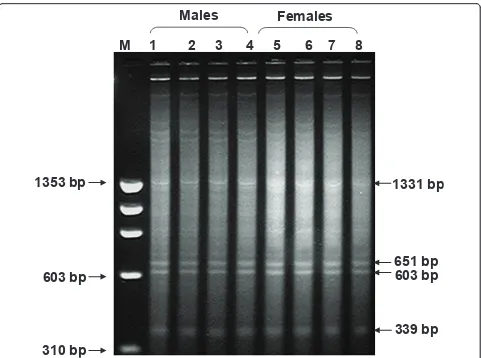
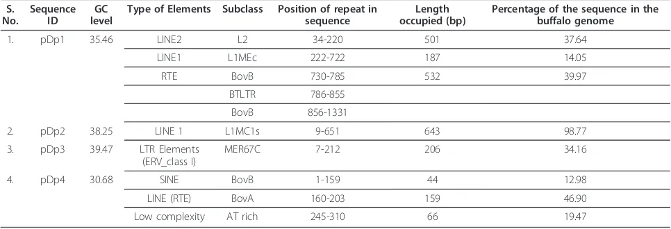
besides minor ones, showed four prominent bands ran-ging from 1331 base pairs, pDp1; 651, pDp2; 603, pDp3; to 339; pDp4 (Figure 1). Approximately, 15-20 recombi-nant clones subjected to restriction digestion and slot blot hybridization screening yielded ten positive clones for each repeat. Five-six clones from each fragment were then sequenced. The accession numbers of the recombinant clones are given in the Additional File 1. All the four major repeat elements were AT rich but sequence-wise, were different from one another (Addi-tional File 2). No inter-clonal variations were detected in these sequences. However, random repeats were pre-sent in the nucleotide sequences (Table 1). Repeat Mas-ker programme revealed presence of LTR LINE, SINE
Males
Females
1331 bp
651 bp
603 bp
339 bp
1353 bp
603 bp
310 bp
M 1 2 3 4 5 6 7 8
elements within the four fragments (Table 2). Blast search for each clone showed 69-98% homology with genomic DNA/contigs and 68-93% with transcribing genes in the database (Additional File 1) mostly in UTRs (Additional File 3).
Buffalo derivedRsaIOpen Reading Frame (ORFs) has
amino acid similarity to LINE reverse transcriptase
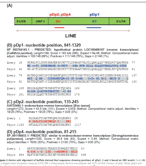
Most striking feature of pDp1 sequence was the presence of 489 bp, +1 ORF (nucleotide position, 841-1329) (Fig-ure 2B). BLASTP search with GenBank sequences using conceptual translation of this ORF (162aa) gave matches to putative reverse transcriptase’s domain. This region corresponds to central position of the Reverse transcrip-tase ORF (Figures 2A and 3). Similarly, pDp2, 111 bp, +3 ORF (nucleotide position, 135-245, 65aa) and pDp4, 135 bp, +3 ORF (nucleotide position, 81-215, 44aa) showed homology to endonuclease reverse transcriptase (Figures 2B and 2C). These ORFs corresponded to central
endonuclease reverse transcriptase of LINE1 ORF2 (Fig-ure 3). pDp3 sequence,112 bp, +3 ORF (nucleotide posi-tion, 492-602, 37 aa) showed no similarity with endonuclease reverse transcriptase.
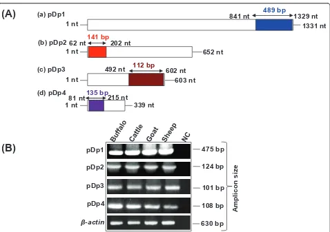
RsaIRepeat status among bovids
Independent cross hybridization of pDp1, pDp2, pDp3 and pDp4 with genomic DNA from different species (mentioned in methods section) under high stringent conditions showed signals only in bovids (Additional File 4). PCR conducted using region specific primers (Table 3) amplified bands in buffalo, cattle, goat and sheep genomic DNA (Figures 3A &3B). Southern hybri-dization of two representativeRsaIsequence pDp1, and pDp2 showed same number of bands within the geno-mic DNA of bovids (Additional File 5). Prominent bands within the smear reflect identical size of the frag-ments dispersed throughout the bovid genomes. Clus-talW alignment of 489 bp pDp1derived fragment with Table 1 Details of repeat clusters present inRsaIsequences identified by repeat finder http://zlab.bu.edu/repfind/ form.html
S. No. Sequence ID Repeat cluster Location
1. pDp1 CGTGAAC 1026,1033
ATGTGAA 485, 666, 721, 734
GTG 23, 41, 69, 161, 190, 198, 487, 668, 723, 736, 746, 780, 786, 800, 817, 832, 838, 879, 988, 1027, 1034, 1200
GTGA 163, 198, 487, 668, 723, 736, 786, 800, 817, 832, 988, 1027, 1034
GTGAA 487, 668, 723, 736, 817, 832, 1027, 1034
TGTGAA 486, 667, 722, 735, 831,
CGTGAA 816, 1026, 1033,
GGATTTT 140,147
2. pDp2 AAACTATGG 209, 375,592
AAACTAT 209, 375, 468, 592
ATACATTTGT 498, 538
3. pDp3 AACATT 469, 481, 488
TAT 15, 123, 139, 446, 451, 454, 464, 474, 583
Table 2 Genome-wide distribution ofRsaIrepeat fragments and their homologies with SINE and LINEs
S.
Type of Elements Subclass Position of repeat in sequence
Length occupied (bp)
Percentage of the sequence in the buffalo genome
1. pDp1 35.46 LINE2 L2 34-220 501 37.64
LINE1 L1MEc 222-722 187 14.05
RTE BovB 730-785 532 39.97
BTLTR 786-855
BovB 856-1331
2. pDp2 38.25 LINE 1 L1MC1s 9-651 643 98.77
3. pDp3 39.47 LTR Elements
(ERV_class I)
MER67C 7-212 206 34.16
4. pDp4 30.68 SINE BovB 1-159 44 12.98
LINE (RTE) BovA 160-203 159 46.90
cattle (AF060172), goat (AF404302) and sheep (AC148038) showed high level of sequence conservation among the bovids (Additional File 6). Phylogenetically, with respect to all the four sequences, cattle and buffalo were found to be closer to each other (Additional File 7), constituting the same monophyletic group.
Differential expression ofRsaIfragments in somatic tissue
and spermatozoa of buffalo
RT-PCR analysis using internal primers of pDp1, pDp2, pDp3 and pDp4 (see Table 3) and cDNA from somatic tissues and spermatozoa of buffalo showed amplification of band across tissues and sperm confirming their
(A)
(B) pDp1- nucleotide position, 841-1329
XP 002764145.1 - PREDICTED: hypothetical protein LOC100406107 (reverse transcriptase) [Callithrix jacchus], Length=188, Score = 193 bits (490), Expect = 6e-48, Method: Compositional matrix adjust. Identities = 100/146 (68%), Positives = 111/146 (76%), Gaps = 2/146 (1%)
Query 18 WKRSLFILIPKKGNAKERSDYHTVTLISHAGKVMLKILQARLQQYVNRELPDVQAGFRKG 77
WK ++FI IPKKGNAKE S+YHTV SHA KVM KILQ RLQQYVNRELPDVQ GFRK
Sbjct 33 WK-AVFIPIPKKGNAKECSNYHTVVFTSHAAKVMPKILQGRLQQYVNRELPDVQTGFRKD 91
Query 78 RGTRDQIANILWIIEKAREFQKSIYFCFTDYAKVFDCLDYNKLWKILKEMGIPAHLICLL 137
RGT Q AN IIEKAREFQK+IY CF D+AK C+D+ KL K+L MGIP H L
Sbjct 92 RGTTGQTANT-CIIEKAREFQKNIYICFIDHAKALHCVDHKKLRKLLIGMGIPDHFTYHL 150
Query 138 RNLYAGQEATVRTGHGTTDCFQIGKG 163
R+LYA Q+ATVRTGHGTTD +I KG
Sbjct 151 RSLYACQQATVRTGHGTTDGSKIRKG 176
(C) pDp2- nucleotide position, 135-245
AAY53484.1- endonuclease reverse transcriptase [Bos taurus]
Length=1272, Score = 43.5 bits (101), Expect = 0.009, Method: Compositional matrix adjust. Identities = 19/25 (76%), Positives = 19/25 (76%), Gaps = 0/25 (0%)
Query 1 MLIAALFIIAKTWKQPGCSLEGEWI 25
M IAALFIIAKTWKQP C EWI
Sbjct 1193 MFIAALFIIAKTWKQPRCPSADEWI 1217
(D) pDp4- nucleotide position, 81-215
XP_001198853.1- PREDICTED: similar to endonuclease-reverse transcriptase [Strongylocentrotus purpuratus], Length=1030, Score = 36.6 bits (83), Expect = 0.84, Method: Compositional matrix adjust.Identities = 16/30 (53%), Positives = 21/30 (70%), Gaps = 0/30 (0%)
Query 1 MSSIKDRNGMDLTEAEDIKKRWQEYTEELY 30
M S+K + G LTE ++KKRW+E EELY
Sbjct 466 MQSVKSKTGRTLTEISEVKKRWKENYEELY 495
Figure 2Amino acid alignment of buffalo derivedRsaIsequences showing positions of pDp1, 2 and 4 based on ORF search. Panel(A)
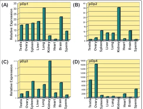
transcriptional potentials (Additional File 8). Quantita-tive Real Time PCR analysis of these sequences showed differential expression ofRsaIrelated transcripts across somatic tissues and spermatozoa (Figure 4). pDp1 showed highest expression in lung, pDp2 and pDp3 in Kidney and pDp4 in ovary. Summary of the relative expression (in folds) derived from 2-∆∆Ctvalues obtained for various transcripts based on Real Time PCR are given in Table 4.
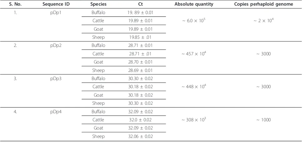
Multiple copies ofRsaIfragments in bovids
The pair of internal primers deduced from pDp1-4 used for expression study was also employed for copy num-ber analysis of these fragments in cattle buffalo, goat and sheep using Real Time PCR. Standard curve slope value was between 3.2-3.5. Single melting peak on dis-sociate curve confirmed primer specificity. pDp1 showed
~2 × 10⁴ copies, pDp2 and pDp3, ~ 3000 copies each
and pDp4 showed ~ 1000, in these species (Table 5 and Additional File 9).
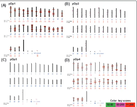
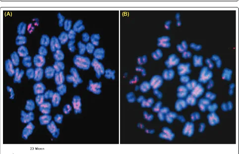
pDp1, pDp2 and pDp3 sequences are dispersed throughout the buffalo genome
In silico analysis of pDp1, pDp2, pDp3 and pDp4 sequences using reference cattle genome revealed its multiple locations on the cow chromosomes (Figure 5). Probing of pDp1 to buffalo genomic DNA digested with
RsaI enzyme detected a strong hybridization signal
(Additional File 10) giving rise to a single isomorphic band in all the samples. FISH mapping of pDp1, 2, 3 spectrum red labeled cloned probes showed ubiquitous discernible signals over buffalo metaphase chromosomes (Figures 6 and 7). All the chromosomes showed dis-persed pattern with all the three probes used indepen-dently. In several chromosomes, signals in the centromeric regions were absent or reduced giving rise to inconsistent pattern. FISH with pDp1 sequence showed more localized signals on the metaphase chro-mosomes as compared to that detected by pDp2 and pDp3. FISH with spectrum red labeled clone pDp4 probe showed background signals even after washing
(A)
the slides under high stringent conditions (60°C in 0.1× SSC). This might be due to the short length (339 bp) of the probe pDp4 used coupled with its dispersed geno-mic organization.
Full length cDNA sequence ofACOT11gene in Buffalo Blast search using reference mRNA sequence revealed
RsaIrepeats to be part of Acyl-coenzyme A thioesterase 11(ACOT11), Vacuolar Protein Sorting 24 (VPS24) and Solute carrier organic anion transporter family member
1A2 (SLCO1A2) genes (Additional File 1) Full length
BuffaloACOT11 cDNA was generated using end point
PCR and gene specific (B. taurus ACOT11) primers
(Table 3). Assembled cDNA sequence of 2592 base pair fragment lacking poly A tail representing six exons (Additional Files 11 and 12) were deposited in the Gen-Bank (HQ848649 and HQ848650).
Discussion
We have studied four (pDp1, pDp2, pDp3 and pDp4)
RsaIfragments from the buffalo genome which are AT
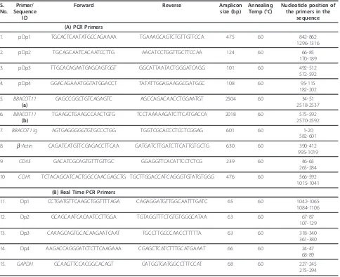
rich, though buffalo genome on the whole is GC rich (40.69%, NC_006295). Database searches with the repeat-maskers revealed the presence of several LTR, LINE and SINE element in the four sequences (Table 2). Apparently, SINEs occupy the (G+C)-rich regions while LINEs are mainly located on the (A+T)-rich regions. Reports suggest that very large number of highly truncated insertions of L1 have occurred in the bovine genome [8]. Full length copies of the human L1 contain two open reading frames, ORF1 and 2. ORF1 encodes a DNA binding protein and ORF2 includes endonuclease and reverse transcriptase domains [27]. Presence of partial reverse transcriptase and endonu-clease domains in pDp1, pDp2 and pDp4 reported Table 3 Details of the primers used for PCR amplification (A) Copy number and Relative expression studies by Real time PCR (B) in buffaloBubalus bubalis
S. the primers in the
sequence (A) PCR Primers
1. pDp1 TGCACTCAATATGCCAGAAAA TGAAAGCAGTCTGTTGTTCCA 475 60 842-862
1296-1316
2. pDp2 TGCAGCAATCACAATCCTTG AACATCCTGGTTGCTTCCAA 124 60 66-85
170-189
3. pDp3 TTGCACAGAATGAGCAGTGGT GGCATTAATACTGGGATCAGG 101 60 492-512
572-592
4. pDp4 GGACAGAAATGGTATGGACCT TATATTGGAGAAGGCGATGGC 108 60 95-115
182-202
5. BBACOT11
(a)
GAGCCGGCTGTCAGAGTC AGCCAGACAACCTGGAATGT 2504 60 34-51
2518-2537
6. BBACOT11
(b)
TGAAGCTGAAGCCAACTGTG TCCTAAAAAGATCTTCATGACCA 2018 60 575-592
2570-2592
7. BBACOT11g AGTGAGGGGGTGTGCCCTGG TGGTCGCACCCTCCTCGGAG 601 60 1-20 582-601
8. b-Actin CAGATCATGTTCGAGACCTTCAA GATGATCTTGATCTTCATTGTGCTG 630 60 390-412 995-1019
9. CD45 GACATCGCAGTGTTTGTTGC GGAGGTTCACATTCCTCTCG 239 60 46-65
265-284
10 CDH1 TCTACAGCATCACTGGCCAACGAGCTG TGCTTGGACCATCAGGGTGTATGTGGG 476 60 566-592
1015-1041
(B) Real Time PCR Primers
11. Dp1 CCTGATGTTCAAGCTGGTTTTAGA CAGAGGATGTTGGCAATTTGATC 65 60 1042-1065
1084-1106
12. Dp2 GCAGCAATCACAATCCTTGGA TGTAGGTTTCTGTGTGGGCATAA 63 60 67-87
107-129
13. Dp3 CAAAGCAGTGCACAAGAATCAAT TGCCTTGCCCAACCTTTTTA 63 60 318-340
361-380
14. Dp4 AAGACCAGGGATCTCTTCAAGAAA CGAGCTCATCTTTGCATGAAAT 66 60 24-47
68-89
herein led to the hypothesis that RsaIrepeats might be related to a novel retrotransposable element.
Interspersed repeats get inserted into a new genomic location through the process of retrotransposition [28,29]. This is reflected by our FISH results of pDp1, 2 and 3 showing signals on all over the chromosomes with varying intensity.In silico analysis of pDp3 on the reference cattle genome showed fewer distribution
suggesting its poor characterization in the cattle gen-ome. This is supported by the fact that using real time PCR, we detected similar copy number of pDp3 in the bovids. Repetitive sequences in centromeric regions are dynamic components, ever prone to mutation, recombi-nation, deletion, and translocation leading eventually to their alterations [30]. Absence of RsaIsequences in the centromere of several chromosomes may be undergoing
0
Kidney
Heart
Brain
Sperm
0
Figure 4Bar diagram based on the Real Time PCR amplification plot showing relative expression of pDp1, pDp2, pDp3 and pDp4 sequence, in different somatic tissues and spermatozoa (A-D).
Table 4 Relative expression analysis of the representativeRsaI mRNA transcripts in different somatic tissues and spermatozoa of buffaloBubalus bubalis
S.No. Transcript ID Relative Expression (in folds) 2-∆∆Ct
Testis Ovary Spleen Liver Lung Kidney Heart Brain Sperm
1. pDp1 14.25 15.02 16.11 17.68 30.49 4.34 Cb 22.06 28.62
2. pDp2 0.99 2.77 10.36 7.88 7.83 10.29 35.09 1.84 Cb
3. pDp3 1.50 44.01 10.30 57.48 18.37 Cb 5.20 1.35 6.56
4. pDp4 841.41 1606.828 107.75 39.39 59.71 16.33 Cb 177.29 404.50
such events. All the four sequences showed no homol-ogy with each other as revealed by clustalW alignment (Additional File 2). Startlingly, these four repeat ele-ments were not detected in any of the non-bovid spe-cies. This suggests that irrespective of their origin and biological significance, their evolutions have been con-fined to limited number of species. Most likely, in non-bovid species, they were not favored evolutionarily and therefore purged slowly and gradually in due course of time. We presume that amplifications of repeat elements could have originated from its discrete blocks. Reinte-gration of extra chromosomal copies of these repeat ele-ments [31] could have allowed its further dissemination in the genome. However, it is not clear whether all the repeats have similar significance in the buffalo genome. It is likely that these repeats are collectively involved in the evolution and sustenance of bovid chromosomes. Reports suggest that new retrotransposons are con-served within the same group of species [32]. This is corroborated by our Slot blot and PCR results. Real time PCR results showed approximately similar copy numbers for pDp1, 2, 3 and 4 in cattle, goat and sheep genomes as mentioned earlier. The copy number assess-ment of these repeats in different known and non-descript breeds of buffalo may enable to establish a cor-relation, if any, towards the delineation of different breeds.
Retrotransposons copies are reportedly involved in the
regulation of transcription [33-36]. Presence of RsaI
repeats in exonic database of various transcribing genes suggests that these sequences function as parts of
mRNA. Since repeat sequences were also present in the introns, it is possible that they are transcribed as pre-mRNA and contribute to the processing of pre-mRNA and splicing. Thus, differential expression of transcripts in somatic tissue and spermatozoa might be under the influence of post transcriptional regulation required for various cellular processes. Presence of transcribing ret-roelements within the buffalo spermatozoa reported here seems to be first such observation. Studies have shown existence of an RT-dependent mechanism oper-ating in the spermatozoa, responsible for the genesis of new biologically active retrogenes [37]. These retrogenes may be delivered to the embryos during fertilization and propagated subsequently in the tissues of adult indivi-duals [37].
RsaIelements were found to be part of the three
func-tional genes (ACOT11, VPS24 and SLCO1A2) mostly
present in 3’ UTR. Our work corroborates recent
reports that most part of retrotransposons inserts them-selves in first and last exons and in untranslated regions (UTRs) [38]. In human, this type of insertion has been shown to create new non-conserved polyadenylation sig-nals [39], influencing the level of gene expression [40]. However, how these insertions affect expression of buf-falo transcriptomes is still a matter of speculation.
Conclusions
Buffalo has several known and non-descript breeds of which a few are considered to be superior with respect
to productivity and economic return. Whether, RsaI
repeat in different breeds of buffalo would show similar Table 5 Absolute quantification of pDp1, pDp2, pDp3 and pDp4 copy number in buffalo, cattle, goat and sheep genomes
S. No. Sequence ID Species Ct Absolute quantity Copies perhaploid genome
1. pDp1 Buffalo 19. 89 ± 0.01
Cattle 19.89 ± 0.01 ~ 6.0 × 10⁵ ~ 2 × 10⁴
Goat 19.89 ± 0.01
Sheep 19.85 ± .01
2. pDp2 Buffalo 28.71 ± 0.01
Cattle 28.71 ± .01 ~ 457 × 104 ~ 3000
Goat 28.70 ± 0.01
Sheep 28.69 ± 0.01
3. pDp3 Buffalo 30.30 ± 0.02
Cattle 30.18 ± 0.02 ~ 448 × 104 ~ 3000
Goat 30.18 ± 0.02
Sheep 30.30 ± 0.02
4. pDp4 Buffalo 32.09 ± 0.02
Cattle 32.0 ± 0.02 ~ 308 × 103 ~ 1000
Goat 32.09 ± 0.02
Sheep 32.06 ± 0.02
organization and expression pattern is not known. How-ever, if informative in breed delineation, these would prove to be useful biomarkers.
Methods
Species sample and DNA extraction
Approximately, 10 ml blood samples from both the sexes of buffalo, goat and sheep were collected from local slaughterhouse, Delhi following the guidelines of institute’s Ethical and Biosafety Committee. Cattle blood sample was procured from the owner of the animal. Blood samples of human, fish, bird, rat, jungle cat, bon-net monkey, were available from other projects in the lab. Tiger, Indian rhinoceros and leopard samples were obtained with due permission from the competent authorities of the state and union government of India following strictly the guidelines of the Institute’s Ethical and Biosafety Committee. Genomic DNA was extracted
according to standard phenol-chloroform procedure [41].
Restriction digestion of buffalo genomic DNA, cloning and sequencing
Approximately, 5μg of buffalo genomic DNA from both
the sexes was digested withRsaIrestriction enzyme fol-lowing supplier’s (NEB) specification. Fragments were separated on 1% agarose gel in 1× TBE. Distinct bands within the smear were sliced from the gel, purified and cloned into dephosphorylated pBluescript II SK+ vector (Stratagene, USA), using standard protocol [42]. Approxi-mately, ten clones, representing each fragment, were screened with restriction enzymeXho1/Nde1 for the pre-sence of insert. Slot-blot hybridization was conducted usingRsaIfragments as probe labeled by random priming (RediprimeTM II kit, Amersham Pharmacia biotech, USA). Finally, positive clones were selected for sequencing.
pDp1
(B)
pDp2(D)
pDp4(C)
pDp3Color key scores
50-80 80-200 >= 200
(A)
12
(a) (b)
(A)
Figure 6Fluorescencein situhybridization (FISH) of pDp1 (A) clone on buffalo metaphase chromosomes (a) and karyotype (b). Note the dispersed signals over the metaphase chromosomes. Scale bar used is given below the figure.
(A)
(B)
In silicoanalysis
Multiple sequence alignment and Phylogenetic tree con-struction were carried out using ClustalW program. Blast search were performed with the sequences in GenBank using BLASTN program (http://blast.ncbi.nlm.nih.gov/ Blast.cgi?PROGRAM=blastn&BLAST_PROGRAMS=me- gaBlast&PAGE_TYPE=BlastSearch&SHOW_DEFAULT-S=on&LINK_LOC=blasthome version 2.2.18+) with default parameters. Repeats were calculated using Repeat masker http://www.repeatmasker.org/cgi-bin/WEBRe-peatMasker. Clustered repeats were found out using http://zlab.bu.edu/repfind/form.html ORFs and amino acid sequence identification was done using http://www. ncbi.nlm.nih.gov/gorf/gorf.html Conserved domain was determined using site http://www.ncbi.nlm.nih.gov/ Structure/cdd/wrpsb.cgi Cattle chromosome map was
constructed using NCBIBos taurusgenome view.
Buffalo genomic DNA analysis
For cross hybridization studies, approximately 500 ng heat denatured genomic DNA from 14 species each mentioned earlier were slot-blotted onto the nylon membrane (Amersham) along with cloned plasmid as positive control and 2× SSC as negative control follow-ing standard protocol [41]. For Southern hybridization, approximately, 4-5μg of buffalo, cattle, goat and sheep
genomic DNA were subjected to restriction digestion usingRsaIenzyme following supplier’s (NEB) specifica-tions. The digested DNA was resolved on 1.5% agarose gel and transferred onto the nylon membrane (Amer-sham) following standard protocol [42]. Membranes were rinsed in 2× SSC, dried and UV cross-linked. Blots
were hybridized at 60°C overnight with a-32P-dCTP
labeled recombinant plasmid (25 ng) using random priming method (rediprimeTM II kit, Amersham Phar-macia biotech, USA). Washing of the membranes was done using standard protocols and signals were recorded by exposure of the blot to X-ray film [41].
PCR
The extent of sequence conservation across bovid gen-omes was further determined by PCR analysis. Primers were designed from ORF using Primer 3 software (Table 3). 50 ng of genomic DNA from buffalo, cattle, goat and sheep were PCR amplified using reaction conditions 95°C for 5 minutes followed by 35 cycles each consisting 95°C for 1 minute, 60°C for 1 minute and 72°C for 1 minute, and final extension at 72°C for 10 minutes for all the four fragments. Amplified fragments were resolved on 1.5% agarose gel in 1% TAE buffer (Table 3).
RNA isolation and synthesis of cDNA
Total RNA was extracted from testis, kidney, liver, spleen, lung, heart, ovary, brain and sperm using
TRIzol (Molecular Research Center, Inc., Cincinnati, OH) following manufacturer’s instructions [43,44]. Tis-sues were procured from the local slaughterhouse, Delhi. Fresh ejaculated semen samples from buffalo bulls were obtained from the animal farm, Lucknow, U.P., India, strictly following the guidelines of the Institutes Ethical and Biosafety Committee. To check the contamination of mRNA from the cells other than spermatozoa, RNA extractions from the sperms were
tested by RT-PCR for both the CDH1 (E-cadherin)
GenBank Accession no. AJ400864 and CD45(tyrosine
phosphatase) GenBank Accession no. NM_001002763 [45]. Similarly, presence of DNA was ruled out by PCR
using b-actin primers, GenBank accession no.
DQ661647 (Table 3). Following this, approximately 10
μg of RNA from different tissues and spermatozoa was
reverse transcribed into cDNA using commercially available high capacity cDNA RT kit (Applied Biosys-tems, USA). The success of cDNA synthesis was con-firmed by 35 cycles of PCR amplification using buffalo derived b-actin primers.
RT-PCR and Relative expression analysis
Expression analysis of pDp1, pDp2, pDp3 and pDp4 transcripts using 50 ng cDNA from different somatic tissues and spermatozoa of buffalo was done with sequence specific internal primers designed by Primer 3 software (Table 3). PCR conditions were same as that mentioned earlier.b-actin was used as positive control. Relative expression analysis for the four transcripts was done using SYBR Green and Real time PCR Sequence Detection System-7500 (ABI, USA). The primers specific to the sequence were designed using Primer Express Software V2.0 (ABI) (Table 3). Primers for the
house-keeping gene GAPDH (Glyceraldehyde 3-phosphate
dehydrogenase) GenBank Accession no. XR_083674.1 was used to normalize the values for each sample. The specificity of each primer pair and efficiency of the amplification were tested by assaying serial dilutions of the cDNA. Each reaction was performed in triplicates and the mean value was used for the analyses [46]. The cyclic conditions comprise 50°C for 2 min and 95°C for 10 min, followed by 40 cycles each of 95°C for 10 s and 60°C for 1 min. Each experiment was repeated three times to ensure consistency of the results. The expression level of the desired sequence in different tissues and spermatozoa was calculated in the form of 2-∆∆Ct
value http://www3. appliedbiosystems.com/cms/groups/mcb_support/docu-ments/generaldocuments/cms_042176.pdf[46].
Copy number calculation
Real-time qPCR assays were performed in a 25 μl
reac-tion volume containing 12.5 μl 2× SYBR Green® PCR
genomic DNA (0.1, 1.0 and 5 ng), forward and reverse primers at final concentration of 100 μM. Copy number
estimation for four RsaI fragments in buffalo, cattle, goat and sheep genomes were done using 10 fold dilu-tions series of recombinant plasmids in the range 30,00,00000 to 3 copies (assuming haploid genome of farm animals =3.3 pg, wt per base pair = 1.096 × 10⁻²¹ gm). Reactions were performed in 96-well MicroAmp Optical Reaction Plates (Applied Biosystems) in tripli-cates using the Real time PCR Sequence Detection Sys-tem-7000 (ABI, USA) and SYBR Green dye. Primers and assay conditions were similar to those used for Relative expression studies (Table 3). Reaction specificity was confirmed with melting curves analysis. The standard curve was prepared using 10 folds dilution series of the recombinant plasmids and buffalo, cattle, goat and sheep genomic DNA [45,46].
Chromosome preparation and fluorescence in situ hybridization (FISH)
Approximately, 200μl of the whole blood from buffalo
was cultured for chromosome preparation following standard protocols [46]. FISH was conducted with spectrum red labeled pDp1, pDp2 and pDp3 and pDp4 cloned probes (Abbott Molecular) on the metaphase chromosomes using Nick Translation Kit, Abbott Molecular, (Illinois, USA). Hybridization was carried
out in 20 μl volume containing 50% formamide, 10%
Dextran sulphate, Cot 1 DNA and 2× SSC, pH 7 for 16 hours at 37°C in a moist chamber. Post hybridiza-tion washes were done in 2× SSC at 37°C (low strin-gent condition) and then at 60°C in 0.1× SSC (under high stringent condition). Slides were counterstained with DAPI, screened under Olympus Fluorescence Microscope (BX51) and images were captured with Olympus U-CMAD-2 CCD camera. Chromosome mapping was done following the International System for Chromosome Nomenclature (ISCND 2000) for Bovids [47].
Generation of full length buffaloACOT11mRNA using
endpoint PCR
Blast search with pDp1 sequences showed 91%
homol-ogy with B. taurus ACOT11 gene from nucleotide
position 730-1331 encompassing 602 bp. Full length
buffalo ACOT11 mRNA was generated using primers
designed from B. taurus ACOT 11 (Accession No.
NM_001103275). Details of Primer sequences and pro-duct size are given in Table 3. PCR amplified propro-ducts were cloned into and pGEMT-easy vector and
sequenced. Finally, buffalo ACOT11 gene sequences
were assembled and full length sequence was deposited in the GenBank.
Additional material
Additional file 1: Details of the Blast search. Details of the Blast search ofRsaIderived repeat sequences of water buffaloBubalus Bubalis.
Additional file 2: Details of ClustalW alignment. ClustalW alignment of buffalo derivedRsaIelement pDp1, pDp2, pDp3 and pDp4, showing each one as separate entity.
Additional file 3: ClustalW alignment with transcribing genes. ClustalW alignment of buffaloRsaIpDp1, pDp2 and pDp4 sequences withBos taurustranscribing genes (A)ACOT11(B)VPS24and(C)
SLCO1A2. Sequences highlighted in yellow indicate UTR.RsaIsequences are marked in blue.
Additional file 4: Details of Cross-hybridization studies. Cross-hybridization ofRsaIrecombinant clones with genomic DNA of different species. Signals were detected only in buffalo, cattle, goat and sheep as shown herein. PC denotes positive control (recombinant plasmids). IDs of the sequences used for hybridization are mentioned on the left.
Additional file 5: Details of Southern blot hybridization across bovids. Representative blots showing distribution of pDp1(A)and pDp2
(B)in buffalo, cattle, goat and sheep genome by Southern blot hybridization. Note discernible bands of 1331 and 652 bp in these species.
Additional file 6: Details of pDp1 alignment across the species. ClustalW nucleotide alignment of buffalo pDp1, 489 bp ORF sequence, with cattle, goat and sheep sequences. Note the close sequence homology among the bovids.
Additional file 7: Phylogenetic analysis. Phylogram based on percent identity of pDp1, pDp2, pDp3 and pDp4(A-D)sequence in different species showing close relationship of buffalo with cattle.
Additional file 8: Details of RT PCR. RT-PCR analysis ofRsaIrepeat sequences using internal primers and cDNA from different somatic tissues and spermatozoa of buffalo, Sequence IDs are indicated on the left and tissues are mentioned on top of the lanes.b-actinwas used a positive control. M denotes 100 base pair marker.
Additional file 9: Details of copy number calculation with Real time PCR. Standard curve based on 10 fold dilution series of pDp1, pDp2, pDp3, pDp4 and genomic DNA from buffalo, cattle, goat and sheep showing the amplification plot(a-d)panel(A), corresponding slopes of -3.3 to -3.5, panel (B)and a single dissociation peak, panel(C), substantiating maximum efficiency of the PCR reaction and high specificity of the primers with target DNA. Arrow indicates genomic DNA from buffalo, cattle, goat and sheep.
Additional file 10: Southern hybridization with pDp1 clone. Southern hybridization ofBubalus bubalis RsaIdigested genomic DNA with pDp1 clone(A). The strongest isomorphic band corresponds to 1331 bp, indicated by an arrow(B).
Additional file 11: Status of Exons inACOT11gene. Pictorial
representation showingBos taurus(A)andBubalus bubalis ACOT11gene
(B)with their representative exons. Nucleotide position 730 to 1331 indicates region of pDp1 showing 92% homology toBos taurus ACOT11. Full length sequence ofBubalus bubalis ACOT11gene lacking poly A tail and exons are given in(C).
Additional file 12: ClustalW alignment ofACOT11gene. ClustalW alignment of buffaloACOT11gene with cattle (NM_001103275.1). Note the high level of sequence homology (92%) between the two species.
Abbreviations
Acknowledgements
This work was supported by a DST Grant No.SR/WOSA/LS-92/2005 to DP, DBT Grants No. BT/PR14102/AAQ/01/438/2010 to SA and a core grant from the Department of Biotechnology, Govt. of India to the National Institute of Immunology, New Delhi. SA thanks Alexander Von Humboldt Foundation, Bonn, Germany for equipment donation and Shri Khem Singh Negi for technical assistance.
Authors’contributions
DP carried out the experiments andin-silicoanalysis, interpreted the data, and wrote the manuscript. SA conceived and designed the study, interpreted the results and revised the manuscript critically. All the authors read and approved the final manuscript
Received: 6 April 2011 Accepted: 1 July 2011 Published: 1 July 2011
References
1. Charlesworth B, Sniegowski P, Stephan W:The evolutionary dynamics of repetitive DNA in eukaryotes.Nature1994,371:215-220.
2. Smit AF:The origin of interspersed repeats in the human genome.Curr Opin Genet Dev1996,6:743-748.
3. Singer MF:SINEs LINEs: highly repeated short long interspersed sequences in mammalian genomes.Cell1982,28:433-434. 4. Rogers J:Origins of repeated DNA.Nature1985,317:765-766. 5. Nikaido M, Okada N:CetSINEs AREs are not SINEs but are parts of
cetartiodactyl L1.Mamm Genome2000,11:1123-1126.
6. Kramerov DA, Vassetzky NS:Short retroposons in eukaryotic genomes.Int Rev Cytol2005,247:165-221.
7. Lindblad-Toh K, Wade CM, Mikkelsen TS, Karlsson EK, Jaffe DB, Kamal M, Clamp M, Chang JL,et al:Genome sequence comparative analysis haplotype structure of the domestic dog.Nature2005,438:803-819. 8. Malik HS, Eickbush TH:The RTE class of non-LTR retrotransposons is
widely distributed in animals is the origin of many SINEs.Mol Biol Evol
1998,15:1123-1134.
9. Ray DA, Xing J, Salem AH, Batzer MA:SINEs of a nearly perfect character.
Syst boil2006,55:928-935.
10. Shedlock AM, Okada N:SINE insertions: powerful tools for molecular systematic.Bioessays2000,22:148-160.
11. Sayah DM, Sokolskaja E, Berthoux L, Luban J:Cyclophilin A
retrotransposition into TRIM5 explains owl monkey resistance to HIV-1.
Nature2004,430:569-573.
12. Clark LA, Wahl JM, Rees CA, Murphy KE:Retrotransposon insertion in SILV is responsible for merle patterning of the domestic dog.Proc Natl Acad Sci USA2006,103:1376-1381.
13. Kobayashi S, Goto-Yamamoto N, Hirochika H:Retrotransposon-induced mutations in grape skin color.Science2004,304:982.
14. Bejerano G, Lowe CB, Ahituv N, King B, Siepel A, Salama SR, Rubin EM, Kent WJ, Haussler D:A distal enhancer an ultraconserved exon are derived from a novel retroposon.Nature2006,441:87-90. 15. Wang T, Zeng J, Lowe CB, Sellers RG, Salama SR, Yang M, Burgess SM,
Brachmann RK, Haussler D:Species-specific endogenous retroviruses shape the transcriptional network of the human tumor suppressor protein p53.Proc Natl Acad Sci USA2007,104:18613-18618. 16. Feschotte C:Transposable elements the evolution of regulatory
networks.Nat Rev Genet2008,9:397-405.
17. Adelson DL, Raison JM, Edgar RC:Characterization Distribution of Retro-transposons Simple Sequence Repeats in the Bovine Genome.Proc Natl Acad Sci USA2009,106:12855-12860.
18. Lev-Maor G, Sorek R, Shomron N, Ast G:The birth of an alternatively spliced exon: 3’splice-site selection inAluexons.Science2003, 300:1246-1247.
19. Sorek R, Ast G, Graur D:Alu-containing exons are alternatively spliced.
Genome Res2002,12:1060-1067.
20. Sorek R, Lev-Maor G, Reznik M, Dagan T, Belinky F, Graur D, Ast G:Minimal conditions for exonization of intronic sequences: 5’splice site formation inAluexons.Mol Cell2004,14:221-231.
21. Wang W, Kirkness EF:Short interspersed elements (SINEs) are a major source of canine genomic diversity.Genome Res2005,15:1798-1808. 22. Cordaux R, Batzer MA:Teaching old dog new tricks: SINEs of canine
genomic diversity.Proc Natl Acad Sci USA2006,103:1157-1158.
23. Almeida LM, Amaral ME, Silva IT, Silva WA Jr, Riggs PK, Carareto CM:Report of a chimeric origin of transposable elements in a bovine-coding gene.
Genet Mol Res2008,7:107-116.
24. Sela N, Mersch B, Gal-Mark N, Lev-Maor G, Hotz-Wagenblatt A, Ast G: Comparative analysis of transposed element insertion within human mouse genomes revealsAlu’sunique role in shaping the human transcriptome.Genome Biol2007,8:127.
25. Gentles AJ, Wakefield MJ, Kohany O, Gu W, Batzer MA, Pollock DD, Jurka J: Evolutionary dynamics of transposable elements in the short-tailed opossumMonodelphis domestica.Genome Res2007,17:992-1004. 26. Gu W, Ray DA, Walker JA, Barnes EW, Gentles AJ, Samollow PB, Jurka J,
Batzer MA, Pollock DD:SINEs evolution genome structure in the opossum.Gene2007,396:46-58.
27. Han JS, Szak ST, Boeke JD:Transcriptional disruption by the L1 retrotransposon implications for mammalian transcriptomes.Nature
2004,429:268-274.
28. Mills RE, Bennett EA, Iskow RC, Luttig CT, Tsui C, Pittard WS, Devine SE: Recently mobilized transposons in the human chimpanzee genomes.
Am J Hum Genet2006,78:671-679.
29. Cordaux R, Batzer MA:The impact of retrotransposons on human genome evolution.Nat Rev Genet2009,10:691-703.
30. Qi L, Friebe B, Gill BS:Complex genome rearrangements reveal evolutionary dynamics of pericentromeric regions in the Triticeae.
Genome2006,49:1628-1639.
31. Hyrien O, Debatisse M, Buttin G, de Saint Vincent BR:The multicopy appearance of a large inverted duplication the sequence at the inversion joint suggest a new model for gene amplification.EMBO J
1988,7:407-417.
32. Sheikh FG, Mukhopadhyay SS, Gupta P:PstIrepeat: a family of short interspersednucleotide element (SINE)-like sequences in the genomes of cattle goat buffalo.Genome2002,45:44-50.
33. Britten RJ:Mobile elements inserted in the distant past have taken on important functions.Gene1997,205:177-182.
34. Brosius J:Genomes were forged by massive bombardments with retroelements retrosequences.Genetica1999,107:209-238.
35. Faulkner GJ, Carninci P:Altruistic functions for selfish DNA.Cell Cycle2009, 8:2895-2900.
36. Faulkner GJ, Kimura Y, Daub CO, Wani S, Plessy C, Irvine KM, Schroder K, Cloonan N, Steptoe AL, Lassmann T, Waki K, Hornig N, Arakawa T, Takahashi H, Kawai J, Forrest AR, Suzuki H, Hayashizaki Y, Hume DA, Orlando V, Grimmond SM, Carninci P:The regulated retrotransposon transcriptome of mammalian cells.Nat Genet2009,41:563-571. 37. Sciamanna I, Vitullo P, Curatolo A, Spadafora C:Retrotransposons reverse
transcriptase the genesis of new genetic information.Gene2009, 448:180-186.
38. Sela N, Mersch B, Gal-Mark N, Lev-Maor G, Hotz-Wagenblatt A, Ast G: Comparative analysis of transposed element insertion within human mouse genomes revealsAlu’sunique role in shaping the human transcriptome.Genome Biol2007,8:127.
39. Gal-Mark N, Schwartz S, Ram O, Eyras E, Ast G:The pivotal roles of TIA proteins in 5’splice-site selection of alu exons across evolution.PLoS Genet2009,5:e1000717.
40. Lee JY, Ji Z, Tian B:Phylogenetic analysis of mRNA polyadenylation sites reveals a role of transposable elements in evolution of the 3’-end of genes.Nucleic Acids Res2008,36:5581-5590.
41. John MV, Ali S:Synthetic DNA-based genetic markers reveal intra-inter-species DNA sequence variability in the Bubalus bubalis related genomes.DNA Cell Biol1997,16:369-378.
42. Sambrook J, Fritschi EF, Maniatis T:Molecular cloning.Cold Spring Harbor Laboratory Press New York1989.
43. Srivastava J, Premi S, Kumar S, Ali S:Organization differential expression of the GACA/GATA tagged somatic spermatozoal transcriptomes in Buffalo Bubalusbubalis.BMC Genomics2008,9:132.
44. Srivastava J, Premi S, Kumar S, Ali S:Expressional dynamics of minisatellite 33.15 tagged spermatozoal transcriptome in Bubalus bubalis.BMC Genomics2009,10:303.
46. Pathak D, Srivastava J, Premi S, Tiwari M, Garg LC, Kumar S, Ali S: Chromosomal localization copy number assessment transcriptional status of BamHI repeat fractions in water buffalo Bubalus bubalis.DNA Cell Biol2006,25:206-214.
47. Cribiu EP, Di Berardino D, Di Meo GP, Eggen A, Gallagher DS, Gustavsson I, Hayes H, Iannuzzi L, Popescu CP, Rubes J, Schmutz S, Stranzinger G, Vaiman A, Womack J:International System for Chromosome Nomenclature of Domestic Bovids (ISCNDB 2000).Cytogenet Cell Genet
2001,92:283-299.
doi:10.1186/1471-2164-12-338
Cite this article as:Pathak and Ali:RsaIrepetitive DNA in Buffalo Bubalus bubalisrepresenting retrotransposons, conserved in bovids, are part of the functional genes.BMC Genomics201112:338.
Submit your next manuscript to BioMed Central and take full advantage of:
• Convenient online submission
• Thorough peer review
• No space constraints or color figure charges
• Immediate publication on acceptance
• Inclusion in PubMed, CAS, Scopus and Google Scholar
• Research which is freely available for redistribution