Breast cancer in Brunei Darussalam – Differential community distribution
and an analysis of common molecular tumour markers
Foo Ren Kang
1, PU Telisinghe
2, Syaffiq bin Abdallah
3, and R. Ramasamy
11Institute of Medicine, Universiti Brunei Darussalam, 2Department of Pathology, RIPAS hospital, 3Department of Oncology,
RIPAS hospital, Brunei Darussalam
abstract
The distribution of breast cancer among women of Chinese and Malay origin of different ages presenting at RIPAS hospital, Brunei Darus-salam and its relative incidence in the Chinese and Malay populations of the country were determined for the years 2004 and 2005. While the incidence rate for Chinese in Brunei is comparable to that seen in Singapore, the incidence rate for Malays in Brunei is significantly lower than in Singapore. The differences are consistent with genetic or culture associated behavioral factors or both playing a role in the development of breast cancer.
The findings also indicate that breast cancer arises more in a slightly younger age group in the South East Asian populations, including Brunei Darussalam when compared to the USA. The difference may be related to genetic, cultural, behavioural, and environmental fac-tors
The presence of the molecular markers, estrogen and progesterone receptors (ER and PR), pS2 protein, HER-2/Neu receptor, Bcl-2, p53 and Ki67 in ductal carcinomas in situ and invasive ductal carcinomas was investigated by immunohistochemistry. While no relationship was observed between tumour stage and the expression of the different molecular markers, the results suggest that the expression of both ER and PR, although not statistically significant, tend to be inversely related to grade of the tumour, consistent with observations made elsewhere. Only 3 of 35 tumours were positive for the Ki67 marker of cell division and all these were of tumour grade 3. The findings did not demonstrate a relationship between p53, Bcl-2 or Her 2 and tumour grade. However the detection of Her 2 in the tumours is important for considering Herceptin therapy.
These findings help establish a baseline for more detailed investigations on factors influencing the incidence of breast cancer, and the use of molecular markers in clinical oncology, in Brunei Darussalam.
Correspondence:
R. Ramasamy, Institute of Medicine, Universiti Brunei Darussalam, Jalan Tungku Link,
Gadong, BE 1410, Brunei Darussalam.
1. introduction
Breast cancer is the most common malignancy among women in many countries [1]. The majority of breast can-cers are adenocarcinomas. They are classified into in situ
carcinomas which are neoplasms that are restricted within the ducts or lobules by the basement membrane, and in-vasive carcinomas that are able penetrate the basement membrane and invade surrounding lymphatic and blood
vasculature and are also able to metastasize to distant re -gions [2]. Insitu carcinomas are classified into intraduct carcinomas (accounting for about 5% of all breast can-cers) and lobular carcinoma (accounting for about 6% of breast cancers). Invasive carcinomas are classified into several types, of which invasive ductal carcinoma is the most common, accounting for about 70% of all breast cancers [3]. We examined the distribution of breast cancer among women of Chinese and Malay origin of different ages presenting at RIPAS hospital, Brunei Darussalam and estimated its relative incidence in the Chinese and Malay populations of the country.
to be useful in prognosis and for devising suitable
treat-ments. Prognostic factors assess the patient’s risk of relapse
based on intrinsic biological characteristics of the tumour and the disease stage at diagnosis. They help to identify the need for adjuvant therapy [4]. Predictive factors are helpful in assessing the responsiveness of the tumour to particular treatment [5]. Several molecular markers ex-hibit both prognostic and predictive values; e.g. estrogen and the HER2/neu (c-erb-2) receptors. The predictive and prognostic values of different molecular markers in breast cancer have been extensively reviewed elsewhere [4-9]. In our study we attempt to relate the presence or absence of the estrogen receptor (ER), the progesterone receptor (PR), pS2, HER2/neu (Her2), p53, Bcl2 and Ki67 to the stage and grade of invasive ductal carcinomas and in situ intraduct breast carcinomas seen in the years 2004-5 at RI-PAS hospital, Brunei Darussalam.
The estrogen receptor (ER) and the progesterone recep-tor (PR) are steroid hormone-activated transcription fac-tors. Estrogens mediate their effects through two specific intracellular ER receptor subtypes, ER α and ER β. Both receptors are expressed form different genes and have similar but not identical ligand binding characteristics [6]. In clinical practice only ER α is currently measured [5]. In terms of its clinical use in breast cancer, overexpres-sion of ER α has been shown to have valuable prognostic and predictive properties [6]. Patients with ER positive tu-mour have shown to have prolonged disease survival rates compared to those with ER negative tumours regardless of the involvement of nodes [7]. They can also be treated with selective ER modulators such as Tamoxifen. The PR is produced when the estrogen-ER pathway is active. It has similar prognostic value as the ER. The pS2 protein is an estrogen regulated secretory protein that is expressed pre-dominantly in ER-positive tumours [9]. The exact function of the protein is uncertain though it seems to be involved in growth regulation, and act as indicator for an intact cel-lular estrogen-processing mechanism [7, 9].
Her2-neu (Her2) proto-oncogene, located on chromosome 17q21-q22 [8], codes for a transmembrane tyrosine kinase growth factor receptor. It is a one of a family of epider-mal growth factor receptors. This gene has been found to be amplified in 15 – 20% of invasive human carcinomas. Her2 overexpression has been associated with poor
prog-nosis of in both primary operable and advanced breast can-cer patients [8]. However, Her2 is the target for treatment of breast cancer with a humanised monoclonal antibody, Herceptin.
p53 is a tumour suppressor gene found on the chromosome 17p13 that acts as a negative regulator of cell proliferation. It does so by binding to DNA and inducing the transcription of p21 protein which goes on to block the entry of the cell into the S phase of the cell cycle [10]. Mutant p53 is the most common genetic deficit in human cancers, including breast cancer [9]. There is a strong relationship between an abnormal p53 phenotype and poor clinical outcome.
Bcl-2 is an anti-apoptotic protein that suppresses the func-tion of Bax, an apoptosis-inducing protein which forms mitochondrial pores. Opening of such pores releases mi-tochondrial cytochrome c which then activates caspases to induce apoptosis [11]. Surprisingly, the loss of Bcl 2 expression correlates with the loss of endocrine sensitivity, unfavourable tumour biology and poor prognosis [3, 7]. The reason for this is not known at present.
The Ki67 marker is a monoclonal antibody reactive nu-clear antigen, detected in dividing cells [7]. High levels of Ki67 are associated with poor histologic differentiation and with metastasis to the lymph nodes, and are capable of predicting 4 year survivability irrespective of the ER and nodal status.
2. Methods and Materials
2.1 Patient details
hospital. Incomplete or missing patient records due to de-ferred follow-ups, etc, were excluded from the analysis. For determination of breast cancer incidence and age on diagnosis, the entire set of patients from 2004-5, totalling 86, were used.
2.2 Statistics
The incidence of breast cancer among females in the popu-lation or ethnic group, and 95% confidence limits, were calculated as follows using the exact binomial test [12]:
No. of patients with breast cancer
Total mid year female population (country or ethnic group) / 100 000 persons
The significance of differing proportions among tumour molecular markers in the varying grades or stages of
tu-mours was determined by the Fisher’s exact test.
2.3 Classification of tumours
From the macroscopic and microscopic results, the size
of the tumour (T), the number nodes exhibiting metasta-sis (N) and the pathological grading were obtained. With these and the metastasis status (M) of patients obtained from the case summaries, the TNM staging of the tumour was determined according to established procedures [13]. The grading of the tumour was based on the microscopic examination of the tumour to assess the level of differen-tiation of the tumour. G1 indicated a well differentiated tumour cells, G2 indicated moderate or poorly differen-tiated tumour cells and G3 indicated undifferendifferen-tiated tu-mour cells [1].
2.4 Immunohistochemical staining data for markers and their relationship to stage and grade of invasive ductal and intraductal carcinomas
Immunohistochemical staining was performed routinely by the Pathology Department of RIPAS hospital, on the bi-opsy specimens, using standard procedures. These showed levels of ER , PR , Her 2, Ki-67, p53, Bcl-2 and pS2 pro-teins. A typical slide of a medullary carcinoma showing the brown immuno-peroxidase reaction for for Her 2 is shown
in Figure 1. The specificity of the reaction is shown by the
absence of staining in adjacent non-tumour cells that serve
as an internal negative control.
Figure 1. Immuno-peroxidase staining for Her2 in
medul-lary carcinoma of the breast
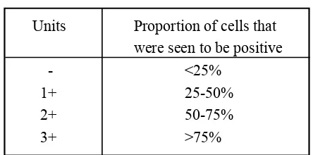
The units for expression levels of the molecular markers
were presented in the form: +1, +2 and +3. The
interpreta-tion of these units were based on the microscopic
examina-tion of the pathologist as follows:
Table1. Interpretation of immunohistochemistry units
The expression levels of each of the molecular markers
were compared with the staging and grading of each of the
tumors. In this study, only invasive ductal carcinomas and
intraduct carcinomas were analyzed due to the relatively
small number of other types of breast carcinomas. Units Proportion of cells that were seen to be positive
- <25%
1+ 25-50%
2+ 50-75%
3. Results
3.1 Incidence of breast cancer in the population and among
ethnic groups
The incidence rates of breast cancer in the country for the
female population of Brunei Darussalam for the period
2004-2005 are shown in Table 2. The results show that the
incidence of breast cancer is higher in the Chinese female
population than in the Malay female population in
Bru-nei Darussalam. There were seven cases of breast cancer
among patients who could not be classified as ethnic
Chi-nese or Malay.
Table 2 Incidence of Breast Cancer (BC) for 2004-2005
3.2 Age at diagnosis of the breast cancer
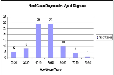
The age at which cancer was first diagnosed in all of the
female breast cancer patients is shown in Figure 1. There
were more patients diagnosed in the 40-49 and 50-59 age
groups than in other age groups in the period 2004-2005,
with equal numbers in both the 40-49 and 50-59 age
(33.7% of all cases each)
ethnicity na incidenceb 95% Ci
Malay 51 22.0 16.4, 28.9
Chinese 28 74.5 49.5, 107.6
Total 86 24.9 19.9, 30.8
a number of breast cancer patients
b number of cases per 100,000 population per year
in the period 2004-2005
CI – confidence interval
No of Cases Diagnosed vs Age at Diagnosis
5 8
29 29
10
4 1 0
5 10 15 20 25 30 35
20-29 30-39 40-49 50-59 60-69 70-79 80-89
Age Group (Years)
No of Cases
Figure 1. Number of cases diagnosed by age group in 2004-2005. The numbers in each group are indicated above the bars in the figure.
3.3 Proportions of tumours with different molecular markers
Because this was a retrospective analysis, results from the determination of molecular markers in tumours were only available for a proportion of the tumours. Of those where results were available, the proportions of stained tumours that gave a positive reaction for the different markers is shown in Table 2.
Marker no. of Tumours no. Positive Percent Positive
examined
ER 59 25 42%
PR 58 25 43%
Her2 56 36 64%
p53 36 15 42%
Ki67 35 03 09%
Bcl2 36 12 33%
pS2 35 10 29%
Table 2. Proportions of positive tumours among those examined for different molecular markers, 2004-2005
3.3. Relationship between the level of markers and tumour stage
3.4 Relationship between the level of markers and tumour
grade
The relationship between the grade of tumour and the
dif-ferent tumour markers was also examined. Figure 2 shows
the relationship between the level of detection of ER and
the grade of the tumour. This relationship was
statisti-cally analysed by comparing the proportions of tumours
expressing the marker at any level, and marker-negative
tumours, in tumours of different grades. The pooling of the
tumours that expressed markers at any level for each grade
of tumour was done to increase the numbers in a given
category for statistical analysis. Although the relationship
did not reach acceptable statistical significance, there
ap-peared to be a tendency for the higher grade tumours to be
ER negative (Fisher’s exact test p=0.097).
Figure 2. Relationship between ER expression levels
and the grade of tumour. The numbers in each group are
indicated above the bars in the figure.
A tendency for a similar relationship between the detection
of PR and the grade of the tumour, although not
reach-ing statistical significance, was also observed (Figure 3,
p=0.112 by Fisher’s exact test).
ER vs Grading
Figure 3. Relationship between PR expression levels and the grade of tumour. The numbers in each group are indi-cated above the bars in the figure.
For p53 the proportions of positive and negative marker reactions in tumours of different grades were not
signifi-cant by the Fisher’s exact test (p=0.889) and no obvious
trend could be discerned (Figure 4). Although a large pro-portion of G3 stage tumours were p53 negative, this was also the case for G1 stage tumours.
Figure 4. Relationship between p53 expression levels and
the grade of tumour. The numbers in each group are indi-cated above the bars in the figure.
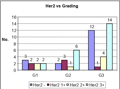
Additionally, no clear trend could be discerned, or
statis-tically significant relationship established, between the expression of Bcl2 or Her2 and the grade of the tumour (Figures 5 & 6). With Ki67, the three tumours where the antigen was detected (two at the 3+ level and one at the
Figure 5. Relationship between Her2 expression levels and the grade of tumour. The numbers in each group are indicated above the bars in the figure.
Figure 6. Relationship between Bcl2 levels and the grade of tumour. The numbers in each group are indicated above the bars in the figure.
3.5 Relationship between ER and pS2 expression in the tumours
Because pS2 is expressed after ER mediated estrogen sig-nalling, a correlation between its expression and that of ER would be expected. It was observed that of 13 ER +ve tumours, 5 were pS2 +ve and of 22 ER –ve tumours, only 5 were pS2 +ve. The relationship between pS2 and ER
ex-pression was not statistically significant by Fisher’s exact
test (p=0.44). However, 49% of the tumours were both pS2 and ER –ve, while only 14% were pS2 +ve and ER –ve.
4.1 Incidence of breast cancer in Brunei
Data from Singapore also show a higher incidence rate in Chinese females compared to Malay females [14]. For the 1998-2002 period in Singapore, the incidence rate for the Chinese female population was 72.7 per 100,000 per year with a 95% CI of 70.64 to 74.8 compared to an incidence rate for the Malay female population of 46.4 per 100,000 per year with a 95% CI between 42.5 and 50.4. While the incidence rates for Chinese in Singapore is comparable to that seen in Brunei in our study, the incidence rate for Ma-lays in Singapore in significantly higher than in Brunei. While this may possibly be due to sociological differences related to seeking medical treatment for breast cancer in the two countries, it may also reflect geographical and other socio-economic factors that are reported to affect the incidence of breast cancer [2, 15]. The differential factors that tend to reduce the incidence of breast cancer among Bruneian Malays merit further investigation.
The differences between the Chinese and Malay popula-tions observed in this study is consistent with genetic or culture associated behavioral factors or both playing a role in the development of breast cancer [3]. It is report-ed for example that the incidence of breast cancer in the USA greater for Caucasians than for African Americans and is least for the Asian/Pacific Islanders/Amerindians [16]. More detailed studies are needed to determine the ethnicity-related factors affecting breast cancer incidence in Brunei.
4.2 Age at diagnosis
The findings therefore suggest that breast cancer arises more in a slightly younger age group in the South East Asian populations, including Brunei Darussalam, than in the USA. The difference may be related to genetic, cul-tural, behavioural, and environmental factors and merits further investigation.
4.3 Relationship between the molecular markers and stag-ing and gradstag-ing
The prognosis is poorer for the more highly staged tumours [13] and for tumours of a higher grade (less differentiated tumours) [7]. The present study could not establish a clear relationship between the staging and any of the molecular markers. On the other hand, the results suggest that the ex-pression of both ER and PR may tend to be inversely re-lated to grade of the tumour which is consistent with other observations [9]. Analysis of a larger number of samples in Brunei for ER and PR will be needed to demonstrate a statistically significant relationship between the expression levels of these two markers and tumour grade. Such a find-ing would assist prognosis.
Mutation in the p53 gene leads to an accumulation of dysfunctional p53 protein that is detectable immunohis-tochemically [18]. Therefore it might be expected that more aggressive tumours of higher grade and stage might have more p53 detectable by immunohistochemistry. However there are conflicting reports in the literature with either a positive [19] or no [20] correlation of p53 detection with grade/stage of breast cancers.
The detection of Ki67 only in the G3 tumours is consistent with it being a marker for dividing cells. It has been report-ed that the expression of Ki67 in >20% of nuclei (approxi-mately > Grade 1 according to the classification used in the present study) correlates significantly with histological grade and the absence of ER and PR, and that it is a simple, inexpensive and reliable measure of proliferation rate in breast cancer compared to flow cytometric measurement of DNA content [21].
Our findings did not demonstrate a relationship between Bcl-2 or Her 2 and tumour grade or stage. However the de-tection of Her 2 in the tumours is important for considering Herceptin therapy.
While not statistically significant, there was a tendency for the absence of the pS2 marker to be correlated with the absence of ER, consistent with pS2 being expressed down-stream of estrogen signalling. However 5/22 ER negative tumours showed detectable pS2, and this may be due to the sensitivity of ER detection or the dysregulation of gene expression associated with malignant transformation. The pS2 positive fraction of ER positive tumours has been re-ported to have a better prognosis [9].
The expression of molecular markers in tumour cells is confounded by the gross genomic instability of tumours, which can result in highly dysregulated expression of proteins. Variable sensitivity of detection in immunocy-tochemistry is also possible. Gene expression profiling using nucleic acid hybridisation techniques is considered to be potentially of great value clinically in predicting sensitivity of tumours to new and established drugs. The findings reported here provide a baseline for more detailed investigations, and investigations using more sensitive techniques, on the use of molecular tumour markers in im-proving the treatment of breast cancer.
5. References
1. Kumar P, Clark M eds. 2005. Clinical medicine.5th
Edi-tion. Saunders. UK
2. Kumar V, Abbas AK, Fausto N 2005. Robbins and Cotran - Pathologic basis of disease. 7th Edition. Elsevier
Saunders
3. Cavalli F, Hansen HH, Kaye SB eds. 2000. Textbook of
medical oncology 2nd Edition. Martin Dunitz, UK
4. Cianfrocca M, Goldstein LJ 2004. Prognostic and pre-dictive factors in early-stage breast cancer. Oncologist 9: 606-616
5. Duffy MJ 2005. Predictive markers in breast and other cancers: a review. Clin Chem 51(3): 494-503.
7. Jones C Lakhani SR 2000. Prognostic and predictive factors in breast cancer. Brunei Int Med J 2: 147-169
8. Ross JS Fletcher JA 1998. The HER-2/neu oncogene in breast cancer: Prognostic Factor, Predictive Factor, and Target for Therapy. The Oncologist 3(4): 237-252
9. Donegan WL 1997. Tumor-related prognostic factors for breast cancer. Cancer J Clin 47: 28-51.
10. Alberts B, Johnson A, Lewis J, Raff M. Roberts K, Walter P 2002. Molecular biology of the cell. 4th Ed.
Gar-land Science, USA.
11. Kurzrock R, Talpaz M.eds. 1999. Molecular biology in cancer medicine 2nd Ed. Martin Dunitz, UK
12. Petrie A, Sabin C 2000. Medical statistics at a glance. Blackwell Synergy, UK
13. Singletary SE, Conolly JL 2006. Breast cancer staging: working with the 6th edition of the AJCC cancer staging manual. Cancer J Clin 56: 37-47.
14. Singapore Cancer Registry 2004. Trends in cancer incidence in Singapore.[Online] Singapore,Stationery Of-fice. Available from:www.hpb.gov.sg/hpb/default.asp?pg_ id=1631
15. Opatt DM, Chung C, Wright MJ, Moroz K, Newsome
RE, 2006. Breast cancer [Internet]. Available from: http:// www.emedicine.com/plastic/topic521.htm
16. American Cancer Society 2008. Breast cancer facts & figures. Available from http://www.cancer.org
17. National Cancer Registry 2004. Cancer incidence in Malaysia 2003. National Cancer Registry, MMA House, Kuala Lumpur. Available from: http://www.crc.gov.my/ ncr
18. Thor AD, Moore DH, Edgerton SM, Kawasaki ES, Reihauss E, et al. 1992. Accumulation of p53 tumour sup-pressor protein: an independent marker of prognosis in breast cancers. J Natl Cancer Inst 8(11):845-855.
19. Temmim L, Baker H, Sinowatz F 2001. Immunohis
-tochemical detection of p53 protein expression in breast can-cer in young Kuwaiti women. Anticancan-cer Res 21: 743-8.
20. Erdem O, Dursan A, Coskun U, Gunel N 2005. The prognostic value of p53 and c-erb-2 expression, prolifera-tive activity and angiogenesis in node-negaprolifera-tive breast car-cinomas. Tumori 91(1): 46-52.
21. Martinez-Arribas F, Martin-Garabato E, Lafuente P, Tejerina A, Lucas R, Sanchez J et al. 2006.