ABSTRACT
Pusher behavior (PB) is a disorder of postural control affecting patients with encephalic lesions. This study has aimed to identify the brain substrates that are critical for the occurrence of PB, to analyze the influence of the midline shift (MS) and hemorrhagic stroke volume (HSV) on the severity and prognosis of the PB. We identified 31 pusher patients of a neurological unit, mean age 67.4±11.89, 61.3% male. Additional neurological and functional examinations were assessed. Neuroimaging workup included measurement of the MS, the HSV in patients with hemorrhagic stroke, the analysis of the vascular territory, etiology and side of the lesion. Lesions in the parietal region (p=0.041) and thalamus (p=0.001) were significantly more frequent in PB patients. Neither the MS nor the HSV were correlated with the PB severity or recovery time.
Key words: pusher behavior, stroke, postural control.
Análise de neuroimagens de pacientes com síndrome do empurrador decorrente de AVC e outras etiologias
RESUMO
A síndrome do empurrador (SE) é um distúrbio de controle postural que acomete indivíduos com lesões encefálicas. Os objetivos deste estudo foram identificar as estruturas encefálicas envolvidas na SE, analisar a influência dos desvios de linha média (DLM) e volume do hematoma (VH) na gravidade e duração da SE. Dentre os pacientes internados na enfermaria de neurologia, foram identificados 31 pacientes com SE, idade média 67,4±11,89, 61,3% homens. Foram realizados exames neurológico e funcional. As análises das neuroimagens incluíram medidas de VH em pacientes com doença cerebrovascular (DC) hemorrágica, DLM, análise do território vascular, etiologia e lado da lesão. Lesão nas regiões parietal (p=0,041) e talâmica (p=0,001) foram significativamente mais frequentes nos pacientes com SE. Não foi observada correlação dos DLM e volume do hematoma com a gravidade e duração da SE.
Palavras-Chave: síndrome do empurrador, doenças cerebrovasculares, controle postural.
Correspondence
João P. Leite
Department of Neuroscience and Behavior Campus Universitário Ribeirão Preto 14049-900 Ribeirão Preto SP - Brasil E mail: [email protected]
Received 1 December 2010 Received in final form 4 July 2011 Accepted 11 July 2011
Department of Neuroscience and Behavior, University of São Paulo School of Medicine at Ribeirão Preto, Ribeirão Preto SP, Brazil.
The pusher behavior (PB) may be the
most intriguing disorder that impairs pos-tural balance after acute encephalic le-sions. Patients with PB lean towards the paretic side actively pushing with the non-paretic arm and leg and resist to any at-tempt of passive correction of their tilted body while sitting or standing1.
Traditionally, the occurrence of PB had been only reported in stroke pa-tients, though this disorder has also been
described in non-stroke conditions2. Pre-vious imaging studies have suggested the posterolateral thalamus as the typically damaged brain structure in pusher pa-tients3,4. Nevertheless, other cortical and subcortical areas such as insular cortex and post-central gyrus5,6 have also been pointed out. Therefore, encephalic
struc-tures essentially affected in PB patients are
still poorly understood.
915 tify brain substrates that are critical for the occurrence
of PB, to analyze the influence of the midline shift and hemorrhagic stroke volume on the signs and duration of the PB over a 3.5-year period of prospective follow-up.
This was a prospective descriptive observational study that was approved by the ethics committee of our institution. Informed consent was obtained from all sub-jects or their legal responsible party. Patients with PB were prospectively identified from inpatients of a neu-rological emergency unit at a tertiary hospital of the University of São Paulo School of Medicine at Ribeirão Preto. All inpatients were screened by a physical thera-pist for any abnormal postural behavior by awaking and placing them in a seated position, as soon as clinically possible. If any instability appeared, they were further assessed for PB.
Control group was composed by acute stroke pa-tients with encephalic lesions confirmed by neuroim-aging study that did not present PB and were matched for age and neurologic deficits with the group of patients with PB. Control group presented more previous ence-phalic lesions than PB group (p=0.027). Nevertheless, 95.66% of the control patients did not present neuro-logic deficits and were completely independent on their activities of daily living (ADL) before the lesion onset an-alyzed in this study.
Patients were investigated by a unique qualified ex-aminer (Santos-Pontelli, TEG). PB was assessed using a previously standardized Scale for Contraversive Pushing (SCP)7,8. The duration of PB was defined as the interval between injury onset and the complete resolution of PB signs (SCP=0). Pusher patients were periodically reeval-uated (minimum 5; maximum 20 days). The reevaluation
intervals were conducted within more than 10 days only after the 90th day post ictus onset.
Severity of neurologic involvement of the patients was assessed by standardized scales such as the National Institutes of Health Stroke Scale (NIHSS)9, Glasgow Coma Scale and Revised Trauma Score. Sensory defi-cits, visual field defects and aphasia were assessed as part of the NIHSS. The degree of paresis of the upper and lower limbs was scored with the usual clinical ordinal scale, where ‘0’ stands for no trace of movement and ‘5’ for normal movement. Patients were classified as having spatial neglect when there was clear evidence of a typ-ical clintyp-ical behavior such as ‘1’ a spontaneous deviation of the head and eyes toward the ipsilesional side, ‘2’ ori-enting toward the ipsilesional side when addressed from the front or the contralesional side, and ‘3’ ignoring of contralesional located people or objects10. If the patient fulfilled these 3 first criteria and was conscious, another
four tests were further assessed: “Coping task”; “Clock Drawing test”; “Cancellation test” and “Line bisection test”10,11. Neglect was considered to be present in disori-ented patients if they fulfilled the three clinical behaviors and, in conscious and oriented patients, if they fulfilled the criterion for spatial neglect in at least two of the four clinical tests, besides the clinical behavior.
Anosognosia was rated by questioning the patient about limb weakness and confirmed only when no ac-knowledgement of motor weakness was obtained even after confrontation12.
ADL function was assessed by the Barthel Index (BI) which evaluates 10 different abilities and ranges a total
score from 0 to 100 points9,13.
Uniform rehabilitation was given to all patients while they were hospitalized. Besides motor and respiratory re-habilitation, inpatients treatment included training and orientation of patients’ families according to passive or assisted mobilization of the paretic limbs, the correct po-sition while lying and sitting and how to transfer the pa-tients. Moreover, patients were asked to stay in vertical position as long as they could during the day. After dis-charge, patients were referenced to several public reha-bilitation centers, where the rehareha-bilitation treatment was conducted at the discretion of the local resources. The
number of physiotherapy sessions per week was obtained from patients’ family.
CT or MRI exams were performed as early as pos-sible according to the accessibility of the scanner and the patients’ clinical conditions. CT examinations were per-formed on a Somatom ARC equipment (Siemens, Er-langen, Germany). MRI examinations were performed on a 1.5-T superconductor system (Siemens, Vision Plus, Erlangen Germany). All images were reported and re-viewed by one neuroradiologist (Santos, AC) and one neurologist (Pontes-Neto, OM).
The vascular topography, etiology and side of the
le-sion were determined by combining clinical and neuro-imaging data. In order to analyze if there was any corre-lation between the location of the lesions and the severity of PB or the recovery time, we considered 1 point to each lesion occurring on thalamus, insula, post-central gyrus and posterior parietal region. These are the brain
struc-tures identified in earlier studies to be responsible for the occurrence of PB3-6. Thalamic compression visual-ized on the neuroimaging scans and confirmed by inter-thalamic adhesion midline shift was also considered as thalamic lesion.
The midline shift (MS) of the pineal, septum
pellu-cidum and interthalamic adhesion was measured using the E-Film Workstation Medical Imaging Software 1.8.3.
The interthalamic adhesion shift was calculated as the
to the perpendicular line connecting the anterior and the posterior insertions of the falx cerebri. The pineal and septum pellucidum shifts were similarly measured, as previously described14 in the available sequential images.
To measure the hemorrhage volume in patients with
hemorrhagic stroke we used the ABC/2 method15 on CT
scans of the acute stroke stage.
Statistical analysis was performed with SPSS (Statis-tical Package for Social Sciences) Software 13.0 for Win-dows. Demographic data were summarized by frequency analysis and descriptive statistics, as appropriate. Chi-square test (χ2) was used to analyze categorical data.
Be-tween groups comparisons for continuous data were carried out with Mann-Whiney U test. Spearman cor-relation coefficient was used to identify correlations be-tween continuous data.
In all tests, the criterion for statistical significance was two-tailed and set at α<0.05.
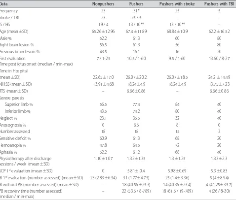
During the 3.5 years of prospective study we iden-tified 31 patients with PB (25 with stroke, 5 with trau-matic brain injury and 1 with brain tumor hemorrhage). Fourteen of them have already been reported2,16,17. The
917
pendent on their activities of daily living before the le-sion onset analyzed in this study. Table 1 describes the demographic and clinical characteristics of the patients.
All patients, except for one, took less than 24 hours from stroke onset to the first CT scan acquisition. This PB patient was excluded only from the analysis of MS and hemorrhagic stroke volume. MRI scan acquisition was taken in 12 PB and 5 control patients within a me-dian time of 35 days.
The middle cerebral artery (MCA) territory was
pre-dominantly injured in ischemic stroke patients with and without PB. A wide range in topography and lesion size was observed in both control and PB patients. Neuro-imaging scans showed a range from no visible lesions to hemispheric lesions affecting more than one related en-cephalic structure in both groups.
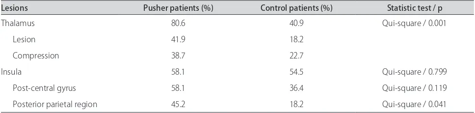
The number of PB related structure lesions was greater in the PB group (p=0.013) but there was no cor-relation between the frequency of the lesions with either the severity or with the prognosis of the PB (Table 2).
Among all control and PB patients with ischemic stroke, there was a positive correlation between MS and NIHSS score (septum pellucidum: p=0.020/r=0.402; in-terthalamic adhesion: p=0.021/r=0.40; pineal: p=0.023/ r=0.394). Nevertheless, there was no correlation between these variables among PB patients only. There was a pos-itive correlation between NIHSS score and HSV in hem-orrhagic stroke PB patients (p=0.039/r=0.732). There were no significant correlations of MS and HSV with severity or prognosis of PB.
Studies comparing neuroimaging data and neurologic deficits in patients with PB have already been conducted. However, this is the first attempt to analyze the relation-ship between neuroimaging data and the severity and prognosis of PB. Additionally, in the present study, for the first time, a neuroimaging analysis included stroke and non-stroke PB patients.
A positive correlation of the NIHSS score with HSV in hemorrhagic stroke PB patients was found. In spite of
this fact, neither the NIHSS score nor HSV were related with the severity or recovery time of PB. Conversely, previous studies showed that the hemorrhagic volume is highly associated with functional and neurologic def-icits18. These data and the fact that the NIHSS score is
a good neurologic outcome predictor19-21 indicate that
the PB evolution and severity may be independent from other neurologic deficits such as those measured by the NIHSS. However, more research is needed to confirm this observation.
No correlation was found between NIHSS score and MS in PB patients, and neither the NIHSS score nor MS were related with the severity or recovery time of PB. In fact, according to Lam et al., MS is not sensitive enough to predict patient’s final outcome of general neurolog-ical deficits22.
The qualitative analysis of each neuroimaging scan
re-vealed many similarities between the PB and control pa-tients. We found lesions on the anterior and posterior crus of the internal capsule, thalamus, frontal, temporal and parietal lobes, insula and also hemispheric lesions in both PB and control patient’s neuroimaging scans. Apparently, similar lesions can result in completely distinct reactions. It is known that slight differences in thalamic lesion locations can cause completely distinct syndromes4,6,23.
This includes, among postural control dysfunctions,
ves-tibular syndromes with disturbances of visual verticality perception and skew deviation23,24, thalamic astasia25
and PB.4,6 The latter has already been associated with
the interoceptive system dysfunction26 but, until now, the
neural network of this system is not well known. Never-theless, it would be possible that distinct neurologic and postural deficits could depend on the location of this net-work’s lesion.
Recently, Ticini et al., found that the posterior thal-amus itself rather than additional malperfusion in distant cortical areas is integral to the occurrence of PB6.
previously related to the PB contribute to the network controlling upright body6.
Johansen et al. analyzed neuroimaging scans of PB patients with a well matched control group5,27. However, these areas were identified with the subtraction tech-nique where the percentage of difference between the PB and control patients neuroimaging scans was not exclu-sively 100% (ranged from 81 to 100%). Although being a meticulous study, this analysis does not exclude the same PB patients’ lesion location existence in control pa-tients. Therefore, the fact that patients with and without PB have apparent similar encephalic lesions is not com-pletely elucidated.
Although the MCA territory was the most common vascular territory affected by ischemic stroke in PB pa-tients, no obvious pattern was observed. There was a substantial heterogeneity of the lesion size and topog-raphy both in the PB and control patients. The present results corroborate with the observations of Pérennou et al.28, Premoselli et al.29 and Pedersen et al.30. The last authors compared the frequency of lesion locations in pusher and control patients and found a significant dif-ference in the posterior crus of the internal capsule. Con-versely, Reding et al.27 described other encephalic struc-tures that seemed to be involved with PB as Paci et al.31 found the PB in a patient with cerebelar ischemic stroke and Karnath et al.32 recently reported a patient with PB due to a right-hemisphere ischeamia clearly affecting the anterior cerebral artery territory. Overlap neuroimaging workups have indicated the posterolateral thalamus, in-sula and post-central gyrus as the critical structures for PB occurrence4-6.
We conducted an additional analysis of the lesion on the thalamus, insula, post-central gyrus and posterior pa-rietal region. Although the posterior papa-rietal region was not previously confirmed as a PB related topography, it was included since this area is part of the sensory-motor network that process multisensory inputs used in motor responses, especially those directed to the extrapersonal space10,33. Therefore, this area could be related to the PB patients’ motor reaction caused by the severe conflict between the perception of body and visual verticality. In fact, we observed that the lesion of this area and the thalamus was significantly more frequent in PB than in control patients. Almost half PB patients presented le-sions on the posterior parietal region and it is consistent in the literature that neglect and aphasia is especially fre-quent in patients with PB 1,5,27,30. Furthermore, since the neural network that processes the postural perception has several participating areas and there was no absolute method that could isolate a strict PB lesion, it is possible that the posterior parietal region was, until now, not rec-ognized. PB does not occur essentially due to neglect or