* corresponding author: [email protected]
Aldose reductase genetic polymorphism is a
risk factor of diabetics retinopathy among
type 2 diabetes mellitus in Yogyakarta,
Indonesia
Jujuk Anton Cahyono1, Ahmad Hamim Sadewa2, Tasmini2 1Program of Health Analyst, Health Polytechnic of Banjarmasin
2Department of Biochemistry, Faculty of Medicine, Gadjah Mada University,
Yogyakarta, Indonesia
ABSTRACT
Diabetes mellitus (DM) is a metabolic syndrome characterized by hyperglycemia and glucose intolerance, due to insulin resistance, insulin deficiency, or both. Diabetics retinopathy (DR) is a DM complication due to retinal abnormality, that causes vision reduction and even blindness. The association between DR and aldose reductase 106T (ALR C-106T) gene polymorphism has been reported in previous studies. This genetic polymorphism increases the sorbitol level inside erythrocyte and pericyte in the retinal membrane that leads to weakness of retinal capillary vessel and microaneurism. The aim of this study was to know the presence of ALR C-106T gene polymorphism and its frequency distribution among diabetics Javanese patients in Dr. Sardjito General Hospital Yogyakarta, Indonesia. In addition, this study also aimed to analyze the difference of erythrocytes osmotic fragility (EOF) among ALR genotypes in type 2 diabetics patients with DR and without DR and to analyze whether ALR genetic polymorphism is a risk factor of DR in type 2 diabetic patients. This was a case control study that involved 40 diabetics patients with DR as case and 40 diabetics patients without DR as control groups. The C-106T ALR gene polymorphism was determined by polymerase chain reaction-restriction fragment length polymorphism (PCR–RFLP) method. Erythrocytes osmotic fragility was analyzed using spectrophotometer. Genotype and allele distributions were analyzed using x2 and other data were analyzed using independent t-test and Mann-Whitney, with p<0.05 was considered as significantly different. The results showed that in type 2 diabetics patients with DR, 33 patients (82.5%) were CC homozygote individuals and 7 patients (17.5%) were CT heterozygote individuals. In type 2 diabetics patients without DR, 27 patients (67.5%) were CC homozygote individuals and 13 patients (32.5 %) were CT heterozygote individuals. The genotype and allele distributions were not significantly different between two groups (p=0.121 for genotype, p=0.151 for allele). Odds Ratio of genotype was 2.270 while allele was 2.023. Erythrocytes osmotic fragility of CC genotype was higher than CT genotype (p=0.047). In conclusion, there was no significant difference between CC and CT genotype distribution among type 2 diabetics patients with and without DR. Erythrocyte osmotic fragility of CC genotype was higher than CT genotype. C-106T gene polymorphism was a risk factor of DR in type 2 diabetic patients.
Key words : ALR genes – polymorphism - type 2 DM - diabetic retinopathy - erythrocytes osmotic fragility
ABSTRAK
kelompok kontrol. Polimorfisme C-106T gena ALR ditentukan dengan metode PCR-RFLP. Fragilitas osmotik di analisa dengan metode spektrofotometri. Distribusi genotype dan alel dianalisa menggunakan uji x2. Data lainnya dianalisa menggunakan uji t independen dan Mann-Whitney dengan nilai p<0,05 untuk menentukan perbedaan secara bermakna hasil analisis. Hasil penelitian menunjukkan distribusi genotipe pasien DM tipe 2 dengan RD adalah CC 33 (82,5 %), CT 7 (17,5 %), dan pada pasien DM tipe 2 tanpa RD adalah CC 27 (67,5 %), CT 13 (32,5 %). Distribusi genotipe dan alel tidak berbeda bermakna antara ke 2 kelompok (p=0,121 untuk genotipe, p=0,151 untuk alel). Rasion Odds genotipe adalah 2,27, sedangkan alel adalah 2,023. Pada genotipe CC fragilitas osmotik lebih tinggi dibandingkan CT (p=0,047). Dari hasil penelititan dapat disimpulkan tidak terdapat perbedaan bermakna antara distribusi genotipe CC dan CT pada pasien DM tipe 2 dengan RD dengan tanpa RD. Fragilitas osmotik eritrosit genotipe CC lebih tinggi dibandingkan CT. Polimorfisme C-106T gena ALR merupakan faktor risiko RD pada DM tipe 2.
Kata kunci : gena ALR – polimorfisme – DM tipe 2 – retinopati diabetika – fragilitas osmotik eritrosit
INTRODUCTION
Diabetes mellitus (DM) is a metabolic disease characterized by hyperglycemia and glucose intolerance, as a result of insulin deficiency, impaired insulin action, or combination of both. The prevalence of DM worldwide is about 4% and is predicted to continue to increase, and by 2025 will reach 5.4%.1 World Health Organization (WHO)
has reported the increase number of people with type 2 DM in Indonesia in 2000 as many as 8.4 millions people and estimated to increase to approximately 21.3 millions in 2030.2 The
prevalence of DM in Yogyakarta, according to the national basic health research (RISKESDAS) Indonesia 2007 was about 1.6 %.
Chronic complications of DM are systemic vascular disease, heart disease, retinal degeneratur disease known as diabetic retinopathy, cataracts, kidney damage and peripheral nerve damage such as diabetic neuropathy.3 Diabetic retinopathy is a
form of DM complication that causes retinal disorder which affect vision convulsion until blindness. Hyperglycemi causes the thickening of retinal blood vessel and leakage. This complication commonly happens if its a diabetic patient has never had a treatment for at least for 15 years.4 In Indonesia,
DR prevalence was 27.2%,5 with 8.7% was in urban
area of Yogyakarta, whereas 7.73% contained in the rural.6 Ethnic or genetic difference plays a role
in the prevalence of DR.
Damage of the retina caused by hyperglycemia involves three existing pathways i.e. activation of the proteins glycation, C kinase protein, and aldose reductase (ALR).7 Aldose reductase catalyzes the
change of glucose to sorbitol through the reduction of aldehyde group of glucose. In hyperglycemia, sorbitol concentration increases and it will be
converted to fructose by sorbitol dehydrogenase (SDH). In hyperglycemia, sorbitol degradation proceeds slow, as of accumulates in cells and causes an increase in osmotic pressure. Excessive accumulation of sorbitol causes pericytes on retinal capillaries weakened and causes microaneurism.8
Hemodynamic disturbance in erythrocytes is one of the causes of blockage and leakage of retinal blood vessels which is a sign of DR.9 Therefore,
erythrocyte osmotic fragility is used as a parameter of pericytes retinal cell damage.10
Aldose reductase is coded by ALR gene which located in the long arm of chromosome 7 (7q35). This gene codes 316 amino acids and has 10 exon. The C-106T polymorphism in ALR gene happens in the basal promoter area and it causes changes in the expression of mRNA of ALR gene.11 Previous
study showed that C-106T polymorphisms in ALR gene is strongly associated with DR incidence in diabetic Chinese. CC genotype carrier strongly related with risk of DR.12
This study aimed to describe the relation of C-106T polymorphism in ALR gene with the risk for DR in diabetic Javanese in Yogyakarta, Indonesia. In addition, this study also aimed to analyze the difference of EOF among ALR genotypes in type 2 diabetics patients with DR and without DR and to analyze whether ALR genetic polymorphism is a risk factor of DR in type 2 diabetic patients.
MATERIALS AND METHODS
Hospital Yogyakarta; 2). Javanesse; 3). aged between 30-65 years old; 4). have been diagnosed for type 2 diabetes at least for 5 years. The exclusion criteria were obesity and hypertension. The patients who fulfilled the inclusion criteria were asked to sign the informed consent form as their willingness to become research subject. The protocol of the study has been approved by the Medical and Health Research Ethics Committee, Faculty of Medicine, Gadjah Mada University, Yogyakarta.
Diagnosis of type 2 DM was established based on fasting blood glucose (FBG) and 2 hours post prandial blood glucose.2 Retinopathy criteria was
according to fundusphoto which showed one of retina disorder symptoms (PDR or NPDR). The acquired data were age, sex, duration of type 2 DM, body mass index (BMI), blood pressure, and lipid profile.
Erythrocyte osmotic fragility was checked using spectrophotometer while lysis percentage and percentage of NaCl solution which cause 50% of erythrocyte lysis. Genotyping of C-106T polymorphism in ALR gene was performed using PCR-RFLP method. Sample used in this study was DNA isolated from blood. The PCR conditions were denaturation at 950C for 2 minutes, followed by 35
cycles of denaturation at 95oC for 60 seconds,
annealing at 67oC for 60 seconds, and extension at
72oC for 60 seconds. The final extension was at
72oC for 5 minutes. The forward primer was:
5’-CCT TTC TGC CAC GCG GGG CGC GGG-3’ (attached on -222 until - 199) and the reverse primer was: 5’-CAT GGC TGC TGC GCT CCC CAG-3’ (attached on +21 until starting codon ATG).
Restriction enzyme used in this study was BfaI
(New England Biolabs). The PCR product was incubated with the BTA for 16 hours. The digestion product the was then electrophoresed on 2% agarose gel and visualized by etidium bromide under UV light. CC genotype had 2 bands: 206 and 57 bp. TT genotype had 3 bands : 147, 59 and 57 bp, whereas genotype of CT had 4 bands : 206, 147, 59 and 57 bp.13
RESULTS
Subject characteristics
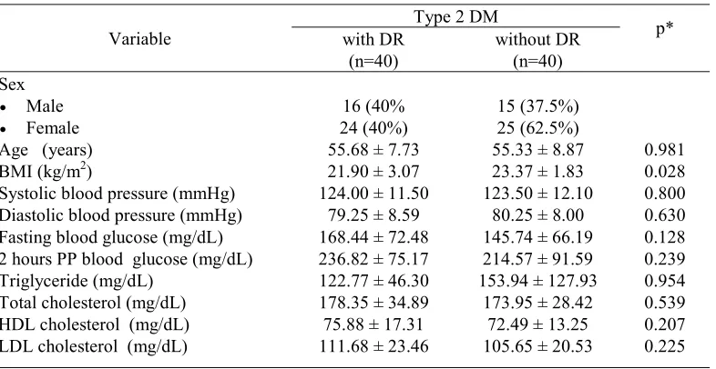
Subject characteristics are shown in TABLE 1. Subject characteristics such as age, blood pressure, fasting blood glucose, 2 hours post prandial blood glucose, serum triglyceride, total cholesterol, HDL and LDL cholesterol were not significantly different between type 2 diabetic patients with DR and without DR (p>0.05). However, the average BMI of type 2 diabetic patients with DR was lower than patients without DR (p<0.05).
TABLE 1. Subject characteristics of type 2 diabetic patients. Data were expressed in mean ± standard deviation except for sex in %
Variable
Type 2 DM
p* with DR
(n=40)
without DR (n=40)
Sex
• Male 16 (40% 15 (37.5%)
• Female 24 (40%) 25 (62.5%)
Age (years) 55.68 ± 7.73 55.33 ± 8.87 0.981
BMI (kg/m2) 21.90 ± 3.07 23.37 ± 1.83 0.028
Systolic blood pressure (mmHg) 124.00 ± 11.50 123.50 ± 12.10 0.800
Diastolic blood pressure (mmHg) 79.25 ± 8.59 80.25 ± 8.00 0.630
Fasting blood glucose (mg/dL) 168.44 ± 72.48 145.74 ± 66.19 0.128
2 hours PP blood glucose (mg/dL) 236.82 ± 75.17 214.57 ± 91.59 0.239
Triglyceride (mg/dL) 122.77 ± 46.30 153.94 ± 127.93 0.954
Total cholesterol (mg/dL) 178.35 ± 34.89 173.95 ± 28.42 0.539
HDL cholesterol (mg/dL) 75.88 ± 17.31 72.49 ± 13.25 0.207
LDL cholesterol (mg/dL) 111.68 ± 23.46 105.65 ± 20.53 0.225
C-106T polymorphism in ALR Gene
The genotyping result of C-106T polymorphism in ALR gene using PCR-RFLP is shown in FIGURE 1. While the distribution of genotype and allele is shown in TABLE 2. Genotype distribution of ALR gene between observed value with expected value Hardy-Weinberg had no significant difference (p= 0.573). The genotype frequency between type 2 diabetic patients with DR and without DR was not different (p = 0.121), so as the allele frequency (p = 0.151). CT genotype carriers had 2.270 times increased risk for DR compared with CC genotype carries, whereas C allele carriers had 2.023 times increased risk for DR than T allele carriers. Therefore, the high risk allele for DR in type a diabetic patients was C allele.
FIGURE 1. Genotyping of C-106T polymorphism in ALR gene
TABLE 2. Genotype and allele of type 2 DM risk with DR
Variable
Type 2 DM
p* OR CI (95%)
with DR (n=40) without DR (n=40)
Genotype CC CT
33 (82.5%) 7 (17.5%)
27 (67.5%)
13 (32.5%) 0.121 2.270 0.794 – 6.488
Alelle C
T
73(91.25%) 7 (8.75%)
67 (83.75%)
13 (16.25%) 0.151 2.023 0.762 – 5.374
*Statistical analysis using Chi square (x2) test with a 95% confidence interval (p<0.05)
Erythrocyte osmotic fragility
The average percentage of NaCl which caused lysis of 50% erythrocyte is shown at TABLE 3 and 4. This study showed a significant difference of erythrocyte osmotic fragility between CC and CT genotype on type 2 diabetic patients (p=0.047). The
erythrocyte osmotic fragility increased in CC genotype individuals (TABLE 3). The percentage of NaCl on type 2 diabetic patients with DR was higher than to those without DR (TABLE 4). However, it was not significantly different (p=0.154).
TABLE 3. The percentage of NaCl (mean ± SD) which caused lysis of 50% erythrocyte based on the genotype of type 2 diabetic patients with and without DR
Variable Genotype p*
CC CT NaCl percentage which caused 50%
lysis of erythrocyte 0.4135 ± 0.0129 0.4067 ± 0.0126 0.047
TABLE 4. The percentage of NaCl (mean ± SD) difference which caused lyses of 50% erythrocyte between type 2 diabetic patients with and without DR
* Statistical analysis using Mann-Whitney test with 95% confidence interval (p<0.05)
DISCUSSION
The subject characteristics of type 2 diabetic patients with DR and without DR were similar except BMI. The BMI of type 2 diabetic patients without DR was higher than those with DR (p=0.028). The BMI of type 2 diabetic patients is influenced by prolonged duration of diabetes and increased insulin resistance which activated lipase-sensitive hormone, therefore the deposite of triglyceride in adipose tissue will be hydrolyzed becomes free fatty acid. This process will decrease the BMI of type 2 diabetic patients.14
Genotype distribution of C-106T polymorphism in ALR gene in type 2 diabetic patients in several
populations has been reported by some authors from different studies (TABLE 5). The studies indicated that different population showed different genotype frequency.7,15,16 Diabetic retinopathy development
depends on environment and genetic factors. This study did not found TT genotype, whereas study in Euro-Brazil, China, and Chilipopulations, TT genotype was found and the number was smaller than CC and CT genotype. Frequency of CC of Javanese was higher than other populations (Euro-Brazil, China and Chili). In type 2 diabetes without DR, the number of individual with CT genotype was lower compared to Euro-Brazil and Chili, but was higher than China.
TABLE 5. Genotype distribution of C106T polymorphism in ALR gene in type 2 diabetic patients in several populations
Population Subject Genotype* n
CC CT TT
* The data were expressed in sum (%); n=total sample
Studies on C-106T polymorphism has showed a strong association of the polymorphism with DR complication, although the case and control subjects had no significant difference.12 For Euro-Brazil
population there was no significant difference between type 2 diabetic patients with DR and without DR. CC genotype and C allele were the
high risk genotype and allele for DR complication. The same result also found in Chili.7
There was correlation between C-106T poly-morphism with (CA) dinucleotide repetition on ALR gene promoter region,17 this might happen because
the location of polymorphism was contiguous with the sequence of ALR gene which causes DR
Variable DM type 2 p*
with DR without DR
NaCl percent which caused lysis of
sensitivity. From the other research which showed that CC was a high risk genotype for DR.13
C-106T polymorphism on basal promoter area of ALR gene will cover the CCAAT promoter element which located between -98 and -94, in cases where that region performs as active functional element. Research showed that CCAAT element was recognized by nuclear factor of Hep G2 cell.11
Other study showed that different allele on location -106 would affect the CCAAT element function by changing the factor binding activity and finally change the basal transcription level of ALR gene.18
The increase of ALR gene expression was identified on retinal epithelial cell of CC homozygote carrier compared with heterozygote genotype CT-106.19
A significant difference of erythrocyte osmotic fragility between CC and CT genotype on type 2 diabetic patient was observed in this study. The erythrocyte osmotic fragility significantly increased in CC genotype individuals (TABLE 3). Previous study showed that higher ALR expression occured in CC genotype individuals compared with CT genotype individuals.7 The increase also happens
on blood and sorbitol level of erythrocyte on type 2 diabetic patients with DR than without DR.20
Sorbitol accumulation will affect osmolality of erythrocyte and causes its fragility to increase.
Higher percentage of NaCl on type 2 diabetic patients with DR indicated a higher erythrocyte fragility compared to those without DR. High level of plasma glucose can affect osmotic pressure and will cause cellular dehydration.21 Sorbitol level in
erythrocyte and ALR in the blood correlates with level of plasma glucose.22 The increase of plasma
glucose level in hyperglycemia will increase the sorbitol level.
Sorbitol is not easy to diffuse through cell membrane, as a consequence sorbitol will accumulate and disturb water entry inside the cell and impair the osmotic.14 Sorbitol concentration raise
significantly in type 2 diabetic patients with DR, compared to those type 2 diabetic patients without complication. The sorbitol accumulation decreases the number of pericyte of retina.23
The beneficial impact of strict glycemic control to prevent diabetic complications has been reported. However, most diabetic patients rarely achieve consistent glucose level. Hence, agents that can
prevent diabetic complications, irrespective of glycemic control, would be an alternative.20 Aldose
reductase inhibitor and protein kinase inhibitor can be included as an alternative treatment to prevent diabetic complications. However, further study is needed in order to obtain optimal benefit of these agents.
CONCLUSION
C-106T polymorphism in ALR gene occured in type 2 diabetic patients with DR on Dr. Sardjito General Hospital Yogyakarta. There was no significant difference on genotype frequency between CC and CT as well as C and T allele between type 2 diabetic patients with or without DR. Significant difference in osmotic erythrocyte fragility was observed between CC and CT genotype of type 2 diabetic patients, but there was no significant difference between type 2 diabetic patients with and without DR. C-106T polymorphism in ALR gene was a risk factor of DR on type 2 diabetic patients.
ACKNOWLEDGMENT
The authors would like to acknowledge all the participants for their coorporation in conduct of this study. We also thank doctors from Dr. Yap Hospital Yogyakarta for expertise on retinal photography, doctors from Department of Internal Medicine, Faculty of Medicine, Gadjah Mada University/Dr. Sardjito General Hospital Yogyakarta for patient recruitment.
REFERENCES
1. American Diabetes Association. Diagnosis and classification of diabetes mellitus. Diabetes Care 2006; 29(1 suppl): 43S-8S.
2. PERKENI. Konsensus pengelolaan dan pencegahan diabetes mellitus Tipe 2 di Indonesia. Jakarta: Pengurus Besar Perkumpulan Endokrinologi Indonesia, 2006. 3. Halliwell B, Getteridge JMC. Free radical in biology and
medicine. 2nd ed. Oxford: Oxford University Press, 1989.
4. Ilyas S. Ikhtisar Ilmu Penyakit Mata. Jakarta: Fakultas Kedokteran Universitas Indonesia, 2009.
5. Refa S, Dewi NA. The correlation between HbA1C and serum lipid level with the severity of diabetic neuropathy. Jurnal Kedokteran Brawijaya 2005;21(3): 138-42. 6. Sudarto N, Agni N. Perbedaan prevalensi retinopati
Istimewa Yogyakarta [Karya Akhir]. Yogyakarta: Fakultas Kedokteran Universitas Gadjah Mada, 2004.
7. Olmos P, Bastian MJ, Vollrath V, Toro L, Trincado A, Salinas P, Claro JC, Lopez JM, Acosta AM, Miquel JF, Castro J. C(-106)T polymorphism of the aldose reductase and the progression rate of diabetic retinopathy. Diabetes Res Clin Prac 2006;(74):175-82.
8. Lorenzi M. The polyol pathway as a mechanism for diabetic retinopathy attractive elusive and resilient. Exp Diab Res 2007; 61038.
9. Chistopher AP, Destylya D, Tambunan R. Retinopati diabetik non proliferatif. Files of Drs Med – FK UNRI [serial online] 2009 [cited 2010 Oct 15]. [16 screens] Available from:http:// yayanakhyar. wordpress. com. 10. Comazzi S, Spagnolo V, Bonfanti U. Erythrocyte changes
in canine diabetes mellitus : in vitro effects of hyperglycaemia and ketoacidosis. Comp Clin Path 2004;(12):199-205.
11. Wang K, Bohren KM, Gabbay KH. Characterization of the human aldose reductase gene promoter. J Biol Chem 1993;(268):16052-58.
12. Qingjie L, Ping X, Jianjun H, Yapeng G, Weimin Z, Huiping S. Polymorphisms and functions of the aldose reductase gena 5’ regulatory region in Chinese patient with type 2 diabetes mellitus. Chin Med J 2002;115(Pt 2): 209-13. 13. Kao YL, Donaghue K, Chan A, Knight J, Silink M. Novel
polymorphism in the aldose reductase the promoter region is strongly associated with diabetic retinopathy in aldolescents with type 1 diabetes. Diabetes 1999;48:1338-40.
14. Bender DA, Mayes PA. The pentose phosphate pathway and other pathways of hexose metabolism. In: Murray RK, Granner DK, Rodwell VW, editors. Harper’s Illustrated Biochemistry. 28th ed. New York: Mc Graw
Hill Companies, 2009:174-83.
15. Santos KG, Tschiedel B, Schneider J, Souto K, Roisenberg I. Diabetic retinopathy in Euro-Brazilian type 2 diabetic patients : relationship with polymorphisms in the aldose reductase, the plasminogen activator inhibitor-1 and the
methylenetetrahydrofolate reductase genes. Diabetes Res Clin Prac 2003;(61):133-6.
16. Wang Y, Maggie CYN, Lee SC, So WY, Tong PCY, Cockram CS, Critchley JAJH, Chan JCN. Phenotypic heterogeneity and associations of two aldose reductase gene polymorphisms with nephropathy and retinopathy in type 2 diabetes. Diabetes Care 2003;(26): 2410-15. 17. Liew G, Klein R, Wong TY. The role of genetics in
susceptibility to diabetic retinopathy. Int Ophthalmol Clin 2009;49(Pt 2) : 35-52.
18. Yang B, Millward A, Demaine A. Functional differences between the susceptibility Z-2/C-106 and protective Z+2/ T-106 promoter region polymorphisms of the aldose reductase gene may account for the association with diabetic microvascular complications. Biochim Biophys Acta 2003; (1639): 1-7.
19. Stevens MJ, Killen P, Wang P, Larkin DD, Greene DA. Overexpression of aldose reductase (AR) in human retinal pigment epithelial (RPE) cell lines is associated with a polymorphism in the AR basal promoter region. Diabetes 2000;49(1 Suppl):167A.
20. Reddy GB, Satyanarayana A, Balakrishna N, Ayyagari R, Padma A, Viswanath K, Petrash JM. Erytrocyte aldose reductase activity and sorbitol levels in diabetic retinopathy. Mol Vis 2008;(14): 593-601.
21. Guyton AC, Hall JE. Textbook of medical physiology. 11th
ed. Philadelphia: Elsevier Saunders, 2006.
22. Hamada Y, Kitoh R, Raskin P. Increased activity of erythrocyte aldose reductase in insulin-independent diabetes with severe diabetic complications. Diabet Med 1991;(8): 226-31.