TeAM
YYePG
Digitally signed by TeAM YYePG
DN: cn=TeAM YYePG, c=US, o=TeAM YYePG, ou=TeAM YYePG, email=yyepg@msn. com
Steven T. Rosen, M.D.,
Series Editor
Sugarbaker, P. (ed): Peritoneal Carcinomatosis: Drugs and Diseases. 1995. ISBN 0-7923-3726-3.
Sugarbaker, P. (ed): Peritoneal Carcinomatosis: Principles of Management. 1995. ISBN 0-7923-3727-1.
Dickson, R.B., Lippman, M.E. (eds.): Mammary Tumor Cell Cycle, Differentiation and Metastasis. 1995. ISBN 0-7923-3905-3.
Freireich, E.J, Kantarjian, H.(eds):Molecular Genetics and Therapy of Leukemia. 1995. ISBN 0-7923-3912-6.
Cabanillas, F., Rodriguez, M.A.(eds): Advances in Lymphoma Research. 1996. ISBN 0-7923-3929-0.
Miller, A.B. (ed.): Advances in Cancer Screening. 1996. ISBN 0-7923-4019-1.
Hait, W.N. (ed.): Drug Resistance. 1996. ISBN 0-7923-4022-1.
Pienta, K.J. (ed.): Diagnosis and Treatment of Genitourinary Malignancies. 1996. ISBN 0-7923-4164-3.
Arnold, A.J. (ed.): Endocrine Neoplasms. 1997. ISBN 0-7923-4354-9.
Pollock, R.E. (ed.): Surgical Oncology. 1997. ISBN 0-7923-9900-5.
Verweij, J., Pinedo, H.M., Suit, H.D. (eds): Soft Tissue Sarcomas: Present Achievements and Future Prospects. 1997. ISBN 0-7923-9913-7.
Walterhouse, D.O., Cohn, S. L. (eds.): Diagnostic and Therapeutic Advances in Pediatric Oncology. 1997. ISBN 0-7923-9978-1.
Mittal, B.B., Purdy, J.A., Ang, K.K. (eds): Radiation Therapy. 1998. ISBN 0-7923-9981-1.
Foon, K.A., Muss, H.B. (eds): Biological and Hormonal Therapies of Cancer. 1998. ISBN 0-7923-9997-8.
Ozols, R.F. (ed.): Gynecologic Oncology. 1998. ISBN 0-7923-8070-3.
Noskin, G. A. (ed.): Management of Infectious Complications in Cancer Patients. 1998. ISBN 0-7923-8150-5.
Bennett, C. L. (ed.): Cancer Policy. 1998. ISBN 0-7923-8203-X.
Benson, A. B. (ed.): Gastrointestinal Oncology. 1998. ISBN 0-7923-8205-6.
Tallman, M.S., Gordon, L.I. (eds): Diagnostic and Therapeutic Advances in Hematologic Malignancies. 1998. ISBN 0-7923-8206-4.
von Gunten, C.F. (ed): Palliative Care and Rehabilitation of Cancer Patients. 1999. ISBN 0-7923-8525-X
Burt, R.K., Brush, M.M. (eds): Advances in Allogeneic Hematopoietic Stem Cell Transplantation. 1999. ISBN 0-7923-7714-1.
Angelos, P. (ed.): Ethical Issues in Cancer Patient Care 2000. ISBN 0-7923-7726-5.
Gradishar, W.J., Wood, W.C. (eds): Advances in Breast Cancer Management. 2000. ISBN 0-7923-7890-3.
Sparano, Joseph A. (ed.): HIV & HTLV-I Associated Malignancies. 2001. ISBN 0-7923-7220-4.
Ettinger, David S. (ed.): Thoracic Oncology. 2001. ISBN 0-7923-7248-4.
Bergan, Raymond C. (ed.): Cancer Chemoprevention. 2001. ISBN 0-7923- 7259-X.
Raza, A., Mundle, S.D. (eds): Myelodysplastic Syndromes & Secondary Acute Myelogenous Leukemia 2001. ISBN: 0-7923-7396.
Talamonti, Mark S. (ed.): Liver Directed Therapy for Primary and Metastatic Liver Tumors. 2001. ISBN 0-7923-7523-8.
Stack, M.S., Fishman, D.A. (eds): Ovarian Cancer. 2001. ISBN 0-7923-75 0-0.
Bashey, A., Ball, E.D. (eds): Non-Myeloablative Allogeneic Transplantation. 2002. ISBN 0-7923-7646-3.
Leong, Stanley P.L. (ed.): Atlas of Selective Sentinel Lymphadenectomy for Melanoma, Breast Cancer and Colon Cancer. 2002. ISBN 1-4020-7013-6.
Andersson , B., Murray D. (eds): Clinically Relevant Resistance in Cancer Chemotherapy. 2002. ISBN 1-4020-7200-7.
Beam, C. (ed.): Biostatistical Applications in Cancer Research. 2002. ISBN 1-4020-7226-0.
Brockstein, B., Masters, G. (eds): Head and Neck Cancer. 2003. ISBN 1-4020-7336-4.
Frank, D.A. (ed.): Signal Transduction in Cancer. 2003. ISBN 1-4020-7340-2.
Figlin, Robert A. (ed.): Kidney Cancer. 2003. ISBN 1-4020-7457-3.
Kirsch, Matthias; Black, Peter McL. (ed.): Angiogenesis in Brain Tumors. 2003. ISBN 1-4020-7704-1.
Keller, E.T., Chung, L.W.K. (eds): The Biology of Skeletal Metastases. 2004. ISBN 1-4020-7749-1.
Kumar, Rakesh (ed.): Molecular Targeting and Signal Transduction. 2004. ISBN 1-4020-7822-6.
Verweij, J., Pinedo, H.M. (eds): Targeting Treatment of Soft Tissue Sarcomas. 2004. ISBN 1-4020-7808-0.
Finn, W.G., Peterson, L.C. (eds.): Hematopathology in Oncology. 2004. ISBN 1-4020-7919-2.
Farid, N., (ed): Molecular Basis of Thyroid Cancer. 2004. ISBN 1-4020-8106-5.
Balducci, L., Extermann, M. (eds.): Biological Basis of Geriatric Oncology. 2004. ISBN
BIOLOGICAL BASIS OF
GERIATRIC ONCOLOGY
edited by
LODOVICO BALDUCCI, MD
Professor of Oncology and Medicine
University of South Florida College of Medicine
Program Leader, Senior Adult Oncology Program
H. Lee Moffitt Cancer Center & Research Institute
Tampa, Florida, USA
MARTINE EXTERMANN, MD, PhD
Associate Professor of Oncology and Medicine
University of South Florida College of Medicine
Senior Adult Oncology Program
H. Lee Moffitt Cancer Center & Research Institute
Tampa, Florida, USA
Print ISBN: 0-387-23961-8
Print ©2005 Springer Science + Business Media, Inc.
All rights reserved
No part of this eBook may be reproduced or transmitted in any form or by any means, electronic, mechanical, recording, or otherwise, without written consent from the Publisher
Created in the United States of America
Boston
©2005 Springer Science + Business Media, Inc.
FOREWORD vii
Lodovico Balducci and Martine Extermann
1.EPIDEMIOLOGY OF CANCER AND AGING 1
Lodovico Balducci and Matti Aapro
2.BIOLOGICAL INTERACTIONS OF AGING
AND CARCINOGENESIS 17
Vladimir N. Anisimov
3.REPLICATIVE SENESCENCE AND CANCER 53
Peter J. Hornsby
4.THE INFLUENCE OF ADVANCED AGE ON CANCER
OCCURRENCE AND GROWTH 75
William B. Ershler
5.AGE AND COMORBIDITY IN CANCER PATIENTS:
A POPULATION BASED APPROACH 89
Lodovico Balducci, Cheryl L. Hardy, and Gary H. Lyman
7. CLINICAL AND BIOCHEMICAL EVALUATION
Angela Abbatecola, B. Gwen Windham, Stefania Bandinelli, Fulvio Lauretani, Giuseppe Paolisso, and Luigi Ferrucci
8. BIOLOGICAL SCREENING AND IMPACT IN ELDERLY
Anne-Chantal Braud and Martine Extermann
9. BIOLOGICAL BASIS OF THE ASSOCIATION OF CANCER
Martine Extermann
10. BIOLOGICAL BASIS OF CANCER IN THE
Claudia Beghe’and Lodovico Balducci
11. DECISION ANALYSIS FOR CANCER PREVENTION AND
Marline Extermann
12. GUIDELINES FOR THE MANAGEMENT OF THE OLDER
Lodovico Balducci
INDEX
CHANGES OVER AGING 135
CANCER PATIENTS 165
AND AGING COMORBIDITY 173
OLDER PERSON 189
CANCER TREATMENT IN THE ELDERLY 223
CANCER PATIENT 233
The population of Western countries is aging, and cancer in older aged persons is becoming increasingly common. The management of these neoplasms is a novel problem. Direct information on the outcome of cancer prevention and of cytotoxic chemotherapy in older individuals is scarce, especially for those aged 80 and over, and it is not clear whether the same process should direct medical decisions in younger and older persons. It is reasonable to assume that the benefits of cancer prevention and treatment diminish and the dangers increase with age. The expected gains from cancer treatment may be lessened by shorter life expectancy. The risk of therapeutic complications may be increased and the consequences of these complications may become more serious due to limited functional reserve of multiple organ-systems, and fading social support and economic resources. In addition, the biology of cancer may change with the age of the patient, due to a series of events that have been clarified only in part. For example, the prevalence of Multidrug Resistance in Acute Myelogenous Leukemia is much higher for patients over 60, which make the treatment less effective and the risk of treatment-related deaths higher. At the same time, the risk of local recurrence of breast cancer after partial mastectomy declines with age, indicating a more indolent disease.
Several publications, including books, review articles and original studies, related to cancer in the elderly have appeared during the last ten years and have highlighted important points that have become widely accepted:
Age by itself is not and should never be a contraindication to cancer management, including prevention and treatment.
cytotoxic chemotherapy and allow the administration of full doses of treatment. These provisions include prophylaxis of neutropenic infections, avoidance of severe anemia, timely management of mucositis, and provision of adequate home care giving.
The practical application of these directions remains somehow controversial however, as the methods to estimate life expectancy, functional reserve, and tumor behavior are poorly defined. The main goal of this book is to provide a simple blueprint enabling the practitioners of oncology, geriatrics, and primary care to decide when a patient may or may not benefit from cancer prevention and treatment. Based on the current knowledge of the biology of aging and cancer, the books examines several facets of patient assessment, including function, comorbidity, physical performance and laboratory tests, as well as the way these different forms of assessment may be integrated in medical decisions. At the meantime, the book explores future possibilities for understanding the interaction of aging and cancer biology and for predicting these interactions, and provides a rationale for clinical trials of chemoprevention of cancer in the older person by unraveling the mechanisms that associate aging and carcinogenesis. Some of these mechanisms, including the genomic changes of age, are predictable, while others, including proliferative senescence, are counter-intuitive, and open new, unsuspected opportunities for intervention. Aware of the rapid evolution of the field, we wanted for this book to become an expandable and adaptable frame of reference, able to accommodate new information and still able to direct the practitioner in the management of older individuals even when the current information will be outdated. The emphasis on current research directions in the biology of aging, of cancer, and of the hemopoietic system that is intimately connected to the management of cancer, should make the reader attuned to new developments and allow the reader to rapidly incorporate these developments into clinical thinking.
Another important goal of this book is to highlight the important lessons coming from the study of aging that may be collapsed into two points:
To a large extent, the study of aging involves a movement from the bedside to the bench, which is directly opposed to the current trend of oncology. As underlined in the initial chapter, epidemiology is the main clue to the biological interactions of cancer and aging: epidemiology and clinical observation are still the main source for experimental hypothesis.
integration of these points in clinical practice and clinical research.
The third and final goal of this book is to provide an updated and practical research handbook for the increasingly large host of young investigators who want to become involved in the field. The need for such handbook is revealed by a number of recent initiatives aimed to promote research in geriatric oncology. Among them we would like to highlight the issuance of a RFA for program grants in geriatric oncology by a combined NCI/NIA effort, and the institution of a number of fellowships in geriatric oncology through a grant of the Hartford Foundation to the American Society of Clinical Oncology.
In addition to all excellent collaborators of the book, we would like to thank the numerous friends and colleagues who have been engaged with us in this adventure of geriatric oncology during the last ten years, and in particular, we would like to acknowledge the leadership of Rosemary Yancik, Ph.D., who single-handedly generated the field more than two decades ago, and the members of the Senior Adult Oncology Program at the H. Lee Moffitt Cancer Center, to whom this book is dedicated.
Lodovico Balducci M.D.
EPIDEMIOLOGY OF CANCER AND AGING
Lodovico Balducci and Matti Aapro
Lodovico Balducci, Program Leader, Senior Adult Oncology Program, H. Lee Moffitt Cancer Center and Research Institute, Tampa, Florida, Professor of Oncology and Medicine, University of South Florida College of Medicine, Tampa, Florida. Matti Aapro, Director, Multidisciplinary Oncology Institute, Clinique de Genolier, Switzerland.
Epidemiology provides the initial clue to causes and mechanisms of diseases. It is well known that age is a risk factor for most common cancer and that incidence and prevalence of cancer increase with age1. In this chapter we explore the epidemiology of cancer and aging, in an attempt to understand the biologic interactions of these processes. In particular, we address the following questions:
1.
2.
3.
4.
Does aging enhance the susceptibility of older individuals to environmental carcinogens?
Is aging associated with increased risk of multiple malignancies?
Does the clinical behavior of cancer change with age?
Does cancer increase the risk of death of older individuals?
1. AGE AND CARCINOGENESIS
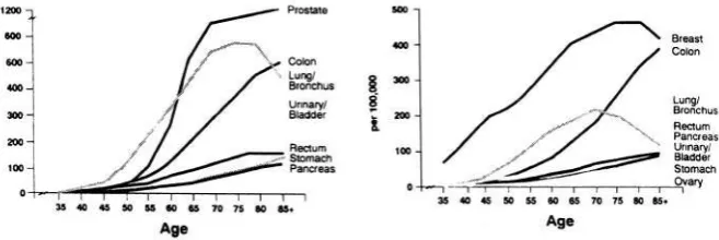
The incidence of common cancers increases with age (Figure 1). This association is universal 2 and is observed with the aging of any population around the world. A clear explanation of this phenomenon is the time-length of carcinogenesis, a stepwise process involving the activation of cellular oncogenes, and the suppression of anti-proliferative genes (anti-oncogenes)3. It is reasonable to assume that the duration of carcinogenesis reflects the number of stages involved in the pathogenesis of different tumors, and that this number be highest for tumors whose incidence peaks late in life, such as adenocarcinoma of the prostate and of the large bowel, or non-melanomatous skin cancer 3. In the era of chemoprevention and recognition and elimination of environmental carcinogens, an alternative possibility should be considered. These interventions may cause the prolongation of one or more carcinogenic steps and, in so doing; they may delay the development of cancer. For example, the incidence of lung cancer has decreased for individuals less than 60, while it has increased for older individuals 4. As a result, the peak incidence of lung cancer has become more and more delayed. Interestingly, these changes have paralleled the incidence of smoking cessation in the Western population. In this case it is reasonable to assume that the length of carcinogenesis has increased as a result of a prolongation of the late carcinogenic stages, from reduced intensity of exposure to tobacco smoke 3. If this hypothesis is correct, one may expect to see a progressive delay in the appearance of common cancer and an increased incidence of neoplasia in advanced ages.
The duration of carcinogenesis may not account completely for association of cancer and aging. . The incidence of some neoplasms, such as prostate and non-melanomatous skin cancer increases more rapidly with age, than it would be expected from the time-length of carcinogenesis alone 3. These findings suggest that the concentration of cells in advanced carcinogenic stages increases with the age of an organism, enhancing the susceptibility of older individuals to environmental carcinogens 3. This possibility is supported by a host of studies of experimental carcinogenesis, summarized in another chapter of this book 3 and also by epidemiologic observations5- 9. Barbone et al reported the risk of lung cancer after exposure to an environmental pollutant in the Italian city of Trieste increased with the age of the subject at the time of exposure 6. Since 1970, the incidence of non-Hodgkin’s lymphoma has increased 80% for individuals 60 and over, and that of malignant brain tumors seven fold (or 700%) for individuals 70 and older 8, 9. It is tempting to infer that older individuals develop cancer after exposure to new environmental carcinogens earlier than the younger ones, because of increased susceptibility to these substances. In other words, older subjects may represent a natural monitoring system for new carcinogens. Unfortunately this hypothesis may have proven true, at least in the case of brain tumors, as the incidence of these neoplasms is now increasing also for individuals aged 50 and older8.
For completeness, other biological changes of aging, beside advanced carcinogenesis, may favor the development of cancer. Immune-senescence may facilitate the growth of highly immunogenic tumors 10, while proliferative senescence may result in loss of cellular apoptosis, and the production of tumor growth factors and proteolytic enzymes that promote the growth and the spreading of cancer respectively 11.
2. AGE AND MULTIPLE NEOPLASMS
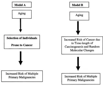
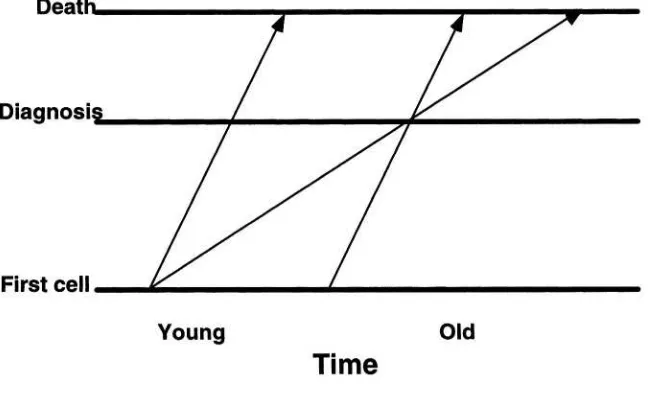
As aging is a risk factor for cancer, it is reasonable to ask whether the incidence of multiple primary malignancies is more common in older persons and in particular whether an aging phenotype of increased cancer risk may be defined. The recognition of such phenotype would have important practical consequences, which include the ability to target certain individuals for cancer prevention and new insight in the molecular pathogenesis of cancer. Luciani and Balducci have considered two alternative hypotheses (Figure 2) 14. According to both hypotheses the incidence of multiple primary malignancies increases with age. In model A this increment reflects only the general risk of cancer associated with aging, whereas in model B previous history of cancer is itself a risk factor for new neoplasms. Model B implies an aging phenotype associated with increased risk of multiple malignancies. After review of the literature, the authors concluded that model A was more likely that model B. Absolute conclusions are not possible, however, due to the limitation of existing data (Table 1). Universal consensus is wanted for the definition of multiple primary malignancies. In the majority of study series the definition of Warren and Gates has been utilized 15. This implies the fulfillment of two conditions: each tumor must present an independent clinical and pathologic picture and the possibility that one neoplasm be a metastasis of the other should be excluded. A number of serious limitations related to this definition are self-evident. First, it fails to distinguish between clinically relevant and irrelevant neoplasms as it is based on autopsy studies. Second it fails to address the issues related to multifocal tumors occurring in the same organ, that are defined by two questions: how can it be established that multifocal tumors are distinct tumors; and should multifocal tumors be considered multiple primary malignancies.
Figure 2.Alternative hypotheses on the increased incidence of multiple primary malignancies with age 14. Model A reflects only the general risk of cancer associated with aging. Model B implies an aging phenotype associated with increased risk of multiple malignancies.
Table 1. Methodological difficulties related to the diagnosis of multiple primary malignancies
Definition
Clinical and pathologic recognition Influence of previous cancer treatment Selection bias
Limitation of existing sources of data Tumor registries
Autopsy series
neoplastic infiltration 17. Though helpful, this criterion appears inadequate on two accounts: it relies on the correctness of individual observations, and it excludes the possibility of surface metastases.
Last but not least, there is an age-specific problem related to the association of age with multiple primary malignancies. This involves the decision whether one should consider that age at which the first or the subsequent tumors did occur. Conceptually, it appears reasonable to consider affected by age-related multiple primary malignancies only those patients whose first cancer was diagnosed during adulthood, but we recognize that this proposal only shifts the problem to the definition of adulthood.
One common problem in the definition of multiple primary malignancies is whether the subsequent neoplasms are metastases of the initial one. This difference can be established with absolute certainty only when the tissue of origin of the original and subsequent tumor is different (for example epithelial and mesenchymal neoplasms). Electron microscopy and immune-histochemistry have also helped to identify tumors of origin from different tissues 14. In the case of some tumors, specific characteristics, such as the presence of hormone receptors in breast cancer allow establishing whether a tumor occurring in different organs is a metastasis of the original neoplasm.
The treatment of cancer may be itself a cause of new cancer, and enhance the risk of a second malignancy in patients who have received antineoplastic treatment. The association of acute myelogenous leukemia with cytotoxic chemotherapy 18 is well known. Cervical cancer has been associated with an increased risk of cancer of the bladder, small intestine, ovary, bones, and of multiple myeloma, but only in patients who had been treated with radiation therapy 19.
The main source of information on multiple primary malignancies is tumor registries and autopsy studies. Tumor registry studies are cohort studies, whose value varies with the quality of the registry as well as with the quality of cancer care provided during the time covered by the registry. For example, studies performed during a time when women received routine mammographic screening are more likely to demonstrate the association of breast cancer with other malignancies, because breast cancer was diagnosed at an earlier stage and associated with a more prolonged survival. In general tumor registry studies showed that the risk of second malignancies increased with the duration of survival since the diagnosis of the initial neoplasm 14. Autopsy studies are by there own nature selective, as they depend on the ability of physicians to obtain autopsy and on the willingness of patients’ family to allow the procedure. These cross-sectional studies showed that the prevalence of multiple malignancies increased with the patient’s age, but it was consistent with the general risk of cancer for that age 14. In conclusion, both autopsy and registry studies demonstrated that the diagnosis of multiple primary malignancy was more likely in patients of advanced age, but age was not a risk factor for increased risk of multiple primary malignancies. These studies favored model B over model A in figure 1. It should be noticed that increased likelihood of association was found between certain types of cancer including smoking related cancer 16, papillary cancer of the kidney and cancer of the bladder and of the prostate 21, and breast and uterine cancer
22
. The latter was observed only in women aged 70 and older.
The increased possibility of multiple primary malignancies in older individuals has important clinical consequences:
The development of a new lesion in patients with history of cancer should be investigated to rule out the possibility of a new and curable malignancy and should not be dismissed as a recurrence off the previous cancer. .
Previous history of cancer should not prevent aggressive treatment of new cancer. It is not unusual for an older individual to carry a diagnosis of two or more primary malignancies, all of which have been curable.
3.AGE AND NATURAL HISTORY OF CANCER
growth of primary breast cancer was inversely related to the degree of mononuclear cell infiltration 10, 28, suggesting that these cells produce a cytokine promoting neoplastic growth. The statement that breast cancer becomes more indolent with age contrasts with some reports that age over 75 is associated with more advanced disease and reduced survival 29-31. The contradiction may be only apparent, as the worst prognosis in women aged 75 and older may reflect lesser utilization of mammographic screening and of adjuvant treatment, and increased risk of mortality from comorbid conditions. Several lines of evidence suggest that breast cancer becomes more indolent with age including reduced risk of life-threatening hepatic and lymphangitic lung metastases, and reduced local recurrence rate after partial mastectomy 32-36.
In the case of non-small cell lung cancer a more indolent course is suggested by reports from different centers that lung cancer presented at an earlier stage in older than in younger individuals 37-39. These reports may be fraught a referral bias, however, as only older patients with resectable tumors might have been referred to the centers for treatment. It is possible that lung cancer after age 70 involved preferentially ex-smokers, in whom reduced exposure to tobacco smoke resulted in more indolent tumors. While several studies have shown that age is associated with decreased treatment response and survival in women with ovarian cancer, the mechanism of this change has not been clarified40.
beneficial to older post-menopausal women 45. Likewise, age does not seem to reduce the benefits of adjuvant chemotherapy in patients with stage III cancer of the large bowel46. The only situations in which the natural history of cancer may suggest to forgo the use of antineoplastic treatment include smoldering AML and early stage prostate cancer in man aged 70 and older. Though smoldering acute leukemia is an obsolete term, this definition may still be helpful to encompass two conditions: hypoplastic acute leukemia, that is AML with a marrow cellularity lower than 10% and AML associated with Myelodysplasia, with a percentage of blasts in the bone marrow between 20 and 30%, that does not undergo any significant change over three months. In both cases the predominant clinical picture is pancytopenia, the incidence of leukostasis is negligible, cytotoxic chemotherapy is associated with low therapeutic response and high risk of early mortality, while supportive treatment with transfusion of blood products and possible erythropoietin may allow months of quality survival 47. The value of local treatment of early prostate cancer in patients aged 70 and over has been debated 48. A study in which patients aged 60 to 75 were randomized to observation and radical prostatectomy demonstrated that surgery was associated with decreased risk of prostate cancer-related deaths, but not overall survival benefits 49, 50.
4.PROFILE OF THE OLDER CANCER PATIENT
Aging is associated with reduced functional reserve of multiple organ systems, increased prevalence of comorbidity, memory disorders, depression, malnutrition, polypharmacy and functional dependence 51. It is legitimate to ask whether these conditions may interfere with the treatment of cancer and may reduce the patient’s life expectancy and tolerance of treatment to the point that treatment is futile or even harmful.
these studies should be highlighted the need to adjust the doses of chemotherapy to the renal function of older individuals, to investigate anemia, that is a risk factor for mortality, functional dependence, and chemotherapy related toxicity, the management of depression, and the provision of a home caregiver in patients at risk to develop functional dependence during cancer treatment.
Another series of study compared the survival and the general function of older cancer patients with that of individuals of same age without cancer. Diab et al review the SEER breast cancer experience and showed that for women aged 75 and older breast cancer was not associated with a change in survival. Unexpectedly, breast cancer was associated with a more prolonged survival in women aged 80 and older. This observation suggests that breast cancer may affect preferentially women in best general condition, who might have lived even longer if they had not developed breast cancer57. This hypothesis is supported by two other studies. Repetto et al compared functional dependence and comorbidity of patients 65 and older with and without cancer admitted to two general hospitals in Italy and found that cancer patients had lower prevalence of both conditions 58. In a retrospective study of the population of Cusumano, Italy, Ferrucci demonstrated that patients who developed cancer had the highest degree of function and the lowest of comorbidity 59. Similar conclusions were drawn by Stanta et al from autopsy studies of elderly persons with and without cancer 13.
It is reasonable to surmise that cancer is preferentially a disease of healthy elderly individuals and that the treatment of cancer in these individuals may result in prolongation of survival and quality of life improvement.
CONCLUSIONS
A review of the epidemiology of cancer and age allows concludes:
Age is a risk factor both for cancer and carcinogenesis, at least up to age 95;
The biological behavior of cancer may be altered with age: in some cases the neoplasm may become more resistant to chemotherapy, in other cases more aggressive and in other cases more indolent;
Cancer is prevalently a disease of healthy elderly individuals whose life expectancy and quality of life may be compromised by cancer.
REFERENCES
Yancik RM, Ries LAG: Aging and cancer in America. Demographic and epidemiologic perspectives. Hematol/Oncol Clin N Am 2000,14,17-23
LaVecchia C, Lucchini F, Negri E et al: Cancer mortality in the elderly, 1960-1998: a worldwide approach. Oncology Spectrums, 2001, 2, 386-394
Anisimov VN: Biological interactions of aging and carcinogenesis. In Balducci L, Extermann M: Biological Basis of Geriatric Oncology
Wingo PA; Cardinez CJ; Landis SH et al: Trends in cancer mortality in the United States, 1930-1998. Cancer, 2003, 97, 3133-3275
Glass AG, Hoover RH: The emerging epidemics of melanoma and squamous cell cancer. JAMA, 1989, 262, 2097-2100
Barbone S, Bovenzi M, Cavallieri F et al: Air pollution and lung cancer in Trieste, Italy. Am J Epidemiol, 1995, 141, 1161-1169
Halpern MT, Gillespie BW, Warner KE: Patterns of absolute risk of lung cancer mortality in former smokers. J Natl Cancer Inst, 1993,17, 457-464
Jukich PJ, McCarthy BJ, Surawicz TS et al: Trends in incidence of primary brain tumors in the United States, 1985-1994. Neuro-Oncology, 2001, 3, 141-151
O’Reilly SE, Connors JM, Klasa R, et al: Malignant lymphomas in the elderly. Clin Ger Med, 1997,13, 251-263
Kurtz JM; Jacquemier J, Amalric R, et al: Why are local recurrences after breast cancer conserving surgery more frequent in younger patients? J Clin Oncol, 1990, 10, 141-152 Hornsby PJ: Replicative senescence and cancer. In Balducci L, Extermann M: Biological Basis of Geriatric Oncology, 2004
Day JC. Population projections in the United States by age, sex, race, and Hispanic origin: 1995-2050. Washington DC, US Bureau of the Census, Current Population Reports, 1996, 25, 1130
Stanta G, Campagner L, Cavallieri F, et al: Cancer in the oldest old: what we have learned from autopsy studies. Clin Ger Med, 1997, 13, 55-68
Luciani A, Balducci L: Multiple primary malignancies. Sem Oncol, 2004, in press Warren S, Gates O. Multiple primary malignant tumors: a survey of the literature and statistical study. Am J Cancer 1932, 16: 1358-1414
Begg CB, Zhanfg Z-F, Sun M, et al: Methodology for evaluating the incidence of second primary cancers with application of smoking-related cancers from the Surveillance, Epidemiology and End-Results (SEER) program. Am J Epidemiol, 1995, 142, 653-664
18.
Pedersen-Bjergaard J, Philip B, Olesen Larsen S, et al: Therapy-related myelodysplasia and acute myeloid leukemia. Cytogenetic characteristics of 115 consecutive cases and risk in seven cohorts of patients treated intensely for malignant diseases in the Copenhagen series. Leukemia, 1990, 7, 1975-1980
Boice TH, Day NE, Andersen A, et al: Second cancers following radiation treatment for cervical cancer: International Collaboration Among Cancer Registries. JNCI monograph, 1985, 74, 955- 975
Anderson CM; Pusztai L; Palmer JL, et al: Coincident renal cell carcinoma and non-Hodgkin’s lymphoma: M.D. Anderson Experience and review of the literature. J Uro, 1998, 159, 714-717
Rabbani, Grimaldi G, Russo P: Multiple primary malignancies in renal cell carcinoma. J Urol, 1998, 160,1255-1259
Adami H-O, Bergkvist L, Krusemo UB, et al: Breast cancer as a risk factor of other primary malignant diseases. A nationwide cohort study. J Natl Cancer Inst, 1984,73, 1049-1055
Lancet JE, Willman CL, Bennett JM: Acute myelogenous leukemia and aging. Hematol Oncol Clin N Am, 2000, 14, 251-267
The International Non-Hodgkin’s Lymphoma Prognostic Factors Project: A predictive model for aggressive non-Hodgkin’s lymphoma. N Engl J med, 1993, 329, 987-994 Wilson CJ, Cohen HJ, Pieper CF: Cross-linked fibrin degradation products (D-Dimer), plasma cytokines, and cognitive decline in community dwelling elderly persons. J Am GerSoc, 2003, 51, 1374-1381
Preti HA, Cabanillas F, Talpaz M, et al: Prognostic value of serum interleukin-6 in diffuse large-cell lymphoma. Ann Intern med, 1997, 127, 186-194
Balducci L, Silliman RA, Diaz N: Breast cancer in the older woman: the oncologist viewpoint. In Balducci L, Lyman GH, Ershler WB: Comprehensive Geriatric Oncology, Harwood Academic Publishers, Amsterdam, 2004
Nixon AJ, Neuberg D, Hayes DF, et al: Relationship of patients age to pathologic features of the tumor and prognosis for patients with stage I or II breast cancer. J Clin Oncol, 1994, 12, 888-894
Adami HO, Malker B, Holmberg L, et al: The relation between survival and age at diagnosis in breast cancer. N Engl J med, 1986, 315, 559-563
Greenfield S, Blanco DM, Elashoff RM, Ganz PA: Patterns of care related to age of breast cancer patients, JAMA, 1987, 257, 2766-2770
Yancik R, Ries LG, Yates JW: Breast cancer in aging women. A population based study of contrasts in stage, surgery, and survival. Cancer, 1989, 63, 976-981
Holmes FF: Clinical course of cancer in the elderly. Cancer Control, 1994, 1, 108-114 Ershler WB: Tumors and aging: the influence of age-associated immune changes upon tumor growth and spread. Adv Exp Med Biol, 1993,330, 77-92
Daidone MG, Luisi A, Silvestrini R, Di Fronzo G: Biologic characteristics of primary breast cancer in the elderly. Comprehensive Geriatric Oncology. L. Balducci, G.H. Lyman, W.B. Ershler. Harwood Academic Publishers, London, pp. 197-200, 1998 Veronesi U, Luini A, Del Vecchio M, et al: Radiotherapy after breast preserving surgery in women with localized cancer of the breast. N Engl J Med, 1993, 328, 1587-1591 Holmberg L, Lindgren A, Norden T, et al: Age as a determinant of axillary node involvement in invasive breast cancer. Acta Oncol, 1992, 31, 533-538
Goodwin JS, Samet JM, Key CR, et al: Stage at diagnosis of cancer varies with the age of the patient. J Am Ger Soc, 1986, 34, 20-26
39.
Guadagnoli E, Weitberg A, Mor V, et al: The influence of patient age on the diagnosis and treatment of lung and colorectal cancer. Arch Intern med, 1990, 150, 1485-1490 Thigpen T: Gynecological cancer in the elderly. In Balducci L, Lyman GH, Ershler WB: Comprehensive Geriatric Oncology, Harwood Academic Publishers, Amsterdam, 2004 Extermann M, Balducci L, Lyman G: What threshold for adjuvant tamoxifen in older breast cancer patients? A decision analysis. European Journal of Cancer 34(suppl 1):S40, 1998
Walter LC, Brand RJ, Counsell RS, et al: Development and validation of a prognostic index for one year mortality in older adults after hospitalization. JAMA 2001, 285,2987-2993
Morrow M, Harris JR, Schnitt SJ: Local control following breast cancer conserving surgery: results of clinical trials. J Natl Cancer Inst, 1995, 87, 1669-1673
Early Breast Cancer Trialists’ Collaborative Group: Tamoxifen for early breast cancer: an overview of the randomized trials. Lancet 351:1451-1467,1998
Early breast cancer Trialists’ Collaborative Group: Polychemotherapy for early breast cancer; an overview of the randomised trials. Lancet, 1998, 352, 930-942
Sargent DJ, Goldberg RM, Jacobson SD, et al: A pooled analysis of adjuvant chemotherapy for resected colon cancer in elderly patients. N Engl J med, 2001, 345,
1091-1097
Vergilio JA, Bagg A: Myelodysplastic syndromes. Contemporary biologic concepts and emerging diagnostic approaches. Am J Clin Pathol, 2003, 119, s58-77
Chodak GW, Thisted RA, Gerber GS, et al: Results of conservative management of clinically localized prostate cancer. N Engl J med, 1994, 230, 242-248
Holmberg L, Bill-Axelson A, Hegelsen F, et al: A randomized trial comparing radical prostatectomy with watchful waiting in early prostate cancer. N Engl J med, 2002, 347, 781-789
Steineck G, Helgesen F, Adolfsson J, et al: Quality of life after radical prostatectomy or watchful waiting. N Engl J med, 2002, 347,790-796
Ferrucci L: Clinical and Biological evaluation of aging. In Balducci L, Extermann M: Biological Basis of Geriatric Oncology, 2004
Extermann M, Overcash J, Lyman GH, et al: Comorbidity and functional status are independent in older cancer patients. J Clin Oncol, 1998, 16, 1582-1587
Repetto L, Fratino L, Audisio RA, et al: Comprehensive geriatric assessment adds information to the Eastern Cooperative Group performance status in elderly cancer patients. An Italian Group for Geriatric Oncology Study. J Clin Oncol, 2002, 20,494-502 Ingram SS, Seo PH, Martell RE, et al: Comprehensive assessment of the elderly cancer patient: the feasibility of self-report methodology. J Clin Oncol, 2002, 20, 770-775 Yancik R, Ganz P, Varricchio CG, et al: Perspectives on comorbidity and cancer in older patients: approaches to expand the knowledge base. J Clin Oncol, 2001, 19, 1147-1151 Balducci L: Guidelines for the management of the older cancer patients. NCCN Journal, in press
58.
59.
Repetto L, Granetto C, Venturino A, et al: Prognostic evaluation of the older cancer patient. In Balducci L, Lyman GH, Ershler WB: Comprehensive Geriatric Oncology, Harwood Academic Publishers, Amsterdam, 1998, 281-286
BIOLOGICAL INTERACTIONS OF AGING
AND CARCINOGENESIS
Vladimir N. Anisimov
Head, Department of Carcinogenesis and Oncogerontology,
N.N. Petrov Research Institute of Oncology, Pesochny-2, St. Petersburg, Russia
It is well documented that the incidence of malignant tumors increases progressively with age, both in animals and humans 1-3. The relationship between aging and cancer is not clear. Considerable controversy surrounds the mechanisms that lead to increased incidence of cancer in the aged. Three major hypotheses have been proposed to explain the association of cancer and age.
1. AGING AND MULTISTAGE MODEL OF CANCER
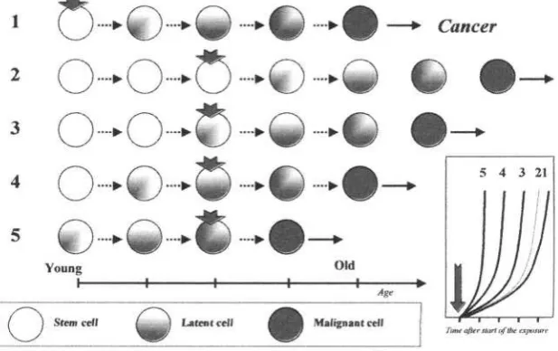
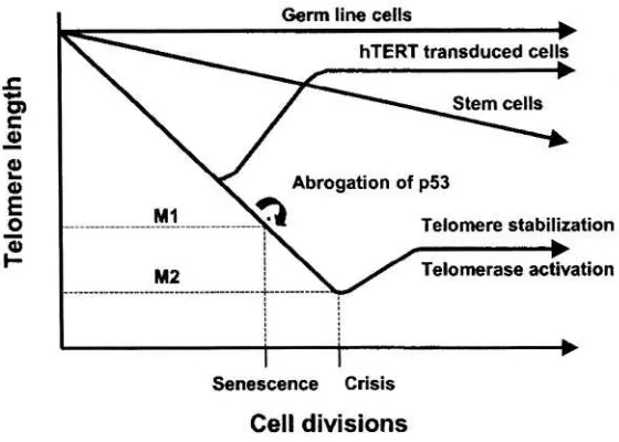
The homeostasis of most tissues is maintained thanks to a pool of stem cells able to reproduce themselves and to differentiate. Cell differentiation is followed by cell death and aging maybe construed as a progressive loss of stem cells to differentiation and death 12. Another possibility involves the immortalization of the stem cell that is associated with a loss of differentiation and apoptosis. These immortalized stem cells may give origin to a clonal population with a survival advantage over the remaining tissues: this process is carcinogenesis 12,13. (Figure 1). Both differentiation and death, and immortalization are multi-stage processes. Many steps of carcinogenesis are well-characterized 5,6,14,15 whereas the steps of aging need better recognition and definition 6,16. Both models of cellular aging and immortalization involve delayed genomic instability that is a transmission of genomic aberrations to distant cellular progenies, accompanied by the occurrence of new aberrations. In one case this process results in cellular death; in the other, in cellular immortalization, and some steps may be shared by the two 16.
Carcinogenesis is a multistage process: neoplastic transformation implies the engagement of a cell through sequential stages, and different agents may affect the transition between contiguous stages 17,18. Several lines of evidence support this conclusion 19:
Histopathology of tumors reveals multiple stages of tumor progression, such as dysplasia and carcinoma in situ
The two-stage model of chemical carcinogenesis in mouse skin shows that different chemicals affect qualitatively different stages in the carcinogenic process
The existence of individuals with genetic traits manifested by an early occurrence of cancer (e.g., familial retinoblastoma, colon and rectum adenomatosis) suggests that one of the carcinogenic steps is a germ-line mutation, but additional somatic effects are required for neoplastic development
Mathematical models based on age-specific tumor incidence curves are consistent with the hypothesis that three to seven independent hits (effects of independent carcinogens) are required for tumor development
Studies with chemical carcinogens in cell cultures reveal that different phenotypic properties of a tumor cell are required for tumor development
Transgenic mice that carry activated proto-oncogenes in their germ-line develop focal tumors, which are apparently monoclonal in origin, suggesting that additional somatic events are required for full malignant progression.
The process of neoplastic development is frequently divided into three operationally defined stages - initiation, promotion and progression. During the first stage of carcinogenesis (initiation) irreversible changes in the genotype of the normal target stem cell leading to its immortality take place. During the initiation the carcinogen or its active metabolite(s) (derived by simple degradation or by active enzymatic process) interacts with nucleic acids leading to mutations in oncogenes and in anti-oncogenes. During the second stage of carcinogenesis (promotion) initiated (latent, immortalized) cell acquires phenotypic features of transformed (malignant) cell, and under the exogenous influence, some of which at least are provided by the neoplastic stroma, tumor progression may occur. A carcinogen affects not only target cells but also influence a lot of factors in the microenvironment of the target cell creating the conditions for promotion of immortalized cell (growth factors, cytokines, immunodepression, biogenic amines, hormonal and metabolic imbalance). Some carcinogens, such as tobacco smoke may effect multiple carcinogenic steps.
Unlike initiation, promotion requires prolonged exposure to the carcinogen and may be reversible to a large extent. The dissection of carcinogenesis into initiation, promotion, and progression is useful as a frame of reference. It should not be assumed, however, that only three carcinogenic stages exist: each stage can be subdivided into multiple substages. Promotion may involve the activation of several enzymes, such as protein kinase C and ornithine decarboxylase; enhanced hexose transport; increased polyamine production, prevention of cell differentiation; and inhibition of cell-to-cell communication 20-21. It was found that 12-O-tetradecanoylphorbol-13-acetate (TPA), a well-known skin tumor promoter, causes free radical-mediated DNA alterations, such as sister chromatid exchanges and expression of proviruses and retroviruses 22.
Discovery of oncogenes and of their function has provided new insight into the carcinogenic process. One may view carcinogenesis as a “cascade” phenomenon, resulting in serial activation of multiple cellular oncogenes and/or inactivation of tumor-suppressing genes (e.g., p53)23.
Figure 2. Integral scheme of carcinogenesis
a minimum of one intermediate stage). Secondly, passage from one stage to another is a stochastic event, the rate of which depends on the dose of a carcinogen that affects the cell. Finally, all cells at any stage of carcinogenesis may enter the next stage independently of each other.
According to this model, the tumor develops only if at least one cell goes through all the necessary stages, and the clonal growth of this cell causes clinical cancer, as a critical volume of neoplastic cells accumulates. In this model, the exact origin of the various stages is ignored and the changes in cell function during the process of carcinogenesis are not assessed. The grade of malignancy is considered to increase with every stage. Various carcinogenic agents (exogenous as well as endogenous) may modulate the process. In addition, some agents act at early stages of carcinogenesis and others at later stages 24. Epidemiological data, analyzed within the framework of a multi-stage model, have helped to estimate the contribution of various factors to the development of cancer. These factors include the time from the start of carcinogenic exposure, and the age of onset of exposure.
Important differences between early and late-stage carcinogens should be highlighted, to illustrate potential interactions of aging and carcinogenesis. Exposure to early stage carcinogens requires a latent period for the development of cancer. During the latent period the transformed cell goes through the subsequent carcinogenic stages. Clearly, elimination of early-stage carcinogens from the environment will not result in immediate cessation in the incidence of cancer. Carcinogens acting at late stages of carcinogenesis cause the tumor incidence rate to rise after a relatively short period of time. The increased rate of tumor incidence will be reversed almost immediately on cessation of exposure 24.
This risk of cancer after exposure to a carcinogen may be calculated as: where I is the risk of cancer, t is the time from initial exposure to the carcinogen, and k is the number of stages that the target cells have undergone before the exposure to the carcinogen. This formula is based on the assumption that with aging there is a progressive accumulation of partially transformed cells primed to the effect of late-stage carcinogens (Figure 3). Age is considered as a variable because older cells may already present in advanced carcinogenic stages, are primed to the effects of environmental carcinogens and consequently may develop cancer more rapidly and at higher rate when exposed to these substances. A number
of factors, including genetic predisposition, oxidative stress, and previous exposure to carcinogens may be responsible of the molecular changes that prime aging cells to environmental carcinogens.
2. EFECT OF AGING ON THE SUSCEPTIBILITY TO CARCINOGENESIS IN VIVO
Animal experiments seem to confirm that there are age related differences in sensitivity to carcinogen in some tissues. Thus, with age, susceptibility to carcinogens in murine mammary gland, small intestine and colon, thyroid, ovarian follicular epithelium decreases, in subcutaneous tissue, cervix uteri and vagina increases and in others (lung, hemopoietic tissues) it remains stable (Table 1). For details see references 1,5-6). Age-related differences in cancer susceptibility have been observed after exposure to the same carcinogens in experimental systems. For example, in female rats exposed to N-nitrosomethylurea (NMU) in doses 10, 20 or 50 mg/kg at the age of 3 month developed mammary carcinomas, tumors of the kidney, ovaries and colon. In contrast to young animals, the rats exposed to the same doses of the carcinogen at the age of 15 months showed a higher frequency of tumors of the corpus and cervix uteri, and a lower frequency of mammary and intestinal adenocarcinomas and tumors of the ovary and kidney 25. Comparison of the results with the data on DNA alkylation, synthesis and O6-methylguanine repair obtained on the same model suggests a critical role of age-related proliferative activity changes occurring in the target tissues in the mechanism of age in modifying the effect on carcinogenesis. Obviously, there are no common patterns of age related changes in DNA synthesis and repair or in proliferative activity of different tissues with age 1,5,6.
Abbreviations:AAF- 2-acetylaminofluorene; BHBNA – N-butyl-N-(4-hydroxybutyl)nitrosamine; BP–benzo(a)pyrene; DBA – 1,2,5,6-dibenzanthracene; DENA– N-diethylnitrosamine; DMNA – N-dimethylnitrosamine; DMH- 1,2-dimethylhydrazine; MAMNA – N-methyl-(acethoxymethyl)nitrosamine; MCA – 20-methylcholanthrene; MNNG – N-methyl-N’-nitro-N-nitrosoguanidine; NMU – N-nitrosomethylurea; TC – tobacco smoke condensate; UV- ultra violet irradiation; X-rays - Roentgen irradiation.
available data concerning age related changes of these parameters have been discussed elsewhere 1-3,23,28. Obviously, there are no common patterns of age related changes in DNA synthesis and repair or in proliferative activity of different tissues with age.
The homeostatic regulation of cell numbers in normal tissues reflects a precise balance between cell proliferation and cell death. Programmed cell death (apoptosis) provides a protective mechanism from cancer, by removing senescent, DNA damaged, or diseased cells that could potentially interfere with normal function or lead to neoplastic transformation 23, 29. Apoptosis plays a substantial role in many other aspects of aging and cancer, including control of the life span of most members of transformed cells, and the rate of growth of tumors 30. p53 mediated apoptosis was suggested as a safeguard mechanism to prevent cell proliferation induced by oncogene activation 31.
3. AGING AND SUSCEPTIBILITY TO CARCINOGENESIS IN VITRO
Some in vitro observations support the suggestion on accumulation in tissues of premalignant cells. Thus, transformed by 24-hours exposure to DMBA, foci in murine bladder epithelium have appeared earlier (on the 40th to the 60th day) and more often (25%) in explants of old (28-30 months) donors in comparison with 100 days and 0.9% in cultures received from five to seven month old mice. A spontaneous transformation of bladder epithelium occurred only in the explants received from old donors 32. The aging of the tissue donor was associated with increased susceptibility of primary cultures of rat fibroblasts to transformation induced by SV-40 33. However rat embryonal fibroblasts were much susceptible to v-scr
transformation than when they were isolated from an adult rat 34. Nettesheim et al. 35 reported that the sensitivity of trachea epithelium explants of old animals to chemical carcinogens was lower in comparison to explants from young animals.
Susceptibility to transformation varies during the different stages of proliferative senescence depending on the carcinogen. Thus, young cells are more susceptible to transformation by chemical carcinogens and by low-dose ionizing radiation, susceptibility to ultraviolet radiation is identical throughout the life span of human fibroblasts, whereas susceptibility to a tumor promoter is identical through the cell life span with exception of the final stage, and susceptibility to SV40 is highest during the final stage 36,37.
changes in susceptibility to carcinogenesis in target tissues, and organ and tissue variability in age distribution of spontaneous tumor incidence. This conclusion generates a critical question: does the aging accompanied by the accumulation of premalignant lesions in target tissues?
4. EFFECT OF AGING ON THE SUSCEPTIBILITY TO TUMOR PROMOTERS IN VIVO
There is evidence of age-related accumulation of cells that are in the late stage of multi-stage process of carcinogenesis. Numerous experiments support this model. Thus, single skin application with 7,13-dimethylbenz[a]anthracene (DMBA) in mice aged 8 and 48 weeks at doses ranging from 10 to 300 caused increased skin papilloma incidence in older mice 38. Also, the average diameter of the tumors was larger in the older animals. Of particular interest are the experiments using skin transplants. TPA failed to induce tumors in the skin of 2-month-old mice grafted to animals of different ages, but caused the same tumor incidence in the skin of 1-year-old donors irrespective of the recipient’s age 39,40. These results indicate that the age of the target tissue, more than the age of the host, determines susceptibility to late-stage agents. Delaying wounding 16 weeks after initiation with a carcinogen led to a more pronounced skin tumor response compared with delay of only 6 weeks in young mice 41. Delaying promotion has also been reported to lead to an increased tumor response with the promoters chrysarobin 42 or mezerein 43. These findings are in agreement with data on age-related decrease in cellular DNA repair capacity in skin 44,45 and increasing p53 mutation frequency with advancing age in human normal skin 46 and in basal-cell skin carcinomas 47,48. Post-ultraviolet DNA repair capacity was found to undergo an age-related decline to which corresponded age-related increase in post-ultraviolet mutability in cultured primary skin fibroblasts from normal donors from the first to the tenth decade of life 44. It was suggested that there was the age-related increase in the number of telomerase positive basal cells in the skin 49. However in some studies the papilloma response either decreased with age or was the same as the response in younger mice 50-52 .
In Tg.AC transgenic (v-Ha-ras) mice, skin tumor incidence and multiplicity were strongly age-dependent, increasing with increasing age of the animal when first treated with TPA, or exposed to wounding, or UV-light
53
hepatocarcinogenesis is very convenient for this purpose because of the availability of strains of animals with different susceptibility to hepatic carcinogenesis. In the liver of highly susceptible mice, the concentration of hepatocytes in advanced stages of carcinogenesis was increased early in life before the exposure to experimental carcinogens 54. In the liver of F344 rats the number of spontaneous proliferative foci is proportional to the animal age 55,56. The incidence of proliferative foci and hepatic tumors induced by
phenobarbital, carbon tetrachloride or peroxisome proliferators in rodents is also a function of age55-57.
Single intravenous injection of NMU at doses of 10, 20 or 50 mg/kg was administered to female rats aged 3 or 15 months 25. The NMU carcinogenic dose dependence in different age groups was considered in the context of a multi-stage model. It was calculated that the number of events necessary for complete malignant transformation in 15-month-old rats under the influence of NMU was lower than in three month-old. In this experiment as well as in another sets of experiments in rats and in mice it was shown that tumors developed earlier in older than in younger animals after exposure to the same doses of NMU 14,59-62. The combined incidences of severe endometrial hyperplasia and adenocarcinomas tended to increase with the increase in intervals between a start of promoting estrasiol treatment after N-nitrosoethylurea initiation in mice 62.
5. EFFECT OF AGING ON TRANSPLANATABLE TUMOR
GROWTH
An important question related to the integrated carcinogenic model (Figure 2) concerns age-related changes in tissue microenvironment as these changes may both favor or oppose carcinogenesis in different circumstances. Should aging alter the environment in which tumor develops, the growth rate of transplantable tumors may vary with the age of the tumor recipient 63. These experiments bypass the effect of age on carcinogenesis itself and explore the role of age-related changes in the organism on the growth and progression of transformed cells. Evaluation criteria for such experiments should include: (a) tumor transplantability, (b) rate of tumor growth, and (c) survival time of tumor bearing animals. The natural history of spontaneous tumors in humans (the rate of tumor doubling, metastasizing potential) and on the survival of cancer patients newly diagnosed at different ages provide information on the effects of age on tumor growth in humans. Available data both in experimental animals and in humans are contradictory and support different effects of age on tumor development (Table 3) l,6. In general, an “age effect” may be recognized both in experimental and in human malignancies.
In another experiment, RA-2 cells from a 3-month-old donor were inoculated into 2-3 or 21-23- month-old recipients and 3 weeks later were separately taken from “young” and “old” hosts and transplanted into 3-month-old recipients. The number of lung colonies was significantly decreased in 3-month-old recipients injected with RA-2 cell passed via “old” host 60. The results obtained suggest the critical role of host and donor microenvironment in lung colony forming potential of RA-2 cells.
McCullough et al. 65 have observed that transformed rat hepatocytic cells lines were only weakly tumorigenic following transplantation into the livers of young adult rats. The tumorigenicity of these cell lines increased progressively with the age of the tumor recipients. These results suggest strongly that the tissue microenvironment represents an important determinant in the age-related tumorigenic potential of transformed cells.
Krtolika and Campisi 66 have shown that senescent stromal fibroblasts can stimulate the hyperproliferation and malignant progression of preneoplastic and neoplastic cells in culture. They also tested the ability of senescent fibroblasts to stimulate epithelial cell growth in vivo by inoculation of preneoplastic epithelial cells with presenescent or senescent human fibroblasts into nude mice 67. None of the tumors when injected alone. Both preneoplastic mouse mammary epithelial cells and preneoplastic human keratinocytes did not form tumors in the presence of presenescent fibroblasts but formed large lethal tumors in the presence of senescent fibroblasts. In the case of human breast cancer cells, senescent fibroblasts markedly stimulated the rate of tumor growth67.
6. MECHANISMS OF INTERACTION OF AGING AND CARCINOGENESIS
Cancer is a common denomination given to a number of different diseases. Common features to all cancers include 23,68
potential immortality of cancer cells due to avoiding apoptosis
ability to invade surrounding tissues due to reduced sensitivity to signals from neighboring cells aimed to offset proliferation
cell de-differentiation with re-appearance of some embryonal proteins
(e.g. in cytoplasm
growth signals autonomy, which allows cancer cells to proliferate in absence of outside signals due to only inner growth signals
release of growth factors and promotion of angiogenesis in tissue, which favor tumor growth and metastasis
Gene mutations, as well as changes in regulation of gene expression, which can produce these typical features, were suggested to be key genetic events leading to cancer development23,68,69. Down regulation of apoptosis gene, p53, as well as upregulation of myc and ras genes, which may favor excessive proliferation, could be examples of such events 69,70.
Both carcinogenesis and aging are associated with genomic alterations, which may act synergistically in causing cancer 23,68-71. In
particular, three age-related changes in DNA metabolism may favor cell transformation and cancer growth. These changes are genetic instability, DNA hypomethylation, and formation of DNA adducts.
Genetic instability involves activation of genes that are normally suppressed, such as the cellular proto-oncogenes, and/or inactivation of some tumor suppression genes (p53, Rb, etc.) 23,31. DNA hypomethylation is characteristic of aging, as well as of transformed cells. Hypomethylation, a potential mechanism of oncogene activation, may result in spontaneous deamination of cytosine and consequent base transition, i.e., substitution of the pair thymine:adenine. Accumulation of inappropriate base pairs may cause cell transformation by activation of cellular proto-oncogenes 23. Age-related abnormalities of DNA metabolism may be, to some extent, tissue- and gene-specific. For example, hypomethylation of the c-myc
proto-oncogene has been found in the hepatocytes, but not in the neurons of old mice 72,73. Within the same cell, different DNA segments express different degrees of age-related hypomethylation. The uneven distribution of hypomethylation may underlie selective overexpression of proto-oncogenes by senescent cells. For example, the transcription of c-myc is progressively increased in the liver but not in the brain of rats between the ages of 4-22 months, whereas the transcription of c-sis and c-src does not appear to be age-related in any tissues 72,73. The different extent of DNA abnormalities among aging tissues may account in part for the different susceptibility of these tissues to carcinogens 74,75.
The damage caused by endogenous oxygen radicals has been proposed as a major contributor to both aging and cancer 76-78. Endogenous oxidative damage to lipids and proteins increases with age 77,78. It was shown that oxygen free radicals may induce active mutations of the human c-Ha-ras
proto-oncogene 78. The level of one oxidized nucleoside,
ascorbic acid), uric acid and the pineal indole hormone melatonin 80-83.
There is evidence of an age-related accumulation of spontaneous mutations in somatic and germ cells 71. Accumulation with age of some spontaneous mutations or mutations evoked by endogenous mutagens can induce genome instability and, hence, increase the sensitivity to carcinogens and/or tumor promoters. It has been shown that clonally expanded mtDNA mutations accumulate with age in normal human tissues as well as in human tumors 84,85. The finding that deleted mtDNA accumulated in human muscle tissue as well as evidence for partially duplicated mtDNA in aged human tissues 85 suggests the important role of clonal expansion of mutant mtDNA in the age-related increase of systemic oxidative stress in the whole organism
86. A significant trend toward increasing p53 mutations frequency with
advancing age was found in some normal and malignant tissues 46,47. Simpson 9 suggested that the aging human body accumulates enough mutations to account for multistep carcinogenesis by selection of preexisting mutations. The evidence showed that both genetics of the selected cellular clone and the epigenetics of the selective environment contribute to tumor development87.
Thus, the data available show that some changes in structure and function of DNA are evolving with natural aging. The character of these changes could vary in different tissues and might cause uneven tissue aging. Dolle et al. 88 using a lacZ plasmid transgenic mouse model, determined spectra of spontaneous point mutations in different organs in young and old mice. While similar at a young age, the mutation spectra among these organs were significantly different in old age. The authors stressed that the replicative history per se is not the underlying causal factor of age-related organ-specific differences in mutations spectra. Rather, differences in organ function, possibly with association with replicative history, may explain the divergence in mutation spectra during aging. In turn, this may explain both age-related increase in spontaneous tumor incidence and age-related changes in susceptibility to carcinogens in various organs.
Crosstalk between mesenchyme and epithelium has been described as a known driver of differentiation and development89,90. It was shown that changes in stromal behavior can promote epithelial transformation 66,89.
Thus, the data available show that some changes in structure and function of DNA are evolving with natural aging. The character of these changes could vary in different tissues and might cause uneven tissue aging. In turn, this may lead to both age-related increases in spontaneous tumor incidence and age-related changes in susceptibility to carcinogens in various organs. Table 4 summarizes the data available in literature and obtained in our experiments on some hormonal metabolic shifts in the organism and disturbances at tissue and cellular levels observed in natural aging and in different types of carcinogenesis in vivo. Despite incomplete data, it can be seen that there is a similarity between the shifts in aging and carcinogenesis. Carcinogens could be supposed to initiate a normal cell, interacting with its elements on the molecular level, on the one hand, and to produce diverse changes in the organism facilitating promotion and progression of tumor growth, on the other hand.
7. THE ROLE OF THE INSULIN/IGF-1 SIGNALING PATHWAY IN AGING AND CANCER
The potential link between aging and insulin/IGF-1 signaling has attracted substantial attention during last years, on the basis of evidence including age-related increase in incidence of insulin resistance and type 2 diabetes in accelerated aging syndromes and life span extension by caloric restriction in rodents. Concomitant reduction in plasma insulin and plasma glucose levels, which implies increased sensitivity to insulin, emerged as a hallmark of increased longevity 91,92. Hyperglycemia is an important aging factor involved in generation of advanced glycosylation endproducts (AGEs)
93,94. There are evidence that hyperinsulinemia favors accumulation of
oxidized protein by reducing its degradation as well as facilitates protein oxidation by increasing steady-state level of oxidative stress 95. Untreated diabetics with elevated glucose levels suffer many manifestations of accelerated aging, such as impaired wound healing, obesity, cataracts, vascular and microvascular damage 8. It was shown that centenarians have a preserved glucose tolerance and sensitivity to insulin as well as lower degree of oxidative stress as compared to aged persons 96. It is worthy to note that hyperinsulinemia is an important factor not only in aging but also in the development of cancer 8,97,98.
molecular mechanisms underlying aging 91,92,99. In D. melanogaster, the mutation of genes operating in the signal transduction from insulin receptor to transcription factor daf-16 (age-1, daf-2, CHICO, InR are strongly associated with longevity 99,100. It was demonstrated that FKHR, FKHRL1
and AFX, which are mammalian homologues of daf-16 forkhead
transcription factor, function downstream of insulin signaling and akt/PKB under cellular conditions 101,102.
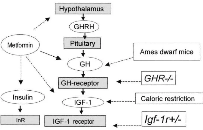
Daf-2 and InR are structural homologues of tyrosine kinase receptors in vertebrates that include the insulin receptor and the insulin-like growth factor type 1 receptor (IGF-1R). It was shown that in vertebrates the insulin receptor regulates energy metabolism whereas IGF-1R promotes growth. At
least three genes Ghr) whose knockout leads to dwarfism
have been identified. The expression of these genes is associated with reduced levels of IGF-1 and insulin and increased longevity 103,104. In Snell and Ames dwarf mice, sexual maturation is delayed, and only few males are fertile, while females are invariably sterile. These mice as well as knockout mice have significantly reduced glucose levels and fasting insulin levels, decreased tolerance to glucose and increased sensitivity to insulin which appears to be combined with reduced ability to release glucose in response to acute challenge 91.
Recently, strong support for the role of insulin/IGF-1 signaling pathway in the control of mammalian aging and for the involvement of this pathway in longevity of IGF-1 deficient mice was provided by Hsieh et al 105,106. It was shown that in the Snell dwarf mice, GH deficiency would lead to reduced insulin secretion and alterations in insulin signaling via IRS-1 or IRS-2 and P13K affects genes involved in the control of longevity. The authors concluded that the Pit1 mutation may result in physiological homeostasis that favors longevity.
Reduction in both glucose and insulin levels as well as an increase in the sensitivity to insulin are a well-documented response to caloric restriction in rodents and monkey 107,108. It is worthy to note that mice have a major increase in the level of insulin receptors 109, while Ames dwarf mice have a smaller increase in insulin receptor and substantially increased amount of insulin receptor substrates IRS-1 and IRS-2 110. The development of tumors in Ames dwarf mice was postponed and the incidence was reduced as compared to the control108.
body size, delayed puberty, and significantly increased sensitivity to insulin action.
Holzenberger et al 113 inactivated the Igf1r gene by homologous recombination in mice. It was shown that mice dead early in life, whereas heterozygous mice live on average 26% longer than wild-type littermates. These mice did not develop dwarfism; their energy metabolism was normal. Food intake physical activity, fertility and
reproduction were also unaffected in mice. These mice and
embryonal fibroblasts derived from them were more resistant to oxidative stress than controls. The spontaneous tumor incidence in the aging cohort of mice was similar to that in wild-type controls. At the molecular level, insulin receptor substrate and the p52 and p66 isoforms of Shc, both main substrates of IGF-1 receptor, showed decreased tyrosine phosphorylation. mediated cellular responses to oxidative stress. Two main pathways -the extracellular-signal related kinase (ERK)/mitogen-activated protein kinase (MAPK) pathway and the phosphatidylinositol 3-kinase (PI3K)-Akt pathway – were downregulated in mice.
The extension of longevity was observed in fat-specific insulin receptor knockout (FIRKO) mice 114,115. These animals have reduced fat mass and were protected against age-related obesity and its subsequent metabolic abnormalities including deterioration in glucose tolerance, although their food intake was normal. Both male and female FIRKO mice had increased mean life span (by 18%) with parallel increases in maximum life span. Extended longevity in FIRKO mice was associated with a higher age threshold beyond which age-dependent increase in mortality risk became appreciable and a decreased age-adjusted mortality rate, especially after 36 months of age. In FIRKO mice, the resistance to obesity, despite normal food intake, suggested that metabolic rate is increased, rather than decreased 115. The authors believe that decreased fat mass could lead to a decrease in oxidative stress. Another possibility is that the increased longevity in these mice is the direct result of altered insulin signaling.
Shimokawa et al. 116 designed a transgenic strain of rats whose GH gene was suppressed by an anti-sense GH transgene. Male rats homozygous for the transgene (tg/tg) had a reduced number of pituitary GH cells, a lower plasma concentration of IGF-1, and a dwarf phenotype. Heterozygous rats