Conocarpus erectus
Leaf Extract for Green Synthesis
of Silver Nanoparticles and Their Antibacterial Activity
Amjed Mirza Oda
*Science Department, College of Basic Education, University of Babylon, Babylon 51002, Iraq
Received July 5, 2017; Accepted October 24, 2017
ABSTRACT
Silver nanoparticles (SNPs) were synthesized by a green method using Conocarpus erectus leaves extract. Leaves were cleaned and used freshly then the extract was obtained by heating leaves in water for 15 min. Silver nitrate used as SNPs precursor and 5 mL of extract were added to silver ion solution at 80 °C and the growth of nanoparticle was monitored by electronic spectra at plasmon resonance absorption of SNPs, where the growth obeys sigmoidal kinetics. SNPs were characterized by SEM, XRD, FTIR, UV-vis absorption and fluorescence, and particle size was estimated. SEM reveals that the SNPs had spherical shape and particle size less than 100 nm. XRD analysis showed that only Ag phase was present and the estimated particle size was 20 nm. FTIR spectra analysis showed similarity of extract and SNPs, indicating the adsorption of active components of the extract on the high surface area of SNPs. Electronic absorption and fluorescence spectroscopy showed that plasmon absorption and fluorescence in the visible region were a characteristic feature of SNPs. Antibacterial activity examined against
Escherichia coli and Staphylococcus aureus indicated that the inhibition zones were equal to 24 and 19 mm, respectively.
Keywords:silver nanoparticles; green synthesis;Conocarpus erectus
ABSTRAK
Nanopartikel perak (SNPs) disintesis dengan metode hijau menggunakan ekstrak daun Conocarpus erectus. Daun segar dibersihkan dan kemudian diekstrak dengan memanaskan daun dalam air selama 15 menit. Perak nitrat digunakan sebagai prekursor SNP dan 5 mL ekstrak ditambahkan ke larutan ion perak pada suhu 80 °C dan pertumbuhan partikel nano dipantau menggunakan spektrum elektronik pada penyerapan resonansi plasmon SNP, dimana pertumbuhannya memenuhi kinetika sigmoid. SNP dikaraterisasi dengan SEM, XRD, FTIR, UV-vis absorpsi dan fluoresensi serta perkiraan ukuran partikel. SEM menunjukkan SNP memiliki bentuk bola dan ukuran partikel kurang dari 100 nm. Analisis XRD menunjukkan adanya fasa Ag saja dan ukuran partikel diperkirakan 20 nm. Analisis FTIR ekstrak dan SNP menunjukkan kemiripan, dan membuktikan adanya adsorpsi komponen aktif ekstrak pada luas permukaan tinggi SNP. Serapan elektronik dan spektroskopi fluoresensi menunjukkan penyerapan plasmon dan fluoresensi di daerah yang terlihat merupakan ciri khas SNP. Aktivitas antibakteri diperiksa terhadap
Escherichia coli dan Staphylococcus aureus, dan menghasilkan zona penghambatan masing-masing sebesar 24 dan 19 mm.
Kata Kunci:nanopartikel perak; sintesis hijau;Conocarpus erectus
INTRODUCTION
Green synthesis opened a wide field for nanoparticles synthesis as an alternative route of the bottom to up method and the production is easy to perform, also good controlling and stabilization. This route is favorable because of it’s ecofriendly and non-toxic, where chemicals usually are expensive, rarely pure, toxicity and not compatible with products that enter in cosmetics and pharmaceuticals [1]. The challenge in green chemistry synthesis is the development of ecological and friendly methods to produce the nanomaterials. The nanomaterials are used in various
ideality [5]. Among these methods are plants and plant extracts, where it exhibits alternative route substituting toxic chemicals and the best choice of synthesis. The natural products of this natural factory are non-toxic metabolites and this variety in compounds work as reducing agent of noble metal to produce nanoparticles. Also, plant extract components are a stabilizing agent, where it is the way for prolonging the lifetime of nanoparticles [6]. Phytochemicals and plant-derived polysaccharides, such as cellulose, starch, dextran, and alginic acid, play dual roles as reducing and stabilizing agents. The oxidation of the hydroxyl groups of polysaccharides to carbonyl groups is most likely involved in the reduction of metal ions to produce metallic NPs [7]. Like silver nanoparticles are traditionally synthesized by chemical methods using ethylene glycol, pyridine and sodium borohydride. All chemicals used in this route are toxic and highly reactive costing a risk to the environment and humans, or expensive to be feasible procedures in the industrial field [8]. As a comparison, the chemical methods involve hazardous chemicals, low conversion and production, consuming high energy and difficult to purifications of the products and need waste treatment [9-10]. Green synthesis has advantages over traditional chemical methods. Many features can be summarized: green synthesis involves a one-pot reaction, giving simplicity and rapid preparation, scale up is under controlling, eliminate toxicity, biocompatibility with normal tissues for in vivo applications, and supplying self-reducing agents and capping agents, providing enhanced colloidal stability [11-12]. Noble metal nanoparticles are the subject of science community due to their unique optical, electronic, mechanical, magnetic and chemical properties that differ from those of bulk materials [13]. Silver nanoparticles have an attraction in biology and medicines as the physiochemical properties are unique. The applications of metallic NPs are wide like antimicrobial applications [14], drug delivery, medical imaging and diagnostics [15-16] artificial implant, HIV inhibition, nanodevice fabrication and medicine [17]. Centuries ago, silver has been used as an antimicrobial agent, this interest for this element is a resurgence, because of increasing threat of microorganisms resistance to antibiotic [18].
Silver nanoparticle produced by biological synthesis using natural products depending on phytoconstituents like Amaranthus dubius, Amaranthus polygonoides, Alternanthera sessilis and Portulaca oleracea [19],Murraya koenigii[8], Moringa oleifera leaf extract [20], Azhadirachta indica [1], Pisonia grandis
[19], Ananas comosus[21], where plant extracts act as potential reducing agents for the green synthesis and also plant extracts act as stabilizing agents [22].
Conocarpus erectus L. (family Combretaceae) is an
evergreen tree and it is a mangrove species also called buttonwood growing in coastal areas in tropical and subtropical regions of the world. The species is usually a shrub of the genus Nerium and family Apocynaceae about 1.5 to 4 m in height but can become a tree up to 20 m or more in height [23-24].The leaves, stem, flowers and fruits of Conocarpus erectus has been studied and the main active components are total phenolic, flavonoids, and tannin contents. Besides that, the tree ofConocarpus erectusL. is extensively spread in Iraq and is evergreen with a lot of flowers and fruits [25-26].
In this research, green synthesis of silver nanoparticle was adapted by using the aqueous extract ofConocarpus erectusleaves that no study had shown the activity of aqueous extract in SNPs synthesis. The active components of extract (the crude) was used for reducing the silver ion to SNPs at 80 °C. The formation of SNPs was monitored by absorption spectra for 15 min to study the mechanism of SNPs nucleation. SNPs were analyzed by SEM, XRD, FTIR and UV-vis absorption and fluorescence spectroscopy. Finally, SNP antibacterial activity was examined against two kinds of bacteria, gram-positive (Staphylococcus aureus) and gram-negative (Escherichia coli).
EXPERIMENTAL SECTION
Materials
Silver nitrate (AgNO3) was supplied by BDH
Company and used without any further purification. Stock solution of silver nitrate was prepared in 0.1 M and diluted to prepared 0.001 M. Conocarpus erectus
leaves were collected at my work from the garden of basic education college, University of Babylon, Babylon, Iraq in May 2nd, 2017.
Instrumentation
SNP solution was dirty red to brown color and electronic spectra were recorded for the first minute of reaction to study the sol growth at wavelength 458 nm. FTIR analysis: the extract and SNPs were tested by FTIR spectrophotometry using Affinity IR instrument (Shimadzu, Japan) by disk method. 1-2 mg of sample was mixed with 10-200 mg of dry KBr and pestle with mortar then pressed like disk and spectra was recorded in the range 400-4000 cm-1. XRD diffraction: SNPs
diffraction angle (2theta). SEM analysis: SNP sol was decanted on aluminum foil and left one day to be ready for SEM analysis. Scanning Electron Microscope Inspect 550, Netherland. SEM was operated at 25 kV. Synchronous fluorescence is tested by fluorometer (FS-2, Scinco Co. Korea) in the range 200-800 nm, slits = 1
nm, resolution = 0.5 nm, interval = 0.1 nm and Δλ = 90
nm.
Procedure
Extract preparation
Conocarpus erectus leaves (10 g) were used freshly and cleaned with tap water for removing dust and dirt then washed with distilled water three times. Leaves were cut into fine pieces then transferred into 50 mL distilled water and heated for 15 min at boiling. The extract was filtrated triple to get a clear yellow color and used freshly.
Synthesis of silver nanoparticle (SNPs)
Green synthesis of SNPs was adopted by using AgNO3as a precursor of SNPs. 100 mL of 1 mM of fresh
silver nitrate solution was heated to 80 °C with continuous stirring, after that 5 mL of freshConocarpus erectus leaves extract was added to the hot solution of silver nitrate. The reaction proceeding was monitored every min and electronic spectrum recorded between 200 to 800 nm until the absorbance was constant for 15 min.
Collection of bacterial strains and diagnosis
Two kinds of clinical microbial isolates were gram positive (Staphylococcus aureus) and gram negative (Escherichia coli) were isolated. These isolates were identified by using conventional biochemical tests and Vitek 2 system (Biomeraux, France), and cultivated in pure culture.
Determination of antimicrobial activity of SNPs The antibacterial activity of silver nanoparticles was tested according to disc method, where disks are 6 mm in diameter was prepared then autoclaved and dried. SNPs are loaded on the discs by impregnation. 0.5 McFarland standard (1.5 × 10⁸ CFU/mL) was
adjusted by using a sterile swab to inoculate Mueller Hinton agar plates. The prepared disks were placed individually on the Mueller Hinton agar surface in Petri dishes (90 mm) and each isolate was tested in triplicate in the same dishes. After incubation at 37 °C for 18 h, the inhibition zone was measured in millimeters.
RESULT AND DISCUSSION
SNPs were synthesized by Conocarpus erectus
leaves to extract as green synthesis. Conocarpus erectus is spread in most region of Iraq, and its active components like phenolic compounds, tannin and alkaloids were studied by several studies. Thus author explores its activity as reducing agent for SNPs synthesis. The study in 2016 reported the use of
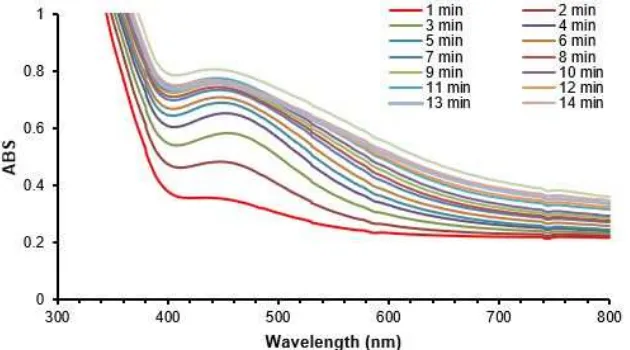
Conocarpus erectus in SNPs synthesis using only methanol extract of leaves as reducing agent [26]. In this study the extract was prepared from fresh leaves and the aqueous solution was obtained after boiling for 15 min. SNPs were tested by electronic spectra between 200–800 nm every 1 min. The absorption between 400–500 nm is attributed to plasmon resonance of electronic behavior of SNPs surface. This feature is a very intrinsic property of silver when prepared at nanoscale level and its electrons in the surface can be easily excited by visible light. This property give SNPs a variety in color according to structure, shape and size. The maximum absorbance is 458 nm and this absorbance is increased with time every minute. This is due to SNPs growth with time,
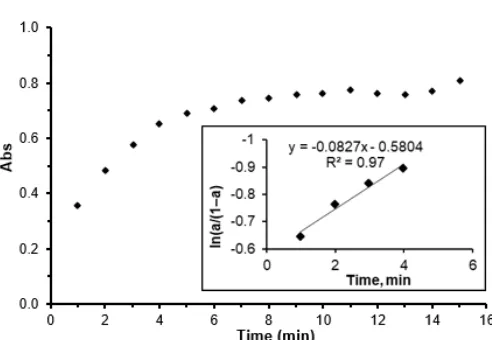
Fig 2.The variation of SNPs absorbance at 458 nm with growth time. The inset graph is the plots of ln(a/(1–a)) as a function of reaction time
Fig 3.Synchronous fluorescence spectra of SNPs
then absorbance is stable at the end of reaction as in Fig. 1 which is show the electronic spectra of SNPs growth with time for 15 min.
The absorbance at 458 nm was monitored with time and the absorbance is increased with sigmoidal kinetics. The kinetics of SNPs formation mechanism is proposed by Watzky and Fink where the first step is slow continuous nucleation and the second step is fast autocatalytic growth. This growth takes place in two different paths: the first is the formation of nuclei and continue joining together in the first stage, the second proposal theory is particles are formed as a solid surface then its enlargement due to accumulation of instant reduced silver ion on big nucleus by collision with freshly reduced metal ions [27]. According to reference [28], the author studied the growth of SNPs and according to this paper, the growth is started slow, then the fast reaction is occurred and finally saturation. This belongs to the autocatalytic reaction. The growth is including formation the first nucleus and works as an adsorbent of silver ion, where silver ion is reduced on the surface by their subsequent reduction. In Fig. 2 the absorbance at 458 nm is increased with time and fast reaction is occurred in the first minute to the fifth minute, after that the reaction is slow. The fast reaction is autocatalytic reaction and changing in absorbance is effective values, where the formation of SNPs last five minutes. After the fast stage, the formation of SNPs is completed and no effect of silver ion on reaction kinetic as the active reducing power is depleted. The insight figure in Fig. 2 shows the kinetic of SNPs growth by plotting of ln(a/(1–a) verse time and high regression is enough at first four minutes and the slope is the apparent kinetic constant ka and it was equal 0.083 min-1.
When noble metal like silver are excited with photons at limited wavelength, the excited state tries to dissipate energy then photons emitted with less energy
called the photoluminescence (PL). This is attributed to the transition of electrons the upper d band and conduction sp band.
Silver nanoparticles have emission peak positions located in a wide range of wavelength between 320 to 520 nm. Using synchronous fluorescence spectroscopy (SFS) is a benefit to minimize broadening and spectral overlap that found in conventional fluorescence spectra and this improves the selectivity. In SFS technique, the
λex and λem scanning are simultaneous. The scan rate
is constant for both excitation and emission monochromators and keeping a constant wavelength
interval (Δλ) between λem and λex. SFS technique is a
convenient technique for analyzing impure materials that consist of multi-component samples without the need to further separation procedures [29].
Synchronous fluorescence spectra appear in Fig. 3, where it was recorded in the range of 200–800 nm,
where Δλ = 90 nm. According to SNPs impurity, the
spectra are synchronous. There are several peaks of SNPs in solution, a shoulder at 360 nm of excitation and 448 nm of emission, originated from the metal– ligand charge transfer absorption [30]. Peaks at 542, 640 and 723 nm are the emission of the multi-scale size of SNPs explained by Kun et al. [31] that a combination of surface plasmon resonance and weak fluorescence under UV irradiation is found. The larger sized SNPs are responsible for the plasmonic extinction peak at 415 nm, while ultra-small SNPs appear weak fluorescence emission around 550 nm and these results close to the value of SNPs in this paper.
The prepared SNPs by green synthesis using
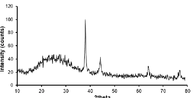
Fig 4.XRD pattern of SNPs prepared by leaves extract ofConocarpus erectus
Fig 5.SEM image of SNPs
was tested by XRD and gives a variety intensity angles, 38, 44.37, 63.9, and 76.8 degrees corresponding to (111), (200), (220) and (311) planes of silver which are agreed with the literature reported (JCPDS silver file # 87–0720). Fig. 4 represents XRD pattern of SNPs synthesized by leaves extract. All angles belong to silver metal only and no trace of any other phases of silver oxides is observed. Angles between 20–30 degrees do not belong to silver metal, but it belongs to the active components of extract adsorbed on SNPs. Thus the extract works as an active reducing agent to produce SNPs and stabilizer to keep silver ion and convert to SNPs. The particle size of SNPs was estimated by using Debye-Scherrer’s equation [1,13]:
k D
cos
k: constant, λ: wavelength of the X-ray, β: full width half maximum of the XRD peak (radians), θ: Bragg’s angle of
the XRD peak. According to the measurement of XRD, the particle size is in nanoscale and was not more than 20 nm. This is a good result showing that the
Conocarpus erectusleaves extract has the ability to the synthesis of SNPs with controlling of particle size.
SNPs characterized by SEM can be seen in Fig. 5, showing SEM image of SNPs in 1-micrometer scale, precipitated on aluminum foil. According to this image, SNPs appear fine powder with high roughness and the diameter was 79 nm and this is a good result compatible with the green synthesis of nanomaterials. The size and shape are controlled, where the SNPs are an agglomeration of the uniform globular nanoparticle. This controlling may be due to the components of extract that work as reducing agent and as well as a stabilizer. Since the high surface area of SNPs, it works as a good adsorbent of the active components of
Conocarpus erectusleaves extract. These components are a good controlling factor of shape and stabilizer that prevents the SNPs to oxidized.
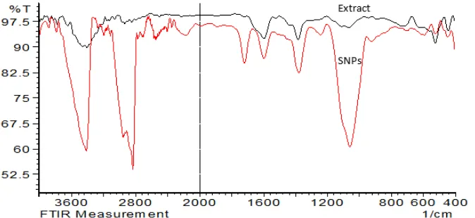
FT-IR spectra in Fig. 6 identify that the biomolecules are responsible for the reduction of the Ag+ions and work as a capping agent.
Fig 6.FT-IR analysis of extract ofConocarpus erectusleaves (dark line) and SNPs (light line)
Fig 7.Photographic picture of two dishes ofEscherichia coli on left and Staphylococcus aureus on right, where
each dish contains 100 μL of SNPs solution was tested
in triplicate and inhibition zone calculated by the average
compounds that reduced Ag ions to SNPs. The main IR bands that are strong in the analysis are 3.462 cm−1 correspond to –OH stretching. The band at 2.922 cm−1 belong to stretching of aliphatic -C-H. The bands at 1.598 cm−1are due to the double bond (C=C) stretching. Also, the IR bands at 1.384 and 1.070 cm−1 may be attributed to -C-O and -C-O-C stretching modes, respectively. It is obvious that the possible compound in
Conocarpus erectus are tannins, steroids, phenols, saponins, flavonoids, glycosides, triterpenoids, and glycerides present in the leaf extract of Conocarpus erectusare the effective compounds potentially active to reduce silver ion and synthesis of SNPs, also these compounds work as stabilizer like capping agent. These results are agreed with a previous similar study [32].
The antibacterial activity of SNPs was tested against gram-positive (Staphylococcus aureus) and gram negative (Escherichia coli) by disk method as in Fig. 7 showing photographic picture of two dishes of
Escherichia coli on left and Staphylococcus aureus on right by disk method, where each disk about 6 mm
contains 100 μL of SNPs solution was tested in triplicate
and inhibition zone was calculated by the average. The inhibition zone was measured and equal to 19 and 24 mm, respectively. This is a good result of this work, where SNPs as an antibacterial agent have been
synthesized by green synthesis using Conocarpus erectus, and there is no study employing the antibacterial activity of SNPs prepared from aqueous extract of this plant. According to literature, SNPs synthesized in action of Bauhinia Acuminata and
Biophytum Sensitivum, the inhibition zone does not exceed 13 mm [33], and not exceed 10 using
Triumfetta Rotundifoliaplant extract for SNPs synthesis [34].
Inhibition zone was clear and each bacteria was tested triplicate in the same dish to minimize errors and the average of inhibition zone was calculated. The mechanism of the SNP activity against bacteria is under study and not very well-known. SNPs may attach the surface membrane of the bacteria wall and damage its function like permeability and respiration. Small size of SNPs provides more surface area available for interaction causing bactericidal effect large [35]. The turbulence of membranous permeability by SNPs increases reducing sugars and proteins in cells. This may attribute to the damage taking place on the lipopolysaccharide or membrane proteins in the outer membrane. SNPs at high concentration decrease the enzyme activity and inhibit respiratory chain dehydrogenase enzymes in E. coli. It is assumed that SNPs destroy respiratory chain dehydrogenases. Silver ion interacted with enzyme thiol (–SH) of E. coli
causing inhibition of respiration. This replaces Ag+
instead of hydrogen atom of cysteine thiol group and forms –S–Ag, and a malfunction the enzymatic activity leads to bacteria growth inhibition [36-38].
CONCLUSION
Silver nanoparticles have been prepared with green synthesis route using Conocarpus erectus
indicated with the similar bands of dry crude extract and SNPs. XRD and SEM revealed that the prepared SNPs were in nanometric scale and uniform globular shape. Antibacterial activity of SNPs showed 24 and 19 mm inhibition zone of Escherichia coli and Staphylococcus aureus, respectively, and this inhibition results from the nanometric scale of SNPs leading the damage of membrane and turbulence of permeability.
REFERENCES
[1] Firdhouse, M.J., and Lalitha, P., 2012, Green synthesis of silver nanoparticles using the aqueous extract of Portulaca oleracea (L.), Asian J. Pharm. Clin. Res., 6 (1), 92–94.
[2] Gross, M., Winnacker, A., and Wellmann, P.J., 2007, Electrical, optical and morphological properties of nanoparticle indium–tin–oxide layers,
Thin Solid Films, 515 (24), 8567–8572.
[3] Parak, W.J., Gerion, D., Pellegrino, T., Zanchet, D., Micheel, C., Williams, S.C., Boudreau, R., Le Gros, M.A., Larabell, C.A., and Alivisatos, A.P., 2003, Biological applications of colloidal nanocrystals,
Nanotechnology, 14 (7), R15–R27.
[4] Ahmad, N., Alam, M.K., Singh, V.N., and Sharma, S., 2009, Bioprospecting AgNPs from wild Desmodium species,J. Bionanosci., 3 (2), 97–104. [5] Ahmad, N., Sharma, S., Alam, M.K., Singh, V.N.,
Shamsi, S.F., Mehta, B.R., and Fatma, A., 2010, Rapid synthesis of silver nanoparticles using dried medicinal plant of basil, Colloids Surf., B, 81 (1), 81–86.
[6] Ankamwar, B., Chaudhary, M., and Sastry, M., 2005, Gold nanotriangles biologically synthesized using tamarind leaf extract and potential application in vapor sensing, Synth. React. Inorg. Met.-Org. Nano-Met. Chem., 35 (1), 19–26.
[7] Park, Y., Hong, Y.N., Weyers, A., Kim, Y.S., and Linhardt, R.J., 2011, Polysaccharides and phytochemicals: A natural reservoir for the green synthesis of gold and silver nanoparticles, IET Nanobiotechnol., 5 (3), 69–78.
[8] Christensen, L., Vivekanandhan, S., Misra, M., and Mohanty, A.K., 2011, Biosynthesis of silver nanoparticles usingMurraya koenigii(curry leaf): An investigation on the effect of broth concentration in reduction mechanism and particle size,Adv. Mater. Lett., 2 (6), 429–434.
[9] Kowshik, M., Ashtaputre, S., Kharrazi, S., Vogel, W., Urban, J., Kulkarni, S.K., and Paknikar, K.M., 2002, Extracellular synthesis of silver nanoparticles by a silver-tolerant yeast strain MKY3,
Nanotechnology, 14 (1), 95–100.
[10] Nabikhan, A., Kandasamy, K., Raj, A., and Alikunhi, N.M., 2010, Synthesis of antimicrobial silver
nanoparticles by callus and leaf extracts from saltmarsh plant, Sesuvium portulacastrum L.,
Colloids Surf., B, 79 (2), 488–493.
[11] Majdalawieh, A., Kanan, M.C., El-Kadri, O., and Kanan, S.M., 2014, Recent advances in gold and silver nanoparticles: Synthesis and applications,J. Nanosci. Nanotechnol., 14 (7), 4757–4780.
[12] Im, A.R., Han, L., Kim, E.R., Kim, J., Kim, Y.S., and Park, Y., 2012, Enhanced antibacterial activities of Leonuri herba extracts containing silver nanoparticles,Phytother. Res., 26 (8), 1249– 1255.
[13] Caroling, G., Tiwari, S.K., Mercy Ranjitham, A., and Suja, R., 2013, Biosynthesis of silver nanoparticles using aqueous broccoli extract characterization and study of antimicrobial, cytotoxic effects,Asian J. Pharm. Clin. Res., 6 (4), 165–172.
[14] Morones, J.R., Elechiguerra, J.L., Camacho, A., Holt, K., Kouri, J.B., Ramírez, J.T., and Yacaman, M.J., 2005, The bactericidal effect of silver nanoparticles, Nanotechnology, 16 (10), 2346– 2353.
[15] De Jong, W.H., and Borm, P.J.A., 2008, Drug delivery and nanoparticles: Application and hazards,Int. J. Nanomed., 3 (2), 133–149.
[16] Thorley, A.J., and Tetley, T.D., 2013, New perspectives in nanomedicine, Pharmacol. Ther., 140 (2), 176–185.
[17] Tolaymat, T.M., El Badawy, A.M., Genaidy, A., Scheckel, K.G., Luxton, T.P., and Suidan, M., 2010, An evidence-based environmental perspective of manufactured silver nanoparticle in syntheses and applications: A systematic review and critical appraisal of peer-reviewed scientific papers,Sci. Total Environ., 408 (5), 999–1006. [18] Gardea-Torresdey, J.L., Gomez, E., Peralta-Videa,
J.R., Parsons, J.G., Troiani, H., and Jose-Yacaman, M., 2003, Alfalfa sprouts, a natural source for the synthesis of silver nanoparticles,
Langmuir, 19 (4), 1357–1361.
[19] Firdhouse, M.J., and Lalitha, P., Competence of different methods in the biosynthesis of silver nanoparticles, 2014,J. Chem. Pharm. Res., 6 (6), 1089–1093.
[20] Prasad, T.N.V.K.V, and Elumalai, E.K., 2011, Biofabrication of Ag nanoparticles using Moringa oleiferaleaf extract and their antimicrobial activity,
Asian Pac. J. Trop. Biomed., 1 (6), 439–442. [21] Ahmad, N., and Sharma, S., 2012, Green
synthesis of silver nanoparticles using extracts of
Ananas comosus, Green Sustainable Chem., 2, 141–147.
utilizing plant extracts, Toxicol. Res., 30 (3), 169– 178.
[23] Bailey, L.H., and Bailey, E.Z., 1976,Hortus Third: A Concise Dictionary of Plants Cultivated in the United States and Canada, New York, Macmillan Gen. Ref. New York, USA, Pp-1290.
[24] Abdel-Hameed, E.S.S., Bazaid, S.A., and Sabra, A.N.A., 2013, Extracts on CCl4-Induced Chronic Liver Injury in Mice,Global J. Pharmacol., 7, 52–60. [25] Hussein, R.A., 2016, Evaluation antioxidant and
antibacterial activities of n-Butanol fraction of
Conocarpus erectusL. leaves extract,Int. J. Pharm. Med. Res., 4 (6), 394–400.
[26] Ahmed, K., Ahmed, N., Siddiqui, M.T., and Aziz, A.A., 2016, Green synthesis of silver nano particles by plant leaf extract,FUUAST J. Biol., 6(1), 61–64. [27] Watzky, M.A., and Finke, R.G., 1997, Transition
metal nanocluster formation kinetic and mechanistic studies. A new mechanism when hydrogen is the reductant: Slow, continuous nucleation and fast autocatalytic surface growth, J. Am. Chem. Soc., 119 (43), 10382–10400.
[28] Papp, S., Patakfalvi, R., and Dékány, I., 2007, Formation and stabilization of noble metal nanoparticles, Croat. Chem. Acta, 80 (3-4), 493– 502.
[29] Píš, L., Májek, P., and Sádecká, J., 2011, Synchronous fluorescence spectroscopy for differentiating between brandies and wine distillates,
Acta Chim. Slov., 4 (1), 47–58.
[30] Xu, J., Han, X., Liu, H., and Hu, Y., 2006, Synthesis and optical properties of silver nanoparticles stabilized by gemini surfactant, Colloids Surf., A, 273 (1-3), 179–183.
[31] Kun, J., Pan, W., Liting, Y., Xuefei, Z., Wenjin, C., and Xiaobo L., 2015, Facile synthesis of
luminescent silver nanoparticles and fluorescence interactions with blue-emitting polyarylene ether nitrile,J. Mater. Chem. C, 3 (15), 3522–3529. [32] Gopinath, K., Gowri, S., and Arumugam, A., 2013,
Phytosynthesis of silver nanoparticles using
Pterocarpus santalinus leaf extract and their antibacterial properties, J. Nanostruct. Chem., 3, 68.
[33] Antony, E., Sathiavelu, M., and Arunachalam, S., 2017, Synthesis of silver nanoparticles from the medicinal plant Bauhinia acuminata and
Biophytum sensitivum–a comparative study of its biological activities with plant extract, Int. J. Appl. Pharm., 9 (1), 22–29.
[34] Ananthi, P., Jeyapaul, U., Anand, A.J.B., and Kala, S.M.J., 2016, Green synthesis and characterization of silver nanoparticles using
Triumfetta rotundifolia plant extract and its antibacterial activities,J. Nat. Prod. Plant Resour., 6 (3) 21-27
[35] Guzmán, M.G., Dille, J., and Godet, S., 2009, Synthesis of silver nanoparticles by chemical reduction method and their antibacterial activity,
Int. J. Chem. Biomol. Eng., 2 (3), 104–111.
[36] Li, W.R., Xie, X.B., Shi, Q.S., Zeng, H.Y., Ou-Yang, Y.S., Chen, Y. Ben, 2010, Antibacterial activity and mechanism of silver nanoparticles on
Escherichia coli, Appl. Microbiol. Biotechnol., 85 (4), 1115–1122.
[37] Rawashdeh, R., and Haik, Y., 2009, Antibacterial Mechanisms of Metallic Nanoparticles, Dyn. Biochem. Process Biotechnol. Mol. Biol., 3, 12–20. [38] Franci, G., Falanga, A., Galdiero, S., Palomba, L., Rai, M., Morelli, G., and Galdiero, M., 2015, Silver nanoparticles as potential antibacterial agents,