Temperature and Salinity Effects on
Bioaccumulation, Gill Structure, and Radiation
Dose Estimation in the Milkfish Chanos chanos
Exposed to
137
Cs
W.R. Prihatiningsih
1,2, H. Suseno
2*, N.P. Zamani
1and D. Soedharma
11
Department of Marine Science and Technology, Bogor Agricultural University, Bogor, West Java, Indonesia 2
Center for Radiation Safety Technology and Metrology, National Nuclear Energy Agency Jl. Lebak Bulus Raya No. 49, Jakarta 12070, Indonesia
A R T I C L E I N F O A B S T R A C T
Article history:
Received 2 November 2015
Received in revised form 1 July 2016 Accepted 17 July 2016
Keywords:
Global warming Bioaccumulation Chanos chanos 137
Cs Erica tools
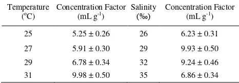
The present trend of global warming has led to an increase in seawater temperature and salinity. The effects of increasing salinity and temperature on the accumulation of 137Cs by milkfish Chanos chanos was studied under laboratory conditions to obtain information on Chanos chanos adaptability under environmental changes. The uptake of radioactive cesium by Chanos chanos increased with temperature of seawater. The concentration factors (CF) of 137Cs for temperatures of 25°C, 27°C, 29°C, and 31°C at steady state period were 5.25, 5.91, 6.78, and 9.98 mL g-1 for the whole-body of Chanos chanos. The concentration factors at steady state (CFss) of 137Cs for salinities of 26‰, 29‰, 32‰, and 35‰ were 6.23, 9.93, 9.24, and 6.86 mL g-1, respectively. After temperature exposure to 31°C, the fish gills showed hyperplasia of epithelial cells in branchial secondary lamellae, congestion of blood vessels, and hypertrophy of pillar cells. The fish from the treatment group exhibited hemorrhage between the branchial secondary lamellae and an abundance of mucous substance in comparison with control group. This study links radionuclide bioaccumulation data and monitoring data obtained in the field and laboratory experiment with radiation dose determined by ERICA Tools, an approach that will enable better linkages to be made between exposure and dose in Chanos chanos
and its marine food web.
© 2016 Atom Indonesia. All rights reserved
INTRODUCTION
Development of nuclear facilities, including nuclear power station in Indonesia, creates a possibility of a release 137Cs within allowable level into aquatic environment [1]. The radionuclide 137Cs is a fission product which may be present in the low-level aqueous radioactive waste discharged from nuclear facilities. Cesium is of considerable environmental concern because of its high mobility in the marine environment and its ability to accumulate in marine organisms. Understanding the
Corresponding author.
E-mail address: [email protected] DOI: http://dx.doi.org/10.17146/aij.2016.539
biological fate and transport of cesium in aquatic environment is essential for a sound risk assessment of marine ecosystem.
Marine ecosystems are very important in the regulation of the climate, and they are very sensitive to climate change [2]. The predicted impacts of climate change on marine ecosystems can directly and indirectly affect species abundance and distributions, habitat quality, community
composition, and changes in population dynamics [2].
The international organization of climate change, the Intergovernmental Panel on Climate Change (IPCC) in its most recent assessment reported that on a global scale, over the period of
Atom Indonesia
1971 to 2010, the upper 75 m of the ocean warmed by 0.11°C per decade. The gradual rise in ocean temperature initiates episodes of water evaporation.
These two indicators of global warming affect two physical marine parameters, temperature and salinity.
Studies concerning the likely impact of this warming on marine habitats have been limited.
Previous study investigated the effects of
temperature [3-5] and salinity [6-8] on aquatic organisms. Moreover, climate change was observed to influence fish reproductive and growth hormones and alter gill structure and genes [9-11]. On the other hand, there have been few studies that investigated how temperature or salinity could affect accumulation of contaminants, especially radioactive contaminants, by marine organisms.
Labarthe et al. [12] showed that the increase of temperature and pCO2 as an effect of
global warming impacts the bioaccumulation of radionuclides and trace elements in the eggs of Sepia officinalis. The study did not comprehensively discuss the effects of temperature on juvenile stage.
The milkfish Chanos chanos is one of the earliest fish species to be domesticated for food. Studies on the accumulation of radioactive substances by juvenile Chanos chanos have been increased. Unfortunately, this study did not consider the effects of environmental factors such as salinity and temperature changes as impacts of global warming. Additionally, a particular investigation
that is missing from the few previous studies of radionuclide bioaccumulation in fish is the
connection between exposure, capability of
accumulation, and delivered dose. The use of the ERICA Tool as a new technique in dosimetry modeling and dose estimations can now be applied to extending the results of bioaccumulation investigation to estimating the radiation dose received by fish and other biotas exposed to radionuclides [13,14]. However, the application of the ERICA Tool to estimate the time variation of dose rates for fish exposed to radionuclides through multiple routes of exposure has not been conducted.
Further research on target organ as an early
damage indicator in fish is also still needed. The gills, which participate in many important
functions in fish, such as osmoregulation, respiration, and excretion, are particularly sensitive
to changes in water quality as they remain in close contact with the external environment [15].
Changes in temperature, salinity, concentration of
heavy metals, and acidity, and other changes in the composition of the environment, as well as
other physical and chemical alterations of the aquatic medium, influence the fish gill as an sensitive organ. Heavy metal and acid
pollution has been reported to induce desquamation to primary lamellar epithelium and epithelium hyperplasia and alter cell structure [15,16]. Moreover, changes in temperature and salinity have also been reported to alter the gill structure in tropical reef fishes [4].
Our objective was to investigate the uptake of
137
Cs in juvenile Chanos chanos exposed via seawater. Data on 137Cs accumulation and its concentration in seawater and sediment is useful for risk assessment in Jakarta Bay. In addition to modeling, we used ERICA Tool to estimate dose rates and accumulated doses received by marine
organisms, including Chanos chanos, to link
radionuclide exposure with dose. Moreover, we also investigated how temperature can affect the structure of gills as sensitive organs dealing with Jakarta Bay, Indonesia, in March 2015. Afterwards, they were shipped to the Marine Radioecology Laboratory, Center for Technology of Radiation
Safety and Metrology in South Jakarta, where they were acclimatized to laboratory conditions. The Chanos chanos were gradually acclimatized to
experimental conditions, including temperature (25°C, 27°C, 29°C, and 31°C) and salinity (26‰, 29‰, 32‰, and 35‰) for one month prior to the
experiment. During this period, the Chanos chanos were fed daily with LJA-3 commercial pellets.
Radiotracers and counting
The uptake kinetics of 137Cs was determined by using high specific activity radiotracers
purchased from Polatom, Poland (137Cs as CsCl, t½ = 30 years). The radioactivity of the tracers was
activities and appropriate geometries were used to determine the radioactivity by comparison.
137
Cs uptake experiments were conducted using six individual animals held in 10 l for each temperature and salinity. Animals of similar size were used to minimize the possibility of size-related differences in radioisotope uptake. The nominal
activity of the 137Cs radiotracer was 2 kBq/l. The seawater was renewed daily during the first
week, then it was changed every second day in order to keep the radioactivities in the seawater at a constant level. The activity of the radiotracers in the seawater was checked daily to calculate the time-integrated activities of the tracer [17]. The Chanos chanos were counted for 15 minutes.
Kinetics
The uptake of the radiotracers from water was expressed as a change in the concentration factor (CF). The CF of 137Cs is defined as the ratio of the activity per unit mass in the fish (Bq g-1) to the activity per unit volume in the water (Bq mL-1), which is calculated as the mean before and after the exposure for each time point. The uptake rate was calculated as the slope of the linear regression line between the CF and the time of exposure multiplied by the concentrations of dissolved Cs. The uptake kinetics was described by using a single-component first-order kinetic model [17]:
Almost similar with 137Cs uptake experiment, the examined fish were divided into two groups (two samples for each group). The control group was left under normal conditions to study the normal structure of gills, while the second group, or the treatment group, was exposed to high concentrations, cleared in xylene, infiltrated with paraffin at 56°C, then embedded in paraffin wax [18]. Thin sections of the selected gill tissues of
about 4 μm thickness were cut by means of a
rotatory microtome, dehydrated, and stained with haematoxylin and eosin (H&E), and mounted with di-n-butyl phthalate in xylene (DPX). The sections were examined and photomicrographed using an Olympus microscope fitted with photographic attachment. The prepared slides were used to describe the normal histological structure and histological alterations in the gills.
RESULTS AND DISCUSSION
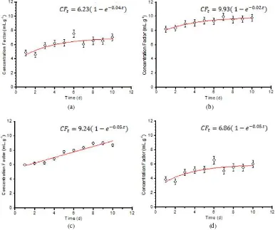
The uptakes of 137Cs under different
temperatures displayed exponential kinetics
reaching a steady state (Fig. 1 and Table 1). The CF of 137Cs for temperatures of 25°C, 27°C, 29°C, and 31°C at steady-state conditions were 5.25, 5.91, 6.78, and 9.98 mL g-1 in whole-body fish, respectively. For different salinities, the uptakes of
137 temperature-enhanced initial uptake rates of 137Cs from water. Increases in accumulation rates at higher temperatures may be partially due to
amplified metabolic activity. Animals were
observed to be much more active at 31°C than at 25°C and the increased internal circulation of water
through the digestive systems and respiratory may result in enhanced in the amounts of all elements bound to internal body surfaces. In addition, production of metal-binding proteins or external mucus may be greater at elevated temperatures.
Our study indicated that the Cs uptake was considerably affected by ambient salinity, consistent with many previous observations on the effects of salinity on bioaccumulation [19]. The concentration factor of cesium increased with salinity at the
salinity range of 26 to 29‰ and started decreasing
with salinity from 32 to 35‰. This study observed
that the optimal salinity for bioaccumulation in
Chanos chanos was 29‰. Various mechanism have
(a) (b)
(c) (d)
Fig. 1. Uptake kinetics of 137Cs by Chanos chanos in different seawater temperatures (a. 25°C, b. 27°C, c. 29°C, d. 31°C).
(a) (b)
(c) (d)
including the physiological conditions of the Chanos chanos such as respiration rate, or change in cell volume and permeability of tissue. Wang (2000) demonstrated that the variability of cesium uptake at different salinities was primarily due to a change in ambient K+ concentration. Further studies are needed to determine the influence of K+ related to salinity effect on Chanos chanos bioaccumulation.
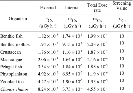
The main project of ERICA Integrated Approach is the quantification of risk or risk assement to the environment for a given release of radioactivity. To attain this aim, the data on environmental dosimetry and transfer characteristics of radioisotopes in the environment are combined to provide an estimate of exposure. Furthermore, the ERICA Integrated Approach recommends the use of expected values (or best estimates) of activity concentrations. The assessment chronology in this study uses normal 137Cs activity concentration in seawater and sediment [20]. On other hand, the distribution coefficient (Kd), CF, radiation weighting
factor, and uncertainty factor in each organism were taken from secondary data which is prepared by ERICA. The screening dose was set at 10 µGy.h-1.
The concentration factor for cesium is a critical value in a risk assessment study of the environmental effect of cesium in coastal and offshore environments. Additional concentration factor data is needed to better describe the distribution of cesium concentration factors in the edible portions of animals used for food. Generic concentration factors are appropriate for use only in a screening-level assessment.
organisms in the food web because all calculated doses listed in Table 2 are lower than the 10 µGy/h screening level at Tier 2. Given the results shown in Table 2, if
137
Cs release actually occurs with activities close to the environmental monitoring data used here, the ERICA Tool would suggest that further investigations are not warranted. A range of decisions will then need to be taken, based on the suggestions given in the generic ICRP exposure situations [13].
Environmental conditions can result in
compensatory mechanisms that deal with
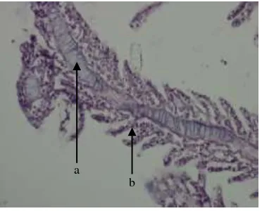
environmental stressors, as in cellular hyperplasia, and in direct toxic effects of the pollutants leading to necrosis and degeneration [15]. In the present case, it seems that the results are in the form of compensatory mechanisms. All histological lesions observed in the
gills of Chanos chanos in this study, are categorized
under cellular atrophy, lamellar cell hyperplasia and
hypertrophy, hemorrhage and bloody congestion,
lamellar damage, and lamellar fusion; these results could be linked with increase in the activities of the test organisms exposed to the changing habitat temperature.
Table 2. ERICA Tool tier-2 assesment for total dose rate
Organism
External Internal Total Dose rate branchial secondary lamellae, congestion of blood vessels, hypertrophy of pillar cells (Fig. 4), abundance of mucous substance, and hemorrhage between the branchial secondary lamellae (Fig. 5), in comparison with control group (Fig. 3).
Under high temperatures, cellular
Fig. 3. Gill section from control Chanos chanos showing epithelial cells (a), secondary lamellae (b), and primary lamellae (c). H&E. ×40.
Fig. 4. Gill section of Chanos chanos exposed to 31°C temperature showing hyperplasia of the epithelial cells (a), hypertrophy of the pillar cells (b), and blood congestion (d). H&E. ×40.
Fig. 5. Gill section of Chanos chanos exposed to 31°C temperature showing structure change in primary lamellae (a) and epithelium rupture with hemorrhage (b). H&E. ×40.
Comprehensive studies that relate the impacts of climate change is greatly needed in the future, as climate changes as mentioned above potentially result in the decrease of ocean pH due to elevated CO2 concentrations; this could also induce
alterations in morphology of gill to keep ion/osmotic and gas exchange balance. This possibility should be considered in future studies.
CONCLUSION
Environmental factors, including salinity and temperature, affect the metabolism of organisms and their growth and consequently their uptake of cesium. Generally, small increases in salinity and temperature tend to increase the cesium uptake and biological activity.The CFss of
137
Cs for temperature 25°C, 27°C, 29°C, 31°C were 5.25, 5.91, 6.78, and 9.98 mL g-1 in the whole body of Chanos chanos. The CFss of
137
Cs for salinities of 26‰, 29‰, 32‰, and 35‰ were 6.23, 9.93, 9.24, and 6.86 mL g-1, respectively. Optimum temperature and salinity for bioaccumulation process of 137Cs by Chanos chanos were 31°C and 29‰. After exposure to 31°C temperature, the fish gills showed hyperplasia of epithelial cells in branchial secondary lamellae, congestion of blood vessels, and hypertrophy of pillar cells. The exposed fish exhibited hemorrhage between the branchial secondary lamellae and an abundance of mucous substance in comparison with control group. From ERICA assessment, it was found that the total dose rates for Chanos chanos other organisms in the marine food web and were lower than the screening dose rate (10 mGy/h), which means that there will be no impact on the
Chanos chanos and other organisms in food web
because the 137Cs concentration was within the allowable level.
ACKNOWLEDGMENT
Sponsorship and financial support were given by the Scholarship Programme, Ministry of Research Technology and Higher Education of Indonesia (KEMENRISTEKDIKTI). The authors are thankful for technical assistance from the Marine Radioecology Group, Center for Radiation Safety Technology and Metrology, BATAN, particularly for the marine ecosystem laboratory support.
REFERENCES
1. W.R. Prihatiningsih and H. Suseno, Mar. Pollut. Bul. In Press, Corrected Proof (2016).
2. O.H. Gudlberg and J.F. Bruno, Science 328
(2010) 1523.
3. R.S. Lugo, C. Mata, A. Oliveros et al., Environ. Toxicol. Pharmacol. 31 (2011) 490.
4. A.J. Bowden, N.M. Gardiner, C.S. Couturier
et al., Comp. Biochem. Physiol. 175
(2014) 64. a b c
a b
c
a
5. S.M. Ahmad, F.A. Shah, F.A. Bhat et al., Therm. Biol. 36 (2011) 492.
6. H. Koyama, S. Okamoto, N. Watanabe et al.,
Comp. Biochem. Physiol. 181 (2015) 59. 7. J.L. Ikerd, K.G. Burnettand and L.E. Burnett,
Comp. Biochem, Physiol. 183 (2015) 97.
8. I.S.A. Anni, A. Bianchini, I. Fernanda et al., Aquaculture 455 (2016) 63.
9. F.P. Arantes, H.B. Santos, E. Rizzo et al., Gen. Comp. Endocrinol. 172 (2011) 400.
10. V.D.J. Keller, P. Llyod, J.A. Terry et al., Environ. Pollut. 197 (2015) 262.
11. G. Levy, D. David, G. Degani et al., Comp. Biochem. Physiol. 160 (2011) 381.
12. T.L. Labarthe, S. Martin, F. Oberhansli et al., J. Exp. Mar. Biol. Ecol. 413 (2012) 45.
13. J.E. Brown, B. Alfonso, R. Avila et al., J. Environ. Radioact. 153 (2016) 141.
14. H.C. Reinardy, J.L. Teyssie, R.A. Jeffree
et al., Sci. Total Environ. 409 (2011) 3771.
15. A.R. Fonseca, L.F.S. Fernandes, A.F. Fernandes et al., Sci. Total Environ. 530 (2016) 972.
16. M. Minghetti, S. Schnell and M.A. Chadwick
et al., Aquat. Toxicol. 154 (2014) 184.
17. M. Metian, M. Warnau, F. Oberhansli et al., J. Exp. Mar. Biol. Ecol. 353 (2007) 58.
18. M. Lai, B. Lu, omprehensive Sampling and
Sample Preparation; Tissue Preparation for Microscopy and Histology, Reference
Module in Chemistry, Molecular Science and Chemical Engineering (2012) 53.
19. Q.C. Wang, W.G. Shen and Z.W. Ma,
Environ. Sci. Technol. 34 (2000) 2711.
20. H. Suseno and W.R. Prihatiningsih, Mar. Pollut. Bull. 88 (2014) 319.
21. N.N. Ortiz, M. Gerdol, V. Stocchi et al., Dev. Comp. Immunol. 47 (2014) 309.
22. W.B. Ameur, Y.E. Megdiche, J.D. Lapuente