Introduction to Pharmaceutical
Microbiology
Chapter · November 2007 DOI: 10.1002/9780470988329.ch1
CITATIONS
9
READS
5,103
3 authors, including:
Stephen P Denyer
University of Brighton
164PUBLICATIONS 3,312CITATIONS
SEE PROFILE
Sean P Gorman
Queen's University Belfast 238PUBLICATIONS 4,604CITATIONS
Hugo and Russell’s
Pharmaceutical
Microbiology
EDITED BY
Stephen P Denyer
B Pharm PhD FRPharmSWelsh School of Pharmacy Cardiff University Cardiff
Norman A Hodges
B Pharm PhD MRPharmSSchool of Pharmacy and Biomolecular Sciences Brighton University
Lewes Road Brighton
Sean P Gorman
BSc PhD MPSSchool of Pharmacy Queen’s University Belfast Medical Biology Centre University Road Belfast
SEVENTH EDITION
Blackwell
Hugo and Russell’s
Hugo and Russell’s
Pharmaceutical
Microbiology
EDITED BY
Stephen P Denyer
B Pharm PhD FRPharmSWelsh School of Pharmacy Cardiff University Cardiff
Norman A Hodges
B Pharm PhD MRPharmSSchool of Pharmacy and Biomolecular Sciences Brighton University
Lewes Road Brighton
Sean P Gorman
BSc PhD MPSSchool of Pharmacy Queen’s University Belfast Medical Biology Centre University Road Belfast
SEVENTH EDITION
Blackwell
© 1977, 1980, 1983, 1987, 1992, 1998, 2004 by Blackwell Science Ltd a Blackwell Publishing company
Blackwell Science, Inc., 350 Main Street, Malden, Massachusetts 02148-5020, USA Blackwell Publishing Ltd, 9600 Garsington Road, Oxford OX4 2DQ, UK
Blackwell Science Asia Pty Ltd, 550 Swanston Street, Carlton, Victoria 3053, Australia The right of the Author to be identified as the Author of this Work has been asserted in accordance with the Copyright, Designs and Patents Act 1988.
All rights reserved. No part of this publication may be reproduced, stored in a retrieval system, or transmitted, in any form or by any means, electronic, mechanical,
photocopying, recording or otherwise, except as permitted by the UK Copyright, Designs and Patents Act 1988, without the prior permission of the publisher.
First published 1977 Second edition 1980 Third edition 1983 Reprinted 1986 Fourth edition 1987 Reprinted 1989, 1991
Italian edition 1991 Fifth edition 1992 Reprinted 1993, 1994, 1995 Sixth edition 1998
Reprinted 1999, 2000, 2002, 2003 Seventh edition 2004
Library of Congress Cataloging-in-Publication Data
Hugo and Russell’s pharmaceutical microbiology / edited by Stephen Denyer, Norman A. Hodges, Sean P. Gorman. — 7th ed.
p. cm.
Rev. ed. of: Pharmaceutical microbiology / edited by W.B. Hugo and A.D. Russell. Includes bibliographical references and index.
ISBN 0-632-06467-6 1. Pharmaceutical microbiology.
[DNLM: 1. Anti-Infective Agents. 2. Technology, Pharmaceutical. QV 250 H895 2004] I. Title: Pharmaceutical microbiology. II. Hugo, W. B. (William Barry) III. Denyer, S. P. IV. Hodges, Norman A.V,. Gorman, S. P. VI. Pharmaceutical microbiology.
QR46.5.P48 2004 615¢.1¢01579 — dc22
2003024264
ISBN 0–632–06467–6
A catalogue record for this title is available from the British Library
Set in Sabon 9.5/12 pt by SNP Best-set Typesetter Ltd., Hong Kong Printed and bound in the United Kingdom by Ashford Colour Press, Gosport
Commissioning Editor: Maria Khan Managing Editor: Rupal Malde Production Editor: Fiona Pattison Production Controller: Kate Charman
Contributors, vii
Preface to Seventh Edition, ix
Preface to First Edition, x
Part 1: Biology of Microorganisms
1. Introduction to Pharmaceutical Microbiology, 3
Stephen Denyer, Norman Hodges and Sean Gorman
2. Fundamental Features of Microbiology, 9
Norman Hodges 3. Bacteria, 23
David Allison and Peter Gilbert 4. Fungi, 44
Kevin Kavanagh and Derek Sullivan 5. Viruses, 59
Jean-Yves Maillard and David Stickler 6. Protozoa, 82
Tim Paget
7. Principles of Microbial Pathogenicity and Epidemiology, 103
Peter Gilbert and David Allison
Part 2: Antimicrobial Agents
8. Basic Aspects of the Structure and Functioning of the Immune System, 117
Mark Gumbleton and James Furr 9. Vaccination and Immunization, 138
Peter Gilbert and David Allison 10. Types of Antibiotics and Synthetic
Antimicrobial Agents, 152 A Denver Russell
11. Laboratory Evaluation of Antimicrobial Agents, 187
JMB Smith
12. Mechanisms of Action of Antibiotics and Synthetic Anti-infective Agents, 202 Peter Lambert
13. Bacterial Resistance to Antibiotics, 220 Anthony Smith
14. Clinical Uses of Antimicrobial Drugs, 233 Roger Finch
Part 3: Microbiological Aspects of Pharmaceutical Processing
15. Ecology of Microorganisms as it Affects the Pharmaceutical Industry, 251
Elaine Underwood
16. Microbial Spoilage, Infection Risk and Contamination Control, 263
Rosamund Baird
17. Chemical Disinfectants, Antiseptics and Preservatives, 285
Sean Gorman and Eileen Scott
18. Non-Antibiotic Antibacterial Agents: Mode of Action and Resistance, 306
Stephen Denyer and A Denver Russell 19. Sterile Pharmaceutical Products, 323
James Ford
20. Sterilization Procedures and Sterility Assurance, 346
Stephen Denyer and Norman Hodges 21. Factory and Hospital Hygiene, 376
Robert Jones
22. Manufacture of Antibiotics, 387 Sally Varian
23. The Manufacture and Quality Control of Immunological Products, 398
Michael Corbel
24. Pharmaceutical Biotechnology, 416 Miguel Cámara
25. Additional Applications of Microorganisms in the Pharmaceutical Sciences, 441
Denver Russell
Contributors
Dr David Allison
School of Pharmacy and Pharmaceutical Sciences
University of Manchester Oxford Road
Manchester M13 9PL UK
Dr Rosamund Baird
Visiting Senior Lecturer
School of Pharmacy and Pharmacology University of Bath
Claverton Down Bath BA2 7AY UK
Dr Miguel Cámara
Senior Lecturer in Molecular Microbiology Institute of Pharmaceutical Sciences School of Pharmaceutical Sciences University of Nottingham Nottingham NG7 2RD UK
Dr Michael Corbel
National Institute for Biological Standards and Control
Blanche Lane South Mimms Potters Bar
Hertfordshire EN6 3QG UK
Professor Stephen Denyer
Welsh School of Pharmacy Cardiff University Cardiff CF10 3XF UK
Professor Roger Finch
Professor of Infectious Diseases Clinical Sciences Building University of Nottingham The City Hospital Nottingham NG5 1PB UK
Dr Robert Jones
School of Pharmacy and Biomedical Sciences University of Portsmouth
St Michael’s Building White Swan Road Portsmouth PO1 2DT UK
Dr Kevin Kavanagh
Head of Medical Mycology Unit Department of Biology National University of Ireland Maynooth
Co. Kildare Ireland
Dr Peter Lambert
Aston Pharmacy School Aston University Aston Triangle Birmingham B4 7ET UK
Dr Jean-Yves Maillard
School of Pharmacy and Biomolecular Sciences
University of Brighton Lewes Rd
Brighton BN2 4GJ UK
Dr Tim Paget
Department of Biological Sciences University of Hull
Hull HU6 7RX UK
Professor A Denver Russell
Welsh School of Pharmacy Cardiff University King Edward VII Avenue Cardiff CF10 3XF Wales
Dr Eileen Scott
School of Pharmacy
The Queen’s University of Belfast Belfast BT9 7BL
Northern Ireland
Professor James Ford
School of Pharmacy and Chemistry Liverpool John Moores University Byrom Street
Liverpool L3 3AF UK
Dr James Furr
Welsh School of Pharmacy Cardiff University King Edward VII Avenue Cardiff CF10 3XF Wales
Professor Peter Gilbert
School of Pharmacy and Pharmaceutical Sciences
University of Manchester Oxford Rd
Manchester M13 9PL UK
Professor Sean Gorman
Professor of Pharmaceutical Microbiology School of Pharmacy
The Queen’s University of Belfast Belfast BT9 7BL
Northern Ireland
Dr Mark Gumbleton
Welsh School of Pharmacy Cardiff University King Edward VII Avenue Cardiff CF10 3XF Wales
Dr Norman Hodges
Principal Lecturer in Pharmaceutical Microbiology
School of Pharmacy and Biomolecular Sciences
Dr Anthony Smith
Department of Pharmacy and Pharmacology University of Bath (5 West — 2.18) Claverton Down
Bath BA2 7AY UK
Professor JMB (Sandy) Smith
Head of Department of Microbiology Otago School of Medical Sciences University of Otago
Dunedin New Zealand
Dr Elaine Underwood
SMA Nutrition Huntercomb Lane South Taplow
Maidenhead Berks SL6 0PH UK
Dr Sally Varian
Consultant Ulverston Cumbria LA12 8PT UK
Dr David Stickler
School of Biosciences Cardiff University Main Building Museum Avenue PO Box 915 Cardiff CF10 3TL Wales
Dr Derek Sullivan
Preface to the Seventh edition
We were much honoured to be recommended by Professor A.D. Russell to act as editors for the 7th edition of Pharmaceutical Microbiology. All three of us have used this textbook in its various editions throughout our careers as teachers and researchers, and we recognize the important role it fulfils.
As might be anticipated when a new editorial team is in place, a substantial number of changes have been made. Well over half the chapters have new authors or co-authors. We also use Chapter 1 to give a rationale for the scope and content of the book, emphasizing the interrelated character of the discipline of pharmaceutical microbiology. In addi-tion, by combining and reorganizing chapters, by introducing new material and through a revised page format we have tried to provide readers with a distinctive 7th edition.
We must thank our contributors for their willing collaboration in this enterprise, especially Professor Russell for his continuing contri-butions, and our publishers for their support and expertise.
Finally, this addition is a tribute to the farsighted-ness of A.D. Russell and W.B. Hugo who took up the challenge in 1977 to produce a popular and con-cise read for pharmacy students required to study pharmaceutical microbiology. We are delighted that this current edition recognizes these origins by continuing the association with Hugo and Russell in its revised title.
When we were first approached by the publishers to write a textbook on pharmaceutical microbiology to appear in the spring of 1977, it was felt that such a task could not be accomplished satisfactorily in the time available.
However, by a process of combined editorship and by invitation to experts to contribute to the various chapters this task has been accomplished thanks to the cooperation of our collaborators.
Pharmaceutical microbiology may be defined as that part of microbiology which has a special bear-ing on pharmacy in all its aspects. This will range from the manufacture and quality control of phar-maceutical products to an understanding of the mode of action of antibiotics. The full extent of microbiology on the pharmaceutical area may be judged from the chapter contents.
As this book is aimed at undergraduate pharmacy students (as well as microbiologists en-tering the pharmaceutical industry) we were under constraint to limit the length of the book to retain it in a defined price range. The result is to be found in
the following pages. The editors must bear respon-sibility for any omissions, a point which has most concerned us. Length and depth of treatment were determined by the dictate of our publishers. It is hoped that the book will provide a concise reading for pharmacy students (who, at the moment, lack a textbook in this subject) and help to highlight those parts of a general microbiological training which impinge on the pharmaceutical industry.
In conclusion, the editors thank most sincerely the contributors to this book, both for complying with our strictures as to the length of their contribu-tion and for providing their material on time, and our publishers for their friendly courtesy and effi-ciency during the production of this book. We also wish to thank Dr H.J. Smith for his advice on vari-ous chemical aspects, Dr M.I. Barnett for useful comments on reverse osmosis, and Mr A. Keall who helped with the table on sterilization methods.
W.B. Hugo A.D. Russell
Part 1
1 Microorganisms and medicines
Despite continuing poverty in many parts of the world and the devastating effects of HIV and AIDS infection on the African continent and elsewhere, the health of the world’s population is progressively improving. This is reflected in the increase in life expectancy that has been recorded for the great majority of the countries reporting statistics to the World Health Organization over the last 40 years. In Central America, for example, the life expectancy has increased from 55 years in 1960 to 71 years in 2000, and the increase in North (but not sub-Saharan) Africa is even greater, from 47 to 68 years. Much of this improvement is due to better nutrition and sanitation, but improved health care and the greater availability of effective medicines with which to treat common diseases are also major contributing factors. Substantial inroads have been made in the prevention and treatment of cancer, cardiovascular disease and other major causes of death in Western society, and of infections and diar-rhoeal disease that remain the big killers in develop-ing countries. Several infectious diseases have been eradicated completely, and others from substantial parts of the world. The global eradication of small-pox in 1977 is well documented, but 2002 saw three of the world’s continents declared free of polio, and the prospects are good for the total elimination of polio, measles and Chagas disease.
The development of the many vaccines and other medicines that have been so crucial to the improve-ment in world heath has been the result of the large investment in research by the major international pharmaceutical companies. This has led to the manufacture of pharmaceuticals becoming one of
the most consistently successful and important in-dustries in many countries, not only in the tradi-tional strongholds of North America, Western Europe and Japan but, increasingly, in Eastern Eu-rope, the Indian subcontinent and the Far East. Worldwide sales of medicines and medical devices are estimated to have exceeded $US 401 billion (ap-proximately £250 billion) in 2002, and this figure is rising by 8% per annum. In the UK alone, the value of pharmaceutical exports is currently £10.03 bil-lion each year, a figure that translates to more than £150 000 for each employee in the industry.
The growth of the pharmaceutical industry in re-cent decades has been paralleled by rising standards for product quality and more rigorous regulation of manufacturing procedures. In order to receive a manufacturing licence, a modern medicine must be shown to be effective, safe and of good quality. Most medicines consist of an active ingredient that is formulated with a variety of other materials (ex-cipients) that are necessary to ensure that the medi-cine is effective, and remains stable, palatable and safe during storage and use. While the efficacy and safety aspects of the active ingredient are within the domain of the pharmacologist and toxicologist, respectively, many other disciplines contribute to the efficacy, safety and quality of the manufactured product as a whole. Analytical chemists and phar-macists take lead responsibility for ensuring that the components of the medicine are present in the correct physical form and concentration, but quality is not judged solely on the physicochemical properties of the product: microorganisms also have the potential to influence efficacy and safety.
It is obvious that medicines contaminated with potentially pathogenic (disease-causing)
micro-Chapter 1
Introduction to pharmaceutical microbiology
Stephen Denyer, Norman Hodges and Sean Gorman
organisms are a safety hazard, so medicines administered by vulnerable routes (e.g. injections) or to vulnerable areas of the body (e.g. the eyes) are manufactured as sterile products. What is less predictable is that microorganisms can, in addition to initiating infections, cause product spoilage by chemically decomposing the active ingredient or the excipients. This may lead to the product being under-strength, physically or chemically unstable or possibly contaminated with toxic materials. Thus, it is clear that pharmaceutical microbiology must encompass the subjects of sterilization and preservation against microbial spoilage, and a pharmacist with responsibility for the safe, hygienic manufacture and use of medicines must know where microorganisms arise in the environment, i.e. the sources of microbial contamination, and the factors that predispose to, or prevent, product spoilage. In these respects, the pharmaceutical microbiologist has a lot in common with food and cosmetics microbiologists, and there is substantial scope for transfer of knowledge between these disciplines.
Disinfection and the properties of chemicals (bio-cides) used as antiseptics, disinfectants and preserv-atives are subjects of which pharmacists and other persons responsible for the manufacture of medi-cines should have a knowledge, both from the per-spective of biocide use in product formulation and manufacture, and because antiseptics and disinfec-tants are pharmaceutical products in their own right. However, they are not the only antimicrobial substances that are relevant to medicine; antibiotics are of major importance and represent a product category that regularly features among the top five most frequently prescribed. The term ‘antibiotic’ is used in several different ways: originally an anti-biotic was defined as a naturally occurring substance that was produced by one microorganism that inhibited the growth of, or killed, other micro-organisms, i.e. an antibiotic was a natural product, a microbial metabolite. More recently the term has come to encompass certain synthetic agents that are usually used systemically (throughout the body) to treat infection. A knowledge of the manufacture, quality control and, in the light of current concerns about resistance of microorganisms, the use of antibiotics, are other areas of knowledge that
contribute to the discipline of pharmaceutical microbiology.
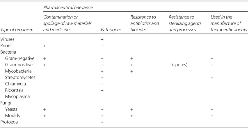
Commercial antibiotic production began with the manufacture of penicillin in the 1940s, and for many years antibiotics were the only significant example of a medicinal product that was made using microorganisms. Following the adoption in the 1950s of microorganisms to facilitate the manu-facture of steroids and the development of recombi-nant DNA technology in the last three decades of the 20th century, the use of microorganisms in the manufacture of medicines has gathered great momentum. It led to more than 100 biotechnology-derived products on the market by the new millennium and another 300 or more in clinical trials. While it is true to say that traditionally the principal pharmaceutical interest in microorgan-isms is that of controlling them, exploiting micro-bial metabolism in the manufacture of medicines is a burgeoning area of knowledge that will become increasingly important, not only in the pharmacy curriculum but also in those of other disciplines em-ployed in the pharmaceutical industry. Table 1.1 summarizes these benefits and uses of microorgan-isms in pharmaceutical manufacturing, together with the more widely recognized hazards and problems that they present.
Looking ahead to the early decades of the 21st century, it is clear that an understanding of the physiology and genetics of microorganisms will also become more important, not just in the pro-duction of new therapeutic agents but in the under-standing of infections and other diseases. Several of the traditional diseases that were major causes of death before the antibiotic era, e.g. tuberculosis and diphtheria, are now re-emerging in resistant form — even in developed countries — adding to the problems posed by infections in which antibiotic re-sistance has long been a problem, and those like Creutzfeldt–Jakob disease, West Nile virus and severe acute respiratory syndrome (SARS) that have only been recognized in recent years.
Introduction to pharmaceutical microbiology
5
The manufacture of: Good manufacturing practice May contaminate non- Non- sterile medicines:
antibiotics Industrial ‘fermentation’ sterile and sterile medicines Enumeration of microorganisms in the steroids technology with a risk of infection manufacturing environment (environmental therapeutic enzymes Microbial genetics monitoring) and in raw materials and
polysaccharides manufactured products
products of recombinant Identification and detection of specific organisms
DNA technology Sterile medicines:
Use in the production of vaccines Quality control of Sterilization methods
immunological products Sterilization monitoring and validation procedures As assay organisms to determine Assay methods Sterility testing
antibiotic, vitamin and amino Assessment and calculation of sterility assurance
acid concentrations Aseptic manufacture
To detect mutagenic or Ames mutagenicity test carcinogenic activity
May contaminate non- Enumeration, identification and detection as above, plus sterile and sterile medicines with Characteristics, selection and testing of
a risk of product deterioration antimicrobial preservatives
Cause infectious and other diseases Immunology and infectious diseases
Characteristics, selection and use of vaccines and antibiotics Use of biocides in infection and contamination control Control of antibiotic resistance
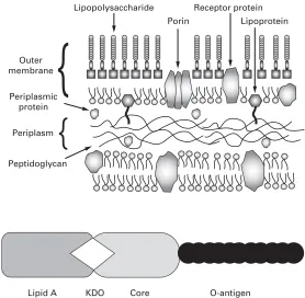
Cause pyrogenic reactions (fever) Bacterial structure
when introduced into the body Pyrogen and endotoxin testing even in the absence of infection
Provide a reservoir of antibiotic Microbial genetics resistance genes
Collection
by
Sagar
the potential to reproduce extremely rapidly; it is quite possible for cell division to occur every 20 minutes under favourable conditions. These characteristics mean that they can adapt readily to a changing environment and colonize new niches. One feature of modern surgery is the ever-increasing use of plastic, ceramic and metal devices that are introduced into the body for a wide variety of purposes, including the commonly encountered urinary or venous catheters and the less common intra-ocular lenses, heart valves, pacemakers and hip prostheses. Many bacteria have the potential to produce substances or structures that help them to attach to these devices, even while combating the immune system of the body. Thus, colonization often necessitates removal and replacement of the device in question — often leading to great discom-fort for the patient and substantial monetary cost to the health-care service. It has recently been estimat-ed that, on average, a hospital-acquirestimat-ed infection results in an extra 14 days in hospital, a 10% in-crease in the chance of dying and more than £3000 additional expenditure on health care. The devel-opment of strategies for eliminating, or at least restricting, the severity or consequences of these device-related infections is a challenge for pharma-cists and microbiologists within the industry, and for many other health-care professionals.
In addition to an improved understanding of the mechanisms of antibiotic resistance, of the links between antibiotic resistance and misuse, and of the factors influencing the initiation of infections in the body, our insights into the role of microorganisms in other disease states have broadened significantly in recent years. Until about 1980 it was probably true to say that there was little or no recognition of the possibility that microorganisms might have a role to play in human diseases other than clear-cut infections. In recent years, however, our perception of the scope of microorganisms as agents of disease has been changed by the discovery that Helicobac-ter pylori is intimately involved in the develop-ment of gastric or duodenal ulcers and stomach cancer; by the findings that viruses can cause cancers of the liver, blood and cervix; and by the suspected involvement of microorganisms in di-verse conditions like parkinsonism and Alzheimer’s disease.
Clearly, a knowledge of the mechanisms whereby microorganisms are able to resist antibiotics, colonize medical devices and cause or predispose humans to other disease states is essential in the development not only of new antibiotics, but of other medicines and health-care practices that miminize the risks of these adverse situations developing.
2 The scope and content of the book
Criteria and standards for the microbiological quality of medicines depend upon the route of administration of the medicine in question. The vast majority of medicines that are given by mouth or placed on the skin are non-sterile, i.e. they may contain some microorganisms (within limits on type and concentration), whereas all injections and ophthalmic products must be sterile, i.e. they contain no living organisms. Products for other anatomical sites (e.g. nose, ear, vagina and bladder) are often sterile but not invariably so (Chapter 19). The microbiological quality of non-sterile medi-cines is controlled by specifications defining the concentration of organisms that may be present and requiring the absence of specific, potentially haz-ardous organisms. Thus the ability to identify the organisms present, to detect those that are prohibited from particular product categories and to enumerate microbial contaminants in the manu-facturing environment, raw materials and finished product are clearly skills that a pharmaceutical microbiologist should possess (Chapters 2–6). So, too, is a familiarity with the characteristics of antimicrobial preservatives that may be a compo-nent of the medicine required to minimize the risk of microbial growth and spoilage during storage and use by the patient (Chapters 16 and 17).
into the body. Strictly speaking, any substance which causes fever following injection is a pyrogen, but in reality the vast majority are of bacterial origin, and it is for this reason that the detection, assay and removal of bacterial pyrogens (endotox-ins) are considered within the realm of microbiology (Chapter 19).
Sterile medicines may be manufactured by two different strategies. The most straightforward and preferred option is to make the product, pack it in its final container and sterilize it by heat, radiation or other means (terminal sterilization, Chapter 20). The alternative is to manufacture the product from sterile ingredients under conditions that do not permit the entry of contaminating organisms (asep-tic manufacture, Chapters 15 and 21); this latter option is usually selected when the ingredients or physical form of the product render it heat- or radiation-sensitive. Those responsible for the manufacture of sterile products must be familiar with the sterilization or aseptic manufacturing pro-cedures available for different product types, and those who have cause to open, use or dispense ster-ile products (in a hospital pharmacy, for example) should be aware of the aseptic handling procedures to be adopted in order to minimize the risk of prod-uct contamination.
The spoilage of medicines as a result of microbial contamination, although obviously undesirable, has as its main consequence financial loss rather than ill health on the part of the patient. The other major problem posed by microbial contamination of medicines, that of the risk of initiating infection, although uncommon, is far more important in terms of risk to the patient and possible loss of life (Chapters 7 and 16). Infections arising by this means also have financial implications, of course, not only in additional treatment costs but in terms of product recalls, possible litigation and damage to the reputation of the manufacturer.
The range of antimicrobial drugs used to prevent and treat microbial infections is large; for example, a contemporary textbook of antimicro-bial chemotherapy lists no fewer than 43 different cephalosporin antibiotics that were already on the market or the subject of clinical trials at the time of publication. Not only are there many antibiotic products, but increasingly, these products really
have properties that make them unique. It is far more difficult now than it was, say, 20 years ago, for a manufacturer to obtain a licence for a ‘copycat’ product, as licensing authorities now emphasize the need to demonstrate that a new antibiotic (or any new medicine) affords a real advantage over estab-lished drugs. Because of this range and diversity of products, pharmacists are now far more commonly called upon to advise on the relative merits of the antibiotics available to treat particular categories of infection than was the case hitherto (Chapters 10, 12 and 14). A prerequisite to provide this informa-tion is a knowledge not only of the drug in quesinforma-tion, but the infectious disease it is being used to treat and the factors that might influence the success of antibiotic therapy in that situation (Chapter 7).
influenc-ing their selection for use in hospital infection control strategies or contamination control in the manufacturing setting are topics with which both pharmacists and industrial microbiologists should be familiar (Chapters 11 and 18).
It has long been recognized that microorganisms are valuable, if not essential, in the maintenance of our ecosystems. Their role and benefits in the carbon and nitrogen cycles in terms of recycling dead plant and animal material and in the fixation of atmospheric nitrogen are well understood. The uses of microorganisms in the food, dairy and brew-ing industries are also well established, but until the late 20th century advances in genetics, immunology and biotechnology, their benefits and uses in the pharmaceutical industry were far more modest. For many years the production of antibiotics (Chapter 22) and microbial enzyme-mediated production of steroids were the only significant pharmaceutical examples of the exploitation of metabolism of microorganisms. The value of these applications, both in monetary and health-care terms has been immense. Antibiotics currently have an estimated world market value of $25 billion and by this crite-rion they are surpassed as products of biotechnolo-gy only by cheese and alcoholic beverages, but the benefits they afford in terms of improved health and life expectancy are incalculable. The discovery of the anti-inflammatory effects of corticosteroids had a profound impact on the treatment of rheumatoid arthritis in the 1950s, but it was the use of enzymes possessed by common fungi that made cortisone widely available to rheumatism sufferers. The syn-thesis of cortisone by traditional chemical methods involved 31 steps, gave a yield of less than 0.2% of the starting material and resulted in a product cost-ing, even in 1950s terms, $200 per gram. Exploiting
microbial enzymes reduced the synthesis to 11 steps and the cost rapidly fell to $6 per gram.
Apart from these major applications, however, the uses of microorganisms in the manufacture of medicines prior to 1980 were very limited. Enzymes were developed for use in cancer chemotherapy (asparaginase) and to digest blood clots (streptoki-nase), and polysaccharides also found therapeutical applications (e.g. dextran — used as a plasma ex-pander). These were of relatively minor importance, however, compared with the products that followed the advances in recombinant DNA technology in the 1970s. This technology permitted human genes to be inserted into microorganisms, which were thus able to manufacture the gene products far more efficiently than traditional methods of extraction from animal or human tissues. Insulin, in 1982, was the first therapeutic product of DNA technology to be licensed for human use, and it has been followed by human growth hormone, interferon, blood clotting factors and many other products. DNA technology has also permitted the development of vaccines which, like that for the prevention of he-patitis B, use genetically engineered surface antigens rather than whole natural virus particles, so these vaccines are more effective and safer than those produced by traditional means (Chapters 9 and 23).
1 Introduction
Microorganisms differ enormously in terms of their shape, size and appearance and in their genetic and metabolic characteristics. All these properties are used in classifying microorganisms into the major groups with which many people are familiar, e.g. bacteria, fungi, protozoa and viruses, and into the less well known categories like chlamydia, rick-ettsia and mycoplasmas. The major groups are the subject of individual chapters immediately follow-ing this, so the purpose here is not to describe any of them in great detail but to summarize their features so that the reader may better understand the dis-tinctions between them. A further aim of this chap-ter is to avoid undue repetition of information in the early part of the book by considering such aspects of microbiology as cultivation, enumeration and genetics that are common to some, or all, of the various types of microorganism.
1.1 Viruses, viroids and prions
Viruses do not have a cellular structure. They are particles composed of nucleic acid surrounded by protein; some possess a lipid envelope and associat-ed glycoproteins, but recognizable chromosomes, cytoplasm and cell membranes are invariably
absent. Viruses are incapable of independent repli-cation as they do not contain the enzymes necessary to copy their own nucleic acids; as a consequence, all viruses are intracellular parasites and are repro-duced using the metabolic capabilities of the host cell. A great deal of variation is observed in shape (helical, linear or spherical), size (20–400 nm) and nucleic acid composition (single- or double-stranded, linear or circular RNA or DNA), but al-most all viruses are smaller than bacteria and they cannot be seen with a normal light microscope; in-stead they may be viewed using an electron micro-scope which affords much greater magnification.
Viroids (virusoids) are even simpler than viruses, being infectious particles comprising single-strand-ed RNA without any associatsingle-strand-ed protein. Those that have been described are plant pathogens, and, so far, there are no known human pathogens in this category. Prions are unique as infectious agents in that they contain no nucleic acid. A prion is an atyp-ical form of a mammalian protein that can interact with a normal protein molecule and cause it to undergo a conformational change so that it, in turn, becomes a prion and ceases its normal function. Prions are the agents responsible for transmissible spongiform encephalopathies, e.g. Creutzfeldt– Jakob disease (CJD) and bovine spongiform en-cephalopathy (BSE). They are the simplest and most
Chapter 2
Fundamental features of microbiology
Norman Hodges
1 Introduction
1.1 Viruses, viroids and prions 1.2 Prokaryotes and eukaryotes
1.2.1 Bacteria and archaea 1.2.2 Fungi
1.2.3 Protozoa 2 Naming of microorganisms 3 Microbial metabolism 4 Microbial cultivation
4.1 Culture media
4.2 Cultivation methods 4.3 Planktonic and sessile growth 5 Enumeration of microorganisms 6 Microbial genetics
6.1 Bacteria 6.2 Eukaryotes
6.3 Genetic variation and gene expression
recently recognized agents of infectious disease, and are important in a pharmaceutical context owing to their extreme resistance to conventional sterilizing agents like steam, gamma radiation and disinfectants (Chapter 18).
1.2 Prokaryotes and eukaryotes
The most fundamental distinction between the various microorganisms having a cellular structure (i.e. all except those described in section 1.1 above) is their classification into two groups — the prokaryotes and eukaryotes — based primarily on their cellular structure and mode of reproduction. Expressed in the simplest possible terms, prokary-otes are the bacteria and archaea (see section 1.2.1), and eukaryotes are all other cellular microorgan-isms, e.g. fungi, protozoa and algae. The crucial dif-ference between these two types of cell is the possession by the eukaryotes of a true cell nucleus in which the chromosomes are separated from the cytoplasm by a nuclear membrane. The prokary-otes have no true nucleus; they normally possess just a single chromosome that is not separated from the other cell contents by a membrane. Other major distinguishing features of the two groups are that prokaryotes are normally haploid (possess only one
copy of the set of genes in the cell) and reproduce asexually; eukaroyotes, by contrast, are usually diploid (possess two copies of their genes) and nor-mally have the potential to reproduce sexually. The capacity for sexual reproduction confers the major advantage of creating new combinations of genes, which increases the scope for selection and evolu-tionary development. The restriction to an asexual mode of reproduction means that the organism in question is heavily reliant on mutation as a means of creating genetic variety and new strains with advan-tageous characteristics, although many bacteria are able to receive new genes from other strains or species (see section 6.1 and Chapter 3). Table 2.1 lists some distinguishing features of the prokary-otes and eukaryprokary-otes.
1.2.1 Bacteria and archaea
Bacteria are essentially unicellular, although some species arise as sheathed chains of cells. They possess the properties listed under prokaryotes in Table 2.1, but, like viruses and other categories of microorganisms, exhibit great diversity of form, habitat, metabolism, pathogenicity and other char-acteristics. The bacteria of interest in pharmacy and medicine belong to the group known as the
Table 2.1 Distinguishing features of prokaryotes and eukaryotes
Characteristic Eukaryotes Prokaryotes
Size Normally >10 µm Typically 1–5 µm
Location of chromosomes Within a true nucleus separated from the In the cytoplasm, usually attached to the cell cytoplasm by a nuclear membrane membrane
Nuclear division Exhibit mitosis and meiosis Mitosis and meiosis are absent Nucleolus Present Absent
Reproduction Asexual or sexual reproduction Normally asexual reproduction Chromosome number >1 1
Mitochondria and chloroplasts May be present Absent Cell membrane composition Sterols present Sterols absent
Cell wall composition Cell walls (when present) usually contain Walls usually contain peptidoglycan cellulose or chitin but not peptidoglycan
Ribosomes Cytoplasmic ribosomes are 80S Ribosomes are smaller, usually 70S Flagella Structurally complex Structurally simple
Pili Absent Present Fimbriae Cilia Present
eubacteria. The other subdivision of prokaryotes, the archaea, have little or no pharmaceutical impor-tance and largely comprise organisms capable of living in extreme environments (e.g. high tempera-tures, extreme salinity or pH) or organisms exhibit-ing specialized modes of metabolism (e.g. by deriving energy from sulphur or iron oxidation or the production of methane).
The eubacteria are typically rod-shaped (bacil-lus), spherical (cocci), curved or spiral cells of approximately 0.5–5.0mm (longest dimension) and are divided into two groups designated Gram-posi-tive and Gram-negaGram-posi-tive according to their reaction to a staining procedure developed in 1884 by Chris-tian Gram (see Chapter 3). Although all the patho-genic species are included within this category there are very many other eubacteria that are harmless or positively beneficial. Some of the bacteria that con-taminate or cause spoilage of pharmaceutical mate-rials are saprophytes, i.e. they obtain their energy by decomposition of animal and vegetable material, while many could also be described as parasites (benefiting from growth on or in other liv-ing organisms without causliv-ing detrimental effects) or pathogens (parasites damaging the host). Rick-ettsia and chlamydia are types of bacteria that are obligate intracellular parasites, i.e. they are inca-pable of growing outside a host cell and so cannot easily be cultivated in the laboratory. Most bacteria of pharmaceutical and medical importance possess cell walls (and are therefore relatively resistant to osmotic stress), grow well at temperatures between ambient and human body temperature, and exhibit wide variations in their requirement for, or toler-ance of, oxygen. Strict aerobes require atmospheric oxygen, but for strict anaerobes oxygen is toxic. Many other bacteria would be described as faculta-tive anaerobes (normally growing best in air but can grow without it) or micro-aerophils (preferring oxygen concentrations lower than those in normal air).
1.2.2 Fungi
Fungi are eukaryotes and therefore differ from bacteria in the ways described in Table 2.1 and are structurally more complex and varied in appear-ance. Fungi are considered to be
non-photosynthe-sizing plants, and the term fungus covers both yeasts and moulds, although the distinction be-tween these two groups is not always clear. Yeasts are normally unicellular organisms that are larger than bacteria (typically 5–10mm) and divide either by a process of binary fission (see section 4.2 and Fig. 2.1a) or budding (whereby a daughter cell aris-es as a swelling or protrusion from the parent that eventually separates to lead an independent exis-tence, Fig. 2.1b). Mouldis an imprecise term used to describe fungi that do not form fruiting bodies vis-ible to the naked eye, thus excluding toadstools and mushrooms. Most moulds consist of a tangled mass (mycelium) of filaments or threads (hyphae) which vary between 1 and >50mm wide (Fig. 2.1c); they may be differentiated for specialized functions, e.g. absorption of nutrients or reproduction. Some fungi may exhibit a unicellular (yeast-like) or mycelial (mould-like) appearance depending upon cultivation conditions. Although fungi are eukary-otes that should, in theory, be capable of sexual reproduction, there are some species in which this has never been observed. Most fungi are sapro-phytes with relatively few having pathogenic poten-tial, but their ability to form spores that are resistant to drying makes them important as contaminants of pharmaceutical raw materials, particularly materials of vegetable origin.
1.2.3 Protozoa
2 Naming of microorganisms
Microorganisms, just like other organisms, are normally known by two names: that of the genus (plural=genera) and that of the species. The former
is normally written with an upper case initial letter and the latter with a lower case initial letter, e.g. Staphylococcus aureusor Escherichia coli. These may be abbreviated by shortening the name of the genus provided that the shortened form is Fig. 2.1 (a) A growing culture of Bacillus megaterium in which cells about to divide by binary fission display constrictions (arrowed) prior to separation. (b) A growing culture of the yeast Saccharomyces cerevisiaedisplaying budding (arrowed). (c) The mould Mucor plumbeusexhibiting the typical appearance of a mycelium in which masses of asexual zygospores (arrowed) are formed on specialized hyphae. (d) The bacterium Streptomyces rimosusdisplaying the branched network of filaments that superficially resembles a mould mycelium. (e) The typical appearance of an overnight agar culture of
unambiguous, e.g. Staph. aureus, E. coli. Both the full and the shortened names are printed initalicsto designate their status as proper names (in old books, theses or manuscripts they might be in roman type but underlined). The species within a genus are sometimes referred to by a collective name, e.g. staphylococci or pseudomonads, and neither these names, nor names describing groups of organisms from different genera, e.g. coliforms, are italicized or spelt with an upper case initial letter.
3 Microbial metabolism
As in most other aspects of their physiology, microorganisms exhibit marked differences in their metabolism. While some species can obtain carbon from carbon dioxide and energy from sunlight or the oxidation of inorganic materials like sulphides, the vast majority of organisms of interest in pharmacy and medicine are described as chemo-heterotrophs — they obtain carbon, nitrogen and energy by breaking down organic compounds. The chemical reactions by which energy is liberated by digestion of food materials are termed catabolic reactions, while those that use the liberated energy to make complex cellular polymers, proteins, car-bohydrates and nucleic acids, are called anabolic reactions.
Food materials are oxidized in order to break them down and release energy from them. The term oxidation is defined as the removal or loss of elec-trons, but oxidation does not invariably involve oxygen, as a wide variety of other molecules can accept electrons and thus act as oxidizing agents. As the oxidizing molecule accepts the electrons, the other molecule in the reaction that provides them is simultaneously reduced. Consequently, oxidation and reduction are invariably linked and such reac-tions are often termed redox reacreac-tions. The term redox potential is also used, and this indicates whether oxidizing or reducing conditions prevail in a particular situation, e.g. in a body fluid or a culture medium. Anaerobic organisms prefer low redox potentials (typically zero to -200 mV or less) while aerobes thrive in high redox potential envi-ronments (e.g. zero to +200 mV or more).
There are marked similarities in the metabolic pathways used by pathogenic bacteria and by mam-mals. Many bacteria use the same process of glycol-ysis that is used by humans to begin the breakdown of glucose and the release of energy from it. Glycol-ysis describes the conversion of glucose, through a series of reactions, to pyruvic acid, and it is a process for which oxygen is not required, although glycolysis is undertaken by both aerobic and anaerobic organisms. The process releases only a relatively small amount of the energy stored in a sugar molecule, and aerobic microorganisms, in common with mammals, release much more of the energy by aerobic respiration. Oxygen is the molecule at the end of the sequence of respiratory reactions that finally accepts the electrons and al-lows the whole process to proceed, but it is worth noting that many organisms can also undertake anaerobic respiration, which uses other final electron acceptors, e.g. nitrate or fumarate.
As an alternative to respiration many micro-organisms use fermentation as a means of releasing more energy from sugar; fermentation is, by defini-tion, a process in which the final electron acceptor is an organic molecule. The term is widely understood to mean the production by yeast of ethanol and car-bon dioxide from sugar, but in fact many organisms apart from yeasts can undertake fermentation and the process is not restricted to common sugar (sucrose) as a starting material or to ethanol and carbon dioxide as metabolic products. Many pathogenic bacteria are capable of fermenting sev-eral different sugars and other organic materials to give a range of metabolic products that includes acids (e.g. lactic, acetic and propionic), alcohols (e.g. ethanol, propanol, butanediol) and other com-mercially important materials like the solvents ace-tone and butanol. Fermentation is, like glycolysis, an anaerobic process, although the term is com-monly used in the pharmaceutical and biotechnolo-gy industries to describe the manufacture of a wide range of substances by microorganisms where the biochemical process is neither fermentative nor even anaerobic, e.g. many textbooks refer to anti-biotic fermentation, but the production vessels are usually vigorously aerated and far from anaerobic.
as foods and the means by which those foods are broken down. Some pathogenic organisms can grow on dilute solutions of mineral salts and sugar (or other simple molecules like glycerol, lactic or pyruvic acids), while others can obtain energy from rarely encountered carbohydrates or by the diges-tion of proteins or other non-carbohydrate foods. In addition to accepting a wide variety of food ma-terials, many microorganisms can use alternative metabolic pathways to break the food down depending on the environmental conditions, e.g. facultative anaerobes can switch from respiration to fermentation if oxygen supplies are depleted. It is partly this ability to switch to different metabolic pathways that explains why none of the major an-tibiotics work by interfering with the chemical reac-tions microorganisms use to metabolize their food. It is a fundamental principle of antibiotic action that the drug must exploit a difference in metabo-lism between the organism to be killed and the human host; without such a difference the antibiot-ic would be very toxantibiot-ic to the patient too. However, not only do bacteria use metabolic pathways for food digestion that are similar to our own, many of them would have the ability to switch to an alterna-tive energy-producing pathway if an antibiotic was developed that interfered with a reaction that is unique to bacteria.
The metabolic products that arise during the pe-riod when a microbial culture is actually growing are termed primary metabolites, while those that are produced after cell multiplication has slowed or stopped, i.e. in the ‘stationary phase’ (see Chapter 3), are termed secondary metabolites. Ethanol is a primary metabolite of major commercial impor-tance although it is only produced in large quanti-ties by some species of yeast. More common than ethanol as primary metabolites are organic acids, so it is a common observation that the pH of a culture progressively falls during growth, and many organ-isms further metabolize the acids so the pH often rises after cell growth has ceased. The metabolites that are found during secondary metabolism are diverse, and many of them have commercial or therapeutic importance. They include antibiotics, enzymes (e.g. amylases that digest starch and proteolytic enzymes used in biological washing powders), toxins (responsible for many of the
symptoms of infection but some also of therapeutic value, e.g. botox — the toxin of Clostridium botulinum) and carbohydrates (e.g. dextran used as a plasma expander and for molecular separations by gel filtration).
4 Microbial cultivation
The vast majority of microorganisms of interest in pharmacy and medicine can be cultivated in the lab-oratory and most of them require relatively simple techniques and facilities. Some organisms are para-sites and so can only be grown inside the cells of a host species — which often necessitates mammalian cell culture facilities — and there are a few (e.g. the organism responsible for leprosy) that have never been cultivated outside the living animal.
4.1 Culture media
A significant number of common microorganisms are capable of synthesizing all the materials they need for growth (e.g. amino acids, nucleotides and vitamins) from simple carbon and nitrogen sources and mineral salts. Such organisms can grow on truly synthetic (chemically defined) media, but many organisms do not have this capability and need a medium that already contains these bio-chemicals. Such media are far more commonly used than synthetic ones, and several terms have been used to describe them, e.g. routine laboratory media, general purpose media and complex media. They are complex in the sense that their precise chemical composition is unknown and is likely to vary slightly from batch to batch. In general, they are aqueous solutions of animal or plant extracts that contain hydrolysed proteins, B-group vitamins and carbohydrates.
cause more amino acid destruction; the term ‘tryp-tic’ denotes the use of the enzyme. Many micro-organisms require B-group vitamins (but not the other water- or fat-soluble vitamins required by mammals) and this requirement is satisfied by yeast extract. Carbohydrates are used in the form of starch or sugars, but glucose (dextrose) is the only sugar regularly employed as a nutrient. Micro-organisms differ in terms of their ability to ferment various sugars and their fermentation patterns may be used as an aid in identification. Thus, other sugars included in culture media are normally present for these diagnostic purposes rather than as carbon and energy sources. Sodium chloride may be incorporated in culture media to adjust osmotic pressure, and occasionally buffers are added to neutralize acids that result from sugar metabolism. Routine culture media may be enriched by the addition of materials like milk, blood or serum, and organisms that need such supplements in order to grow are described as ‘exacting’ in their nutritional requirements.
Culture media may be either liquid or solid; the latter term describes liquid media that have been gelled by the addition of agar, which is a carbohy-drate extracted from certain seaweeds. Agar at a concentration of about 1–1.5% w/v will provide a firm gel that cannot be liquefied by the enzymes nor-mally produced during bacterial growth (which is one reason it is used in preference to gelatin). Agar is unusual in that the melting and setting tempera-tures for its gels are quite dissimilar. Fluid agar solutions set at approximately 40°C, but do not reliquefy on heating until the temperature is in excess of 90°C. Thus agar forms a firm gel at 37°C which is the normal incubation temperature for many pathogenic organisms (whereas gelatin does not) and when used as a liquid at 45°C is at a sufficiently low temperature to avoid killing microorganisms — this property is important in pour plate counting methods (see section 5).
In contrast to medium ingredients designed to support microbial growth, there are many materi-als commonly added to selective or diagnostic media whose function is to restrict the growth of certain types of microorganism while permitting or enhancing the growth of others. Examples include antibacterial antibiotics added to fungal media to
suppress bacterial contaminants, and bile to sup-press organisms from anatomical sites other than the gastrointestinal tract. Many such additives are used in media for organism identification purposes, and these are considered further in subsequent chapters. The term enrichment sometimes causes confusion in this context. It is occasionally used in the sense of making a medium nutritionally richer to achieve more rapid or profuse growth. Alterna-tively, and more commonly, an enrichment medium is one designed to permit a particular type of organ-ism to grow while restricting others, so the one that grows increases in relative numbers and is ‘enriched’ in a mixed culture.
Solid media designed for the growth of anaerobic organisms usually contain non-toxic reducing agents, e.g. sodium thioglycollate or sulphur-con-taining amino acids; these compounds create redox potentials of -200 mV or less and so diminish or eliminate the inhibitory effects of oxygen or oxidiz-ing molecules on anaerobic growth. The inclusion of such compounds is less important in liquid media where a sufficiently low redox potential may be achieved simply by boiling; this expels dissolved oxygen, which in unstirred liquids, only slowly re-saturates the upper few millimetres of liquid. Redox indicators like methylene blue or resazurin may be incorporated in anaerobic media to confirm that a sufficiently low redox potential has been achieved.
Media for yeasts and moulds often have a lower pH (5.5–6.0) than bacterial culture media (7.0–7.4). Lactic acid may be used to impart a low pH because it is not, itself, inhibitory to fungi at the concentrations used. Some fungal media that are in-tended for use with specimens that may also contain bacteria may be supplemented with antibacterial antibiotics, e.g. chloramphenicol or tetracyclines.
4.2 Cultivation methods
Fig. 2.1a) until finally it is broken and the daughter cells separate. In bacteria this pattern of division may take place every 25–30 minutes under optimal conditions of laboratory cultivation, although growth at infection sites in the body is normally much slower owing to the effects of the immune sys-tem and scarcity of essential nutrients, particularly iron. Growth continues until one or more nutrients is exhausted, or toxic metabolites (often organic acids) accumulate and inhibit enzyme systems. Starting from a single cell many bacteria can achieve concentrations of the order of 109cells ml-1 or more following overnight incubation in common liquid media. At concentrations below about 107 cells ml-1culture media are clear, but the liquid becomes progressively more cloudy (turbid) as the concentration increases above this value; turbidity is, therefore, an indirect means of monitoring cul-ture growth. Some bacteria produce chains of cells, and some elongated cells (filaments) that may ex-hibit branching to produce a tangled mass resem-bling a mould mycelium (Fig. 2.1d). Many yeasts divide by budding (see section 1.2.3 and Fig. 2.1b) but they, too, would normally grow in liquid media to produce a turbid culture. Moulds, however, grow by extension and branching of hyphae to produce a mycelium (Fig. 2.1c) or, in agitated liquid cultures, pellet growth may arise.
When growing on solid media in Petri dishes (often referred to as ‘plates’) individual bacterial cells can give rise to colonies following overnight in-cubation under optimal conditions. A colony is sim-ply a collection of cells arising by multiplication of a single original cell or a small cluster of them (called a colony-forming unit or CFU). The term ‘colony’ does not, strictly speaking, imply any particular number of cells, but it is usually taken to mean a number sufficiently large to be visible by eye. Thus, macroscopic bacterial colonies usually comprise hundreds of thousands, millions or tens of millions of cells in an area on a Petri dish that is typically 1–10 mm in diameter (Fig. 2.1e). Colony size is lim-ited by nutrient availability and/or waste product accumulation in just the same way as cell concen-tration in liquid media. Colonies vary between bac-terial species, and their shapes, sizes, opacities, surface markings and pigmentation may all be characteristic of the species in question, so these
properties may be an aid in identification proce-dures (see Chapter 3).
Anaerobic organisms may be grown on Petri dishes provided that they are incubated in an anaer-obic jar. Such jars are usually made of rigid plastic with airtight lids, and Petri dishes are placed in them together with a low temperature catalyst. The cata-lyst, consisting of palladium-coated pellets or wire, causes the oxygen inside the jar to be combined with hydrogen that is generated by the addition of water to sodium borohydride; this is usually contained in a foil sachet that is also placed in the jar. As the oxygen is removed, an anaerobic atmosphere is achieved and this is monitored by an oxidation-reduction (redox) indicator; resazurin is frequently used, as a solution soaking a fabric strip.
Yeast colonies often look similar to those of bac-teria, although they may be larger and more fre-quently coloured. The appearance of moulds growing on solid microbiological media is similar to their appearance when growing on common foods. The mould colony consists of a mycelium that may be loosely or densely entangled depending on the species, often with the central area (the oldest, most mature region of the colony) showing pigmentation associated with spore production (Fig. 2.1f). The periphery of the colony is that part which is actively growing and it is usually non-pigmented.
4.3 Planktonic and sessile growth
Bacteria growing in liquid culture in the laboratory usually exist as individual cells or small aggregates of cells suspended in the culture medium; the term planktonic is used to describe such freely suspended cells. In recent years, however, it has become recog-nized that planktonic growth is not the normal situ-ation for bacteria growing in their natural habitats. In fact, bacteria in their natural state far more com-monly grow attached to a surface which, for many species, may be solid, e.g. soil particles, stone, metal or glass, or for pathogens an epithelial surface in the body, e.g. lung or intestinal mucosa. Bacteria attached to a substrate in this way are described as sessile, and are said to exhibit the biofilm or microcolony mode of growth.
assess the activity of antimicrobial chemicals and processes, but the recognition that planktonic growth is not the natural state for many organisms prompted investigations of the relative susceptibili-ties of planktonic- and biofilm-grown cells to antibiotics, disinfectants and decontamination or sterilization procedures. In many cases it has been found that planktonic and sessile bacteria exhibit markedly different susceptibilities to these lethal agents, and this has prompted a reappraisal of the appropriateness of some of the procedures used (see Chapters 11 and 13).
5 Enumeration of microorganisms
In a pharmaceutical context there are several situa-tions where it is necessary to measure the number of microbial cells in a culture, sample or specimen: • when measuring the levels of microbial contami-nation in a raw material or manufactured medicine • when evaluating the effects of an antimicrobial chemical or decontamination process
• when using microorganisms in the manufacture of therapeutic agents
• when assessing the nutrient capability of a growth medium.
In some cases it is necessary to know the total number of microbial cells present, i.e. both living and dead, e.g. in vaccine manufacture dead and
living cells may both produce an immune response, and in pyrogen testing both dead and living cells in-duce fever when injected into the body. However, in many cases it is the number or concentration of livingcells that is required. The terminology in mi-crobial counting sometimes causes confusion. A total count is a counting procedure enumerating both living and dead cells, whereas a viable count, which is far more common, records the living cells alone. However, the term total viable count (TVC) is used in most pharmacopoeias and by many regu-latory agencies to mean a viable count that records all the different species or types of microorganism that might be present in a sample.
Table 2.2 lists the more common counting meth-ods available. The first three traditional methmeth-ods of viable counting all operate on the basis that a living cell (or a small aggregate or ‘clump’ of cells) will give rise to a visible colony when introduced into or onto the surface of a suitable medium and incubat-ed. Thus, the procedure for pour plating usually in-volves the addition of a small volume (typically 1.0 ml) of sample (or a suitable dilution thereof) into molten agar at 45°C which is then poured into empty sterile Petri dishes. After incubation the resultant colonies are counted and the total is multi-plied by the dilution factor (if any) to give the con-centration in the original sample. In a surface spread technique the sample (usually 0.1–0.25 ml) is spread over the surface of agar which has
Table 2.2 Traditional and rapid methods of enumerating cells
Traditional methods
Viable counts Total counts Rapid methods (Indirect viable counts)
1 Pour plate (counting colonies 1 Direct microscopic counting 1 Epifluorescence (uses dyes that give
inagar) (using Helber or haemocytometer characteristic fluorescence only in living
2 Surface spread or surface drop counting chambers) cells) often coupled to image analysis (Miles Misra) methods (counting 2 Turbidity methods (measures 2 Adenosine triphosphate (ATP) methods colonies on agar surface) turbidity (opacity) in suspensions (measures ATP production in living cells
3 Membrane filter methods or cultures) using bioluminescence)
(colonies growing on membranes on 3 Dry weight determinations 3 Impedance (measures changes in agar surface) 4 Nitrogen, protein or nucleic acid resistance, capacitance or impedance in
4 Most probable number (counts determinations growing cultures)
based on the proportion of liquid 4 Manometric methods (measure oxygen cultures growing after receiving low consumption or CO2production by
previously been dried to permit absorption of the added liquid. The Miles Misra (surface drop method) is similar in principle, but several individ-ual drops of culture are allowed to spread over dis-crete areas of about 1 cm diameter on the agar surface. These procedures are suitable for samples that are expected to contain concentrations in ex-cess of approximately 100 CFU ml-1so that the number of colonies arising on the plate is sufficient-ly large to be statisticalsufficient-ly reliable. If there are no clear indications of the order of magnitude of the concentration in the sample, it is necessary to plate out the sample at each of two, three or more (deci-mal, i.e. 10-fold) dilutions so as to obtain Petri dish-es with conveniently countable numbers of colonidish-es (usually taken to be 30–300 colonies).
If 30 is accepted as the lowest reliable number to count and a pour plate method uses a 1.0-ml sam-ple, it follows that the procedures described above are unsuitable for any sample that is expected to contain<30 CFU ml-1, e.g. water samples where the count may be 1 CFU ml-1or less. Here, membrane filter methods are used in which a large, known vol-ume of sample is passed through the membrane which is placed, without inversion, on the agar sur-face. Nutrients then diffuse up through the mem-brane and allow the retained cells to grow into colonies on it just as they would on the agar itself.
Some of the relative merits of these procedures are described in Table 2.3.
Most probable number (MPN) counts may be used when the anticipated count is relatively low, i.e. from <1 up to 100 microorganisms per ml. The procedure involves inoculating multiple tubes of culture medium (usually three or five) with three different volumes of sample, e.g. three tubes each inoculated with 0.1 ml, three with 0.01 ml and three with 0.001 ml. If the concentration in the sample is in the range indicated above, there should be a pro-portion of the tubes receiving inocula in which no microorganisms are present; these will remain ster-ile after incubation, whster-ile others that received inocula actually containing one or more CFU show signs of growth. The proportions of positive tubes are recorded for each sample volume and the results are compared with standard tables showing the MPN of organisms per ml (or per 100 ml) of origi-nal sample. The procedure is more commonly used in the water, food and dairy industries than in the pharmaceutical industry, nevertheless it is a valid technique described in pharmacopoeias and appro-priate for pharmaceutical materials, particularly water.
Turbidity measurements are the most common means of estimating the total numbers of bacteria present in a sample. Measuring the turbidity using a
Table 2.3 The relative merits of the common viable counting procedures
Counting method Advantages Disadvantages
Pour plate Requires no pre-drying of the agar surface Very small colonies of strict aerobes at the base of the agar Will detect lower concentrations than surface may be missed
spread/surface drop methods Colonies of different species within the agar appear similar — so it is difficult to detect contaminants
Surface spread Surface spread often gives larger colonies than Agar surface requires pre- drying to absorb sample and surface drop pour plates—thus they are easier to count Possibility of confluent growth, particularly with moulds, methods Easier to identify contaminants by appearance masking individual colonies
of the colonies
Membrane If necessary, will detect lower concentrations Viscous samples will not go through the membrane and filtration than other methods particulate samples may block the membrane thereby
spectrophotometer or colorimeter and reading the concentration from a calibration plot is a simple means of standardizing cell suspensions for use as inocula in antibiotic assays or other tests of anti-microbial chemicals. Fungi cannot readily be handled in this way because the suspension may not be uniform or may sediment in a spectrophotometer cuvette. Consequently, dry weight determinations on known volumes of culture are an alternative means of estimating fungal biomass. Direct micro-scopic counting may be an appropriate method for bacteria, yeasts and fungal spores but not for moulds, and indirect measures of biomass like as-says of insoluble nitrogen, protein or nucleic acids are possible for all cell types, but rarely used outside the research laboratory.
Most of the traditional methods of viable count-ing suffer from the same limitations:
• relatively labour intensive • not easy to automate
• slow, because they require an incubation period for colonies to develop or liquid cultures to become turbid
• may require relatively large volumes of culture media, many Petri dishes and a lot of incubator space.
For these reasons much interest and investigative effort has been invested in recent years in the use of so-called ‘rapid’ methods of detecting and counting microorganisms (see also Chapter 3). These methods enumerate viable organisms — usually bacteria and yeasts rather than moulds — in a mat-ter of hours and eliminate the 24–48-hour (or longer) incubation periods that are typical of tradi-tional procedures. The rapid methods employ various means of indirect detection of living cells, but the following operating principles are the most common:
• Epifluorescent techniques use fluorescent dyes that either exhibit different colours in living and dead cells (e.g. acridine orange) or appear colour-less outside the cell but become fluorescent when absorbed and subjected to cellular metabolism (e.g. fluorescein diacetate).
• Living cells generate adenosine triphosphate (ATP) that can readily be detected by enzyme assays, e.g. luciferin emits light when exposed to firefly luciferase in the presence of ATP; light
emission can be measured and related to bacterial concentration.
• The resistance, capacitance or impedance of a culture medium changes as a result of bacterial or yeast growth and metabolism, and these electrical properties vary in proportion to cell concentration. • Manometric techniques are appropriate for monitoring the growth of organisms that consume or produce significant quantities of gas during their metabolism, e.g. yeasts or moulds producing carbon dioxide as a result of fermentation.
These methods are fast, readily automated and eliminate the need for numerous Petri dishes and incubators. On the other hand they require expen-sive equipment, have limitations in terms of detec-tion limits and may be less readily adapted to certain types of sample than traditional methods. Furthermore, there are problems in some cases with reconciling the counts obtained by rapid methods and by traditional means. The newer techniques may detect organisms that are metabolizing but not capable of reproducing to give visible colonies, so may give values many times higher than traditional methods; this has contributed to the caution with which regulatory authorities have accepted the data generated by rapid methods. Nevertheless, they are becoming more widely accepted and are likely to become an integral part of enumeration procedures in pharmaceutical microbiology in the foreseeable future.
6 Microbial genetics
The nature of the genetic material possessed by a microbial cell and the manner in which that genetic material may be transferred to other cells depends largely upon whether the organism is a prokaryote or a eukaryote (see section 1.2).
6.1 Bacteria
circumstances may also be contained upon plas-mids; these are usually similar in structure to chro-mosomes but much smaller and replicate independently (Chapters 3 and 13). The total com-plement of genes possessed by a cell, i.e. those in the chromosome, plasmid(s) and any received from other sources, e.g. bacteriophages (bacterial viruses), is referred to as the genome of the cell.
Typically bacterial chromosomes are 1 mm or more in length and contain about 1000–3000 genes. As many bacterial cells are approximately 1mm long, it is clear that the chromosome has to be tightly coiled in order to fit in the available volume. Although all the genes are contained on a single chromosome (rather than being distributed over two or more), it is possible for a cell to contain severalcopiesof that chromosome at any one time. Usually there are multiple copies during periods of rapid cell division, but some species seem to have many copies all the time. The mechanisms by which bacterial genes may be transferred