Agung Pranoto, Department of Dermatovenereology, Faculty of
The combination effect of triamcinolone
acetonide and tamoxifen citrate on fibroblast
populated collagen lattice contractions
Agung Pranoto, Satiti Retno Pudjiati, Yohanes Widodo Wirohadidjojo
Department of Dermatovenereology,
Faculty of Medicine, Gadjah Mada University, Yogyakarta
ABSTRACT
Agung Pranoto, Satiti Retno Pudjiati, Yohanes Widodo Wirohadidjojo - The combination effectof triamcinolone acetonide and tamoxifen citrate on fibroblast populated collagen lattice contractions
Background: Keloid is caused by fibroblast hyperproliferation stimulated by transforming growth factor-β1 (TGF-β1), and it is usually treated with triamcinolone acetonide (TA), which has the ability to inhibit
TGF-β1 synthesis. However, the clinical results is still unsatisfied. Another drug that may inhibit keloid fibroblast TGF-β1 synthesis is tamoxifen citrate (TC), but the effect of the combination on keloid fibroblast activities has never been published.
Objective: To find out the effect of combined triamcinolone acetonide and tamoxifen citrate on fibroblast keloid activities in vitro.
Methods: It was a parallel post-test only study. The third passage keloid fibroblasts were isolated from a patient with keloid, cultivated in collagen lattice, and treated with several combinations of 5, 10, and 20
μM TA and 10, and 20 μM TC. Lattice contractions were measured based on digital image using scion image.
Results: Among TA groups, the best inhibition of lattice contraction was found among 20 μM treated group and among TC groups. The best inhibition of lattice contraction was found among 20 μM TC. The best combination was found in the combination of 20 μM TA plus 20 μM TC.
Conclusion: The result indicated that a combination of triamcinolone acetonide and tamoxifen citrate had a significant role in suppressing fibroblast activity, better than triamcinolone acetonid or tamoxifen citrate alone.
Key words: tamoxifen - triamcinolone - collagen lattice - keloid fibroblast.
ABSTRAK
Agung Pranoto, Satiti Retno Pudjiati, Yohanes Widodo Wirohadidjojo - Efek kombinasi triamsinolon asetonid dan tamoksifen sitrat terhadap kontraksi kisi kolagen fibroblast.
Latar belakang: Keloid terjadi karena hiperproliferasi fibroblas akibat stimuli TGF-β1. Biasanya keloid diobati dengan triamsinolon asetonid (TA), karena obat ini dapat menghambat sintesis TGF-β1, meskipun hasilnya belum memuaskan. Penelitian telah membuktikan bahwa tamoksifen sitrat (TS) juga dapat menghambat sintesis TGF-β1 oleh fibroblas keloid. Efek kombinasi kedua macam obat terhadap proliferasi fibroblas keloid dan sintesis kolagen belum pernah diteliti.
Tujuan: Menguji pengaruh kombinasi TS dan TA terhadap aktivitas fibroblas keloid secara in vitro.
Bahan dan cara: Dengan rancangan parallel post-test only design, fibroblas passage 3 yang diisolasi dari penderita keloid dibiakkan dalam kisi kolagen tipe I dan diterapi dengan kombinasi TA 5, 10, 20 μM dengan TS 10, 20 μM. Kontraksi kisi kolagen dinilai secara digital dengan program Scion Image for Windows.
Hasil: Pada kelompok TA, hambatan kontraksi kisi kolagen terbaik dijumpai pada dosis 20 μM, sedangkan pada kelompok TS dijumpai pada 20 μM. Kombinasi terbaik dijumpai pada TA 20 μM + TS 20 μM.
INTRODUCTION
Keloids are benign well-demarcated tumours of fibrous tissue overgrowth that extend beyond the original defect. These are characterized by firm, mildly tender tumours, occurring more frequently on shoulders, chest, neck, upper arms and cheeks.1
Aside of pain and itching in keloid lesion, the resulting cosmetic disfigurement often leads to patient depression.2,3
The etiology of keloids is unknown, but there are a number of precipitating factors, e.g. surgery, tattoos, bites, vaccination, burns, and lobular piercing. They may also occur spontaneously.4 The
role of the transforming growth factor-α (TGF-α) as the main factor that induces collagen gene expression leading to tissue fibrosis had been
suggested. Observation had shown that TGF-α
expression often parallels with the increase in type I collagen gene expression in fibrotic lesions, and TGF-α is a potent activator of extracellular matrix
gene expression, both in vitro and in vivo.5
Transforming growth factor could also stimulate keloid fibroblast proliferation in response to epidermal growth factor. Major sources of
TGF-β1 are platelets, macrophages, fibroblasts and
smooth muscle cells.6
One of the potential cellular targets for estrogen is fibroblasts located in the skin or in the connective tissue. It had been shown that estrogen receptor-β
(ER-β) and estrogen receptor-α (ER-α) were expressed in human dermal fibroblasts. It strongly suggests that estrogen mediated its effects on the dermis through direct regulation of fibroblast function, mediated by Ers-α and direct hormonal action mediated by ERs in fibroblasts; this may increase the incorporation of proline into newly synthesized collagen molecules that could reverse the declining synthesis of collagen after menopause.7
Earlier investigations had demonstrated the occurrence of ER-α in human skin fibroblasts, and recently the presence of ER-α had been shown in cultured human skin fibroblasts. However, the precise mechanisms of estrogen-induced increase in collagen content are still poorly known. Regulation of the levels of TGF-β, a growth factor known to promote collagen production, seems to play a role.8
Chau D reported that tamoxifen (TC) decreased
keloid fibroblast collagen synthesis by decreasing TGF-β production.9
Various treatment modalities with variable success had been reported, which included compress-ion therapy, intralescompress-ional steroids, cryotherapy, surgical excision, interferons, 5-fluorouracil, bleomycin, silicon gel, and laser therapy.6 Intralesional injection with
triamcinolone acetonide, used alone or in combinat-ion with surgical exciscombinat-ion, is the most common treatment for keloid. Studies have demonstrated that intralesional triamcinolone acetonide produces symptomatic relief with lesion flattening in a significant proportion of patients.10 The effects of
triamcinolone acetonide were ascribed to decrease collagen in the extracellular matrix of treated lesions,
increase the production of TGFβ and decrease
the production of TGF-β1 by human dermal
fibroblasts.11
However, the management of keloidal scars and scientific analysis of the treatment options remain seriously hampered by suboptimal study design of researches on this phenomenon. Among the problems with existing research are lack of treatment regiment. Our objective was to examine the treatment of keloidal scars for future studies. We have investigated the potential combination of triamcinolone acetonide and tamoxifen citrate as an inhibitor of wound contraction, using fibroblast populated collagen lattices as in vitro model.
METHODS
Culture of human fibroblast
Cell Plating in Serum-Free Media
Experiments were performed using passage 3 keloid fibroblasts. At the time of experiment, confluent fibroblasts were released from the culture flask with 0.05% trypsin. After visualization with light microscopy, which demonstrated cell release from the flask wall, DMEM was placed into each flask in a 3:1 ratio to inactivate the trypsin. The concentration of each cell suspension was determined using microscopy and a hemocytometer. Keloid fibroblasts were seeded into the wells of 24-well plates at a density of 6x10⁴ cells/ml.
Collagen solution preparation
The collagen used in the present work was obtained by an extraction technique with acetic acid of collagen type I rat tail tendons.
Triamcinolone and tamoxifen modulation
After cell seeding, the 24-well plates were placed in a humidified incubator containing a 5% CO2 atmosphere at 37°C for 24 hours in order to allow the cells to attach to the bottom of each well. After 24 hours, each well underwent a single wash with 0.5 ml of PBS in order to remove any dead or poorly attached cells. After aspiration of the PBS from each well, 1 ml of Ultra CULTURE containing either 5, 10, or 20 µM triamcinolone acetonide, 10 or 20 µM tamoxifen citrate and all combinations were added to each well. Control wells received 1 ml of UltraCULTURE containing no triamcinolone acetonide and no tamoxifen citrate. Each
concentration was tested in triplicate for each cell type. Blank wells lacking cells and containing 1 ml of UltraCULTURE were also tested at each time point.
Macroscopic evaluation of the gel contraction
The area of collagen gel populated with fibroblasts was photographed with a digital camera at 0, 24, 48, and 72 hours after the experiment was begun, the gel area was calculated with Scion Image for Windows software.
The percentage of gel contraction in each interval studied was calculated with the following formula: Percentage of contraction = (A1 – A2) / A1 X 100% where: A1 was initial gel area and A2 was the area at the observed intervals.
Statistical analysis
The gel contraction data were analyzed with ANOVA and are expressed as mean percentage of contraction and standard deviation. AP-value < 0.05 was considered to indicate statistical significance.
RESULTS
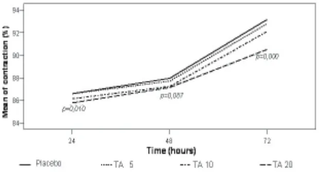
There was no significant difference between 5 µ M group and 10 µ M TA and control group (p>0.05). The significant result was showed by 20 µ M TA group at the 24th hour and 72nd hour
FIGURE 2. Comparison of means of contraction between control group and tamoxifen citrate group of various dosages.
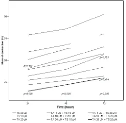
FIGURE 3. Comparison of the means of contractions by each combination of triamcinolone acetonide and tamoxifen citrate.
The groups of 10 µ M and 20 µ M TC concentration could significantly inhibit collagen lattice contraction since the 24th hour until the end
of observation, compared to control group (all ps were 0.000) (FIGURE 2)
The combination of 5 µM TA + 10 µM TC inhibited collagen lattice better than 20 µM TA or 10 µM TC (P<0.05). The 20 µM TC suppressed collagen lattice contraction better than the combination of 5 µM TA + 10 µM TC. The inhibition power of the combination of 10 µM TA+10 µM TC at the 72nd hour observation was almost equal with
the 20 µM TA+ 10 µM TC (p>0.05). The 20 µM TA + 20 µM TC group had the greatest power to inhibit collagen lattice contraction, but at the 72nd
DISCUSSION
Advances in biotechnology can improve the understanding of the phenomenon involved in pathologic scar formation, allowing the management of growth and function of the cell, and also the exploration of new tools for the prevention and treatment of keloid scars. We have investigated the potential of triamcinolone and tamoxifen as an inhibitor of wound contraction, using fibroblast populated collagen lattices as in vitro model. The model described by Bell et al. had been accepted
in the literature as adequate for the study of wound contraction, because it included the two fundamental dermal participants in scar formation: the extracellular matrix and fibroblasts. Fibroblast keloid was embedded within type I collagen, then medium either with or without drugs was added to the collagen lattices.12
Although results of intralesional triamcinolone acetonide could be highly variable, studies had also shown that triamcinolone acetonide inhibited cellular proliferation. The effect of triamcinolone acetonide on dermal fibroblast mitogenesis and collagen production might be mediated through a change in the levels of certain growth factors.13 The doses
were chosen based on a study by Cruz and Korchin, which found that 10 µm triamcinolone acetonide significantly inhibited the growth of keloid and fetal fibroblasts.14 We chose to evaluate 5 and 20 µM in
order to know the efficacy of concentrations above and below this effective dose. FIGURE 1 shows that TA suppressed fibroblast proliferation by inhibition of collagen lattice contraction at 20 µM. The inhibition of TGF-β1 synthesis by TA on keloid fibroblast would normalize fibroblast activity in fibroblast populated collagen lattices (FPCL) and inhibited collagen lattice contraction. This study demonstrated that 20 µM TA inhibited collagen lattice contraction of 20 µM dosage and it was not significantly inhibited in 5 and 10 µM dosages. This means that greater dosage of TA is needed to get a significant result in inhibiting collagen lattice contraction.
Tamoxifen, an estrogen receptor antagonist and known to be a drug of choice for hormone-sensitive breast cancer over the last 20 years, had recently been shown to inhibit the proliferation of fibroblast cultured from keloid biopsies.15 From the
other study by Miculec et al., TC had been shown
to decrease the overall level of TGF-β in keloid fibroblast; they evaluated the effect of tamoxifen on autocrine growth factor expression in keloid and fetal dermal fibroblast and showed increasing
TGF-β1 production keloid fibroblast compared with fetal fibroblast. Tamoxifen appeared to decrease per cell
TGF-β1 production at each of the time points
evaluated.9 This study demonstrated that 10 and 20
µM TC inhibited collagen lattice contraction. Higher concentrations of tamoxifen had a trend toward progressive inhibition of collagen lattice contraction. This study also demonstrated that the combination of the 10 µM TA + 10 µM TC could inhibit greater collagen lattice contraction compared to TA or TC alone, and the best inhibition was caused by 20 µM TA + 20 µ M TC. This meant that these two substances worked synergically inhibiting collagen lattice contraction, and the addition of TC may lead to improve keloid wound healing by reducing the
level of autocrine TGF-β production more than
monotherapy.
CONCLUSION
This study showed that the best combination to suppress collagen lattice contraction was TA 20 µM + TC 20 µM. From this study, we postulate that combination of triamcinolone acetonide and tamoxifen citrate may have potential clinical significance in the treatment of abnormal scarring. Further study is needed to address the role of this combination on collagenase activity, along with stability test in all preparations.
REFERENCES
1. Alexander G. Maneros, Krieg T. Keloid-clinical, pathogenesis, and treatment options. JDDG 2004;2:905-13.
2. Lee SS, Yosipovitch G, Chan YH, Goh CL. Pruritus, pain, and small nerve fiber function in keloid: a control study. J Am Acad Dermatol 2004; 51: 1002-1006,. 3. Joseph J. Shaffer, Taylor SC, Bolden FC Keloidal scars:
a review with a critical look at therapeutic options. J Am Acad Dermatol 2002;46:63-97.
5-Induced Type I Collagen Gene (COL1A2) Expression in Human Fibroblasts via c-Jun NH2-Terminal Kinase/ Activator Protein-1Activation. Mol Pharmacol 2003;64: 707–13.
6. Berman B, Villa A. M, Ramirez CC. Novel opportunities in the treatment and prevention of scarring. J Cutan Med Surg 2005; 32-36.
7. Haczynski J, Tarkowski R, Jarzabek K, Wolczynski S, Magoffin DA, Czarnocki KJ. Human cultured skin fibroblast express estrogen receptor alpha and beta. Int J mol Med 2004,10: 149-53.
8. Chau D, Mancoll JS, Lee S, Zhao J, Phillips LG. Tamoxifen downregulates TGF-beta production in keloid fibroblasts. Ann Plast Surg, 1998; 40:490-93. 9. Mikulec AA, Hanasono MM, Lum J, Kadleck JM. Effect
of tamoxifen on TGF-beta1 production by keloid and fetal fibroblasts. Arch Facial Plast Surg 2001;3(2):111-14.
10. Shejbal D, Badecovic V, Ivkic M, Kalogjera L, Aleric Z, Drvis P. Strategies in the treatment of keloid and hypertrophic scars. Acta Clin Croatia 2004;43:417-22.
11. Kelly AP. Medical and surgical therapies for keloid. Dermatologic Therapy 2004;17: 212-18.
12. Kamamoto F, Paggiaro AO, Rodas A, Herson MR, Mathor MB, Ferreira M.C. 2003. A wound contraction experimental model for studying keloid and wound-healing modulators. Intl Soc Artificial Organs. 27: 701-705.
13. Lisa A, Carroll, Matthew M, Hanasono, Anthony AM. Triamcinolone stimulates bFGF production and inhibits TGF-beta 1 production by human dermal fibroblasts. Dermatol Surg 2002;28: 704-9.
14. Cruz NI, Korchin L. Inhibition of human keloid fibroblast growth by isotretinoin and triamcinolone acetonide in vitro. Ann Plast Surg 1994;33: :401–5.