ILMU PENYAKIT TUMBUHAN
•
Fitopatologi
–
phyton (bhs.Yunani)
= tumbuhan
–
pathos (bhs.Yunani)
= penderitaan, penyakit
–
logos (bhs.Yunani)
= pengetahuan, ilmu
•
Bagian dari ilmu tumbuhan (botani) yang
mempelajari penyakit tumbuhan
–
Ilmu Penyakit Tumbuhan
3
ILMU YANG MENDUKUNG
FITOPATOLOGI
BOTANI MIKOLOGI BAKTERIOLOGI VIROLOGI
MIKROBIOLOGI
BIOKIMIA
KIMIA FISIKA
EKONOMI
KASUS I
KASUS II: PENJELASAN SEORANG
PENYULUH LAPANGAN
6
• Memetik daun kedelai
• Menunjuk bercak-bercak pada daun
• Mengatakan
• Bercak ini adalah PENYAKITNYA
• Mencabut tanaman tomat
• Menunjuk busuk pada akar
• Mengatakan
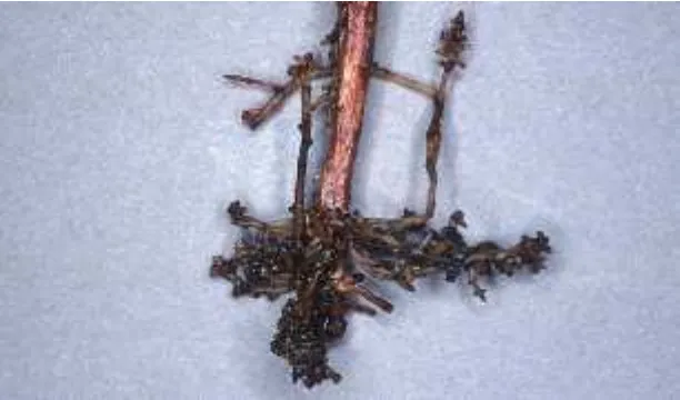
KASUS III: PENJELASAN PETUGAS DISBUN
7
Pohon kelapa sawit yang mati
Pada pangkal batang kelapa sawit
• Bentuk seperti tapal kuda
8
APAKAH BENAR
PENJELASAN PETUGAS PENYULUH ITU
• JAWABANNYA:
TIDAK BENAR !!!
9
•
Layu tanaman, bercak pada daun
•
Busuk pada akar
tetapi
GEJALA
PENYAKIT
• Struktur seperti tapal kuda
adalah
TANDA
PENYAKIT
(Bagian tubuh penyebab penyakit)
PENYAKIT
•
Adalah suatu proses interaksi
–
Oleh karena itu penyakit tidak dapat dilihat
•
Interaksi antara apa?
– Interaksi antara tumbuhan dengan patogen (penyebab penyakit)
•
Apa yang mempengaruhi interaksi itu?
– Lingkungan
•
Apa akibat interaksi itu?
– Terjadinya gangguan/penyimpangan fisiologi atau struktur tumbuhan
DEFINISI
•
Penyakit adalah proses interaksi antara
tumbuhan dengan patogen yang
dipengaruhi oleh lingkungan dan
mengakibatkan gangguan/ penyimpangan
fisiologi dan atau struktur tumbuhan
13
A. PENYAKIT BIOTIK
•
Interaksi antara tumbuhan dgn agens hayati (jasad
hidup) & virus
– Jamur – Bakteri
– Fitoplasma – Protozoa
– Tumbuhan parasit – Nematoda
•
Patogen dapat menular / ditularkan dari tanaman sakit
•
Patogen dapat mengalami variabilitas
14
VARIABILITAS PATOGEN
•
Perubahan sifat genetik patogen
–
Pembentukan varian (
forma speciales
dan ras
jamur, patovar bakteri, strain virus, dll.)
–
Sifat patogen berubah (patogenisitas, virulensi)
•
Disebabkan oleh cekaman faktor lingkungan
•
Menimbulkan masalah dalam upaya
pengendaliannya
CONTOH PENYAKIT BIOTIK
• Nama penyakit Penyebab
– Bercak daun Jamur
– Tepung Jamur
– Karat Jamur
– Layu Jamur / bakteri
– Busuk basah umbi Bakteri
– CVPD (jeruk) Bakteri
– Mosaik Virus
– Keriting Virus
– Tungro (padi) Virus
– Puru akar Nematoda
– Siste keemasan (kentang) Nematoda
B. PENYAKIT ABIOTIK
•
Interaksi antara tumbuhan dgn agens abiotik (bukan
jasad/agens hayati)
– Ketidakseimbangan hara – Suhu (tinggi atau rendah) – Lengas rendah
– Pencemar udara – Hujan asam
•
Patogen tidak ditularkan dari tanaman sakit ke
tanaman sehat
1. KETIDAK SEIMBANGAN HARA
•
Kahat (defisiensi) hara
–
Kekurangan hara utama
–
N, P, K, S, Ca, Mg
•
Keracunan
–
Kelebihan unsur mikro atau unsur runut
–
Al, Fe, B, Cu, Co
2. SUHU TINGGI
•
Radiasi panas
–
Panas sinar matahari
• Pemaparan langsung tanpa naungan (lahan terbuka)
–
Panas api (pembakaran sampah, api unggun)
•
Gejala daun “terbakar” (
scorching
)
3. SUHU RENDAH
•
Chilling injury
– Suhu rendah di atas 0O C (= sejuk) – Buah tropika di rak lemari es
•
Freezing injury
– Suhu rendah di bawah 0O C (=beku) kerusakan jaringan lunak/muda tumbuhan
– Buah & sayuran dlm freezer almari es
• Diperparah oleh freezing and thawing (keluar-masuk) – ‘Embun upas’ di dataran tinggi
4. LENGAS RENDAH
•
Kehilangan air dari jaringan tumbuhan
–
Layu
• Layu sementara (temporary wilt) • Layu permanen (permanent wilt)
–
Kerusakan permukaan jaringan
• Keriput
• Retak/pecah
5. PENCEMAR UDARA
•
Jenis
–
Gas
–
Partikel
•
Pengaruh
–
Langsung (sebagai Patogen Abiotik)
• Kerusakan jaringan/sel • SO2 ===> hujan asam • NOX ===> PAN & ozon
–
Tidak langsung (sebagai Predisposisi)
• Tumbuhan lebih rentan thd. serangan patogen biotik • Efek rumah kaca (ERK)
• Lubang Ozon
23
A. Merugikan
A. MERUGIKAN
•
Nilai ekonomi turun
– Penyusutan produksi
• Bobot, jumlah, ukuran
– Penurunan mutu produk
• Bentuk, warna, tekstur, aroma, rasa
•
Gangguan kesehatan konsumen
– Zat alergen dalam patogen
– Senyawa racun dlm bahan pangan
•
Penurunan mutu lingkungan hidup
– Pengurangan oksigen – Suaka satwa
1. PENURUNAN MUTU PRODUK
•
Bentuk
– Tidak teratur, mengecil, bisul, benjol
•
Warna
– Menjadi kusam, menjadi coklat kehitaman
•
Tekstur
– Mudah menjadi hancur, mudah robek, menjadi lembek
•
Aroma
– Bau menyengat, bau busuk
•
Rasa
– Menjadi masam, pahit, hambar
PENURUNAN MUTU PRODUK
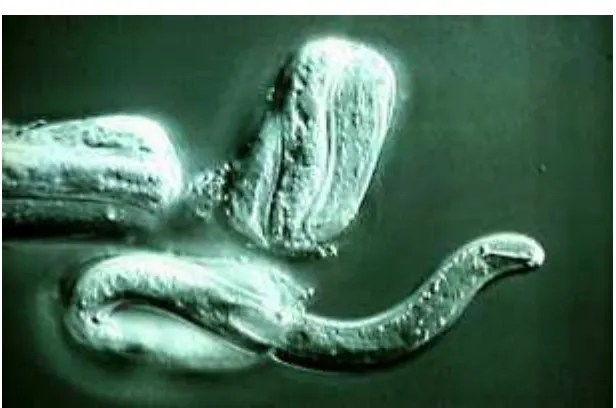
Diagnosing Nematode Problems
• It can be very difficult to decide if nematodes are causing or are likely to
cause significant crop injury.
• If a particular nematode pest was previously found in a site, it probably is
still present. Plan to continue to take steps to manage it.
• In a location for which a complete history is lacking, the identities and
population densities of nematode pests, hence the severity of damage which might be expected can usually be determined by laboratory assay of soil and/or plant samples.
• Another common diagnostic problem is determining the role of
nematodes when established plants are making unsatisfactory growth.
• This task is often difficult because few nematodes cause distinctive
diagnostic symptoms. A sound diagnosis should be based on as many as possible of symptoms above and below ground, field history, diagnostic nematicide tests, and laboratory assay of soil and/or plant samples.
Above-Ground Symptoms
• These are rarely, if ever, sufficient evidence to diagnose a root nematode
problem. However, they are important because possible nematode
problems are almost always first noticed because of abnormal top growth.
• Certain kinds of symptoms are typical of nematode injury to roots, and
should always make one consider nematodes as a possible cause for the inferior performance.
• They can also be used to help locate the most severely affected areas in
the planting after the problem is diagnosed.
• Since most plant nematodes affect root functions, most symptoms
associated with them are the result of inadequate water supply or mineral nutrition to the tops: chlorosis (yellowing) (Figure 14) or other abnormal coloration of foliage, stunted top growth (Figure 15), failure to respond normally to fertilizers, small or sparse foliage (Figure 16), a tendency to wilt (Figure 17) more readily than healthy plants, and slower recovery from wilting.
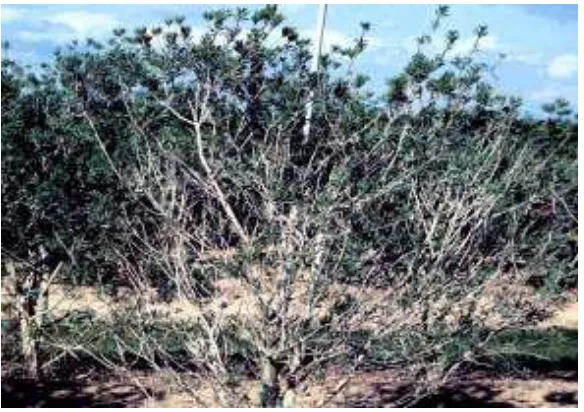
• Woody plants in advanced stages of decline incited by nematodes will
have little or no new foliage when healthy plants have substantial flushes, and eventually exhibit dieback of progressively larger branches. “Melting out” (Figure 18) or gradual decline is typical of nematode-injured turf and pasture.
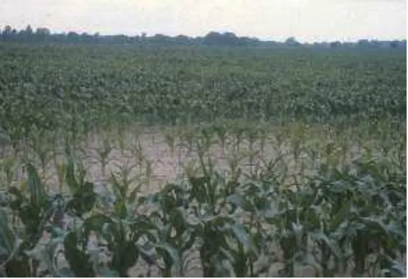
• Plantings that are stunted by nematodes often have worse weed problems
(Figure 19) than areas without nematode injury because the crop is less able to compete with weeds than it should be.
• The distribution of nematodes within any site is very irregular. Therefore, the shape, size, and distribution of areas showing the most severe effects of nematodes will be highly irregular (Figure 20 and Figure 21) within the field.
• Nematodes move very few feet per year on their own.
• In the undisturbed soil of groves, turf, and pastures, visible symptoms of
nematode injury normally appear as round, oval, or irregular areas which gradually increase in size year by year.
• In cultivated land, nematode-injured spots are often
elongated in the direction of cultivation because nematodes are moved by machinery.
• Erosion, land leveling, and any other force which moves masses of soil or lant parts can also spread a nematode infestation much more rapidly than it will go by itself.
• Nematode damage is often seen first and most pronounced in areas under special stresses, such as heavy traffic, excessive drainage because of slope or soil and dry areas outside
regular irrigation patterns
Below-Ground Symptoms
•
These may be more useful than top symptoms
for diagnosing many nematode problems.
•
Galls, abbreviated roots, necrotic lesions in
the root cortex, and root rotting may all help
in diagnosing nematode problems.
Plant Parasitic Nematodes
• Plant nematodes attack all crops grown in Florida, causing farmers millions of dollars in crop loss annually.
• Nematodes also are of great concern to the home owner, since they cause severe damage to turfgrasses, ornamentals and home gardens.
• We are often unaware of losses caused by nematodes since they are hidden from sight and much of the damage caused by them goes unreported or is attributed to other causes.
• both may be required for reproduction. However,
reproduction without males is common, and some species are
• hermaphroditic (“females” produce both sperm and eggs).
Life Cycle and Reproduction
• Plant parasitic nematodes have a simple life cycle of six stages: egg, four juvenile stages, and adult.
• The embryo develops inside the egg to become the first-stage juvenile.
• The first-stage juvenile molts inside the eggshell to become a second-stage juvenile, which hatches (Figure 3) from the egg, and in most species must feed before continuing to develop.
• The nematode molts three more times to become a fully developed adult.
Life Cycle and Reproduction
(Lanjutan…. )
• Egg production by the individual completes the cycle. • The number of eggs deposited by a female varies among
species and is affected by their habitat.
• Most species produce between 50 and 500 eggs, but a few occasionally produce several thousand eggs per female.
• The length of the life cycle varies considerably depending on nematode species, host plant, and the temperature of the habitat.
• During summer months when soil temperatures are in the 80’s, many plant nematodes complete their life cycles in about 30 days.
Nematode Feeding and Host-Parasite
Relationships
• Plant parasitic nematodes feed on living plant tissues.
• All have some form of oral stylet (Figure 4) or spear, which is used somewhat like a hypodermic needle to puncture the host cell wall.
• Many (probably all) plant nematodes inject enzymes into the host cell before feeding.
• These enzymes partially digest the cell contents before they are sucked into the gut.
• Most of the injury that nematodes cause to plants is related in some way to the feeding process.
• Nematodes may feed on plant tissues from outside the plant (ectoparasitic) or inside the tissues (endoparasitic).
• If the adult female moves freely through the soil or plant tissues, the species is said to be “migratory.”
• Species in which the adult females become swollen and
permanently immobile in one place in or on a root are termed “sedentary”.
• The feeding/living relationships that nematodes have with their hosts affect sampling methods and the success of
management practices.
• Ectoparasitic nematodes that never enter roots may be recovered only from soil samples.
• Endoparasitic nematodes often are detected most easily in samples of the tissues in which they feed and live (burrowing and lesion nematodes), but some occur more commonly as migratory stages in the soil (root-knot and reniform
nematodes).
• Those stages of endoparasites, which are inside root tissues, may be protected from nematicides that do not penetrate into roots, such as some soil fumigants. Root tissues may also shield them from many micro-organisms which attack
nematodes in the soil.
• Ectoparasites are fully exposed to pesticides and natural control agents in the soil.
Ectopara sitic Nema todes
• Ectoparasitic nematodes are generally migratory.• Most feed superficially at or very near the root tip or on root-hairs, but a few have stylets long enough to enable them to feed deeper in the root.
• Those which cause the most widespread and severe plant
injury in Florida are the sting (Belonolaimus spp.), stubby root (Trichodorus spp.), and awl (Dolichodorus spp.) nematodes. These feed at or near root tips and usually inhibit root
elongation.
• Ectoparasites (Figure 5) that rarely cause severe injury to their plant hosts include ring (Mesocriconema spp.) and spiral
(Helicotylenchus spp.) nematodes. They apparently feed primarily on root-hairs and superficial cortical tissues and
cause serious injury only to plants that are especially sensitive
• Among plant nematodes, only stubby-root nematodes and their close relatives, the dagger (Xiphinema spp.) and needle (Longidorus spp.) nematodes, are known to transmit plant viruses.
• The corky ringspot disease of potatoes, a problem in the Hastings area, is caused by a virus which is carried and transmitted by stubby-root nematodes.
Migra tory Endoparasites
• Migratory endoparasites can move into, through, and out from host tissues at any stage of development (except the egg).
• Migratory endoparasites generally live and feed in tender tissues such as the root cortex.
• They burrow (Figure 6) through the tissue, breaking open many cells after feeding on them.
• Cells surrounding the feeding area are often killed by toxic materials from the disrupted cells.
• The relatively large areas of dead cells usually turn brown, to become small spots or lesions (Figure 7) big enough to see, and are often easily colonized by fungi.
• Root rot (Figure 8) diseases are often associated with infestations of migratory endoparasitic nematodes.
• The most important examples in Florida are species of burrowing and lesion nematodes. The citrus burrowing
nematode, Radopholus similus, causes the spreading decline
disease of citrus.
• It is the subject of strict (and expensive) quarantine
regulations for ornamentals, nursery stock, and other growing plants being exported from the state, and can severely limit growth of many ornamental plants.
• Various species of lesion nematodes live in the roots of most crops grown in the state and may be especially damaging to citrus, peanuts, and commercial fern nurseries.
• The foliar (Aphelenchoides spp.) nematodes are the most
important representatives in Florida of a group of migratory nematodes that attack plants at the soil line or above the ground.
• They feed on or inside the leaves and buds of ferns,
strawberries, chrysanthemums, and many kinds of foliage ornamentals, and cause distortion or death of buds, leaf
distortion, or yellow to dark brown lesions (Figure 9) between major veins of leaves.
• Other nematodes that attack plants above ground cause leaf or seed galls and still others cause deterioration of the bulbs and necks of onions and their relatives, but are not common here.
• Ectoparasitic and migratory endoparasitic nematodes generally deposit their eggs singly as they are produced, wherever the female happens to be in the soil or plant.
Sedentary Endoparasitic Nema todes
• Sedentary endoparasitic nematodes are typified by the
rootknot (Meloidogyne spp.), cyst (Heterodera spp.), reniform
(Rotylenchulus spp.), and citrus (Tylenchulus semipenetrans)
nematodes.
• In most of these species, the second-stage juvenile is the “infective” stage, which moves through the soil.
• Second-stage juveniles locate host roots and enter them
(Figure 10). They then establish a suitable feeding site within the root tissues. Once a feeding site is selected, the nematode injects growth regulating substances into the cells near its
head, causing some of those cells to enlarge.
• These “giant” or “nurse” cells become specialized food
sources for the nematode. At the same time, the nematode becomes immobile, and the body swells (Figure 11) to a
• Mature females of the sedentary endoparasitic nematodes generally
produce large numbers of eggs that remain in their bodies (Figure 12) or accumulate in masses (Figure 13) attached to their bodies.
• The nematodes and the giant cells on which they feed are very dependent
on each other: if the nematode dies, the giant cells die or lose their highly active condition; if the giant cells die, the nematode dies of starvation, because it cannot move to a new site.
• Sedentary endoparasites damage their hosts by redirecting use of large
amounts of energy and nutrients from normal activities to development of the nematodes and their special feeding sites. The altered tissues of the feeding site also disrupt the vascular system.
• Roots severely galled by root-knot nematodes also usually deteriorate much earlier from root rots than roots that are not galled: gall tissues are succulent, poorly protected from invasion, and rich in nutrients to help the fungi grow rapidly.
• Sedentary endoparasites are the nematodes for which host resistance has
most often been identified. Relatively small differences in heritable
• Plants in which giant cells cannot be established, or in which they degenerate before a nematode can complete its life
cycle, are resistant to that nematode. The attempted infection by the nematode may injure the plant, but the nematode
cannot complete its life cycle in it.
47
Figure 1. Size comparison of a typical plant-parasitic nematode to a cotton thread.
Figure 2. Diagram of a typical plant-parasitic nematode.
Figure 4. A
plant-parasitic nematode stylet is used to puncture plant cells, to inject enzymes, and to ingest cell contents.
48
Figure 2. A ring nematode (thicker nematode) and spiral nematodes (thinner nematodes) feeding on a plant root. These nematodes are ectoparasitic nematodes.
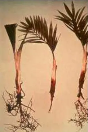
Figure 1. Rotting palm roots resulting from infection with migratory
endoparasitic nematodes.
Figure 3. Root lesions caused by migratory endoparasitic nematodes
49
Figure 6. Second-stage juvenile nematodes entering a root tip. Figure 5. Symptoms of foliar nematode damage are often
angular shaped leaf spots
Figure 7. Root tissue pulled-back to reveal a swollen root-knot
nematode within.
Figure 8. Cyst nematodes retain most of their eggs within their
bodies
50
Figure 9. Yellowing of pepper plants resulting from nematode damage to roots.
Figure 10. Healthy lettuce (left) and stunted lettuce that has been damaged by nematodes.Healthy lettuce (left) and stunted lettuce that has been damaged by nematodes.
Figure 12. Patch of St. Augustinegrass declining from nematode damage.
51
Figure 19. Spurge is a weed often associated with nematode damage to turf grasses.
Figure 17. Wilting bean plants resulting from nematode damage to roots.
Figure 20. Irregular patches of declining turf illustrate the uneven distribution of nematodes.
52
Figure 22. Bead-like galls on cotton roots result from root-knot nematode infection.
Figure 23. Massive lumpy galls caused by root-knot nematodes on passion flower roots.
Figure 24. Root-knot nematode galls (left) are an integral part of the
root and cannot be easily removed. Nitrogen-fixing nodules
53
Figure 26. The tiny white spots on these soybean roots are female cyst nematodes.
2. GANGGUAN KESEHATAN
• Apabila manusia atau hewan
– Terpapar bahan sakit
– Mengkonsumsi bahan sakit
• Tidak terjadi pada bahan yang rusak oleh hama • Penyebab: senyawa yang dihasilkan patogen
– Zat alergen : menyebabkan alergi
• Pengelola gudang biji-bijian: mata merah, batuk, gatal
– Mikotoksin (racun jamur dlm bahan pangan/pakan)
• Aflatoksin : dihasilkan Aspergillus flavus
• Okratoksin : dihasilkan A. ochraceus
• Toksin beras kuning : dihasilkan Penicillium islandicum
• Toksin T-2 : dihasilkan Fusarium tricinctum
CONTOH TOKSIN DLM PANGAN
•
AFLATOKSIN
– Kematian anak-anak kalkun di Inggris (1969) Turkey-X
Disease
– Ransum unggas campuran bungkil kacang tanah impor dari Brasil
– Bungkil kacang berjamur (Aspergillus flavus) – Toksin yang dihasilkan diberi nama AFLATOKSIN
•
TOKSIN DALAM PANGAN
– Asam bongkrek : dlm tempe bongkrek – Salmonellosis : dlm makanan kaleng
AFLATOKSIN
56
Dalam produk yg ditumbuhi
Aspergillus flavus
Pangan resiko tinggi:
• Kacang tanah & olahannya • Produk yg mengandung
RACUN DALAM PANGAN ?
3. PENURUNAN MUTU LINGKUNGAN
B. MENGUNTUNGKAN
•
Hubungan simbiosis antara tumbuhan dengan
“patogen”
– Mikoriza : antara akar dgn jamur – Bintil akar pada legum : antara akar dgn bakteri
•
Nilai ekonomi meningkat
– Tanaman/bagian tanaman eksotik
• Mahkota bunga belang-belang
• Daun belang
• Kerdil/bonsai
– Patogen sebagai bahan farmasi – Kelapa kopyor ?
1. SIMBIOSIS MIKORIZA
MIKORIZA
61
Akar Pinus
2. BUNGA EKSOTIK
62
Tulip sehat
3. BAHAN FARMASI
•
Ganoderma lucidum
(=
ling zhi
)
– Tubuh buah jamur patogen tanaman tahunan – Produk obat/jamu
• DXNR
• Sido Muncul
•
Ergot
Claviceps purpurea
– Sklerosium jamur pada gandum Secale cereale (rye) – Produk farmasi
• ErgodrylR (ergotamine tartrat)
Lingzhi (Ganoderma lucidum)
Ergot (Claviceps purpurea)
66
• Menghentikan pendarahan
G
EJALA PADA DAUN
G
EJALA PADA BATANGP
ENGAMATAN PADA PANGKAL BATANG
KONSEP
PENYAKIT TUMBUHAN
Telah berkembang dari waktu ke waktu
Jawaban thd. pertanyaan: “APAKAH PENYAKIT ITU?”
Tergantung konsep/pengertian yang diyakini/dianut
Mempengaruhi upaya atau tindakan yg dilakukan
PENYAKIT
6
KONSEP PENYEBAB CARA MENGATASI
1. Kekuatan gaib Dewa murka Pesta pora, kenduri Makhluk halus Sesaji
2. Dosa manusia Manusia berdosa Bertaubat
3. Tempat tumbuh Tanah jelek Pemilihan tempat
4. Cairan tubuh Cairan kotor Pelukaan, pemotongan 5. Lingkungan Lingkungan jelek Perbaikan lingkungan 6. Jasad hidup Jasad renik Pemberantasan
1. KONSEP KEKUATAN GAIB
7
• Masyarakat kuna - POLITEISME • Masyarakat primitif - ANIMISME
A. DEWA MURKA
– Romawi kuna :
• Karat tanaman Gandum = kemurkaan DEWA ROBIGUS
Supaya panen baik
==>
PESTA
ROBIGALIA– Jawa kuna
• Padi = titisan (reinkarnasi) DEWI SRI
• “HAMA” tanaman padi = Balatentara BETARA KALA yang
menyerang
B. MAKHLUK HALUS
Pengganggu Perusak
Jawa : Hama MENTHEK pada tanaman padi . Makhluk kerdil, gundul, bugil
. Bermain-main di sawah
==> dilakukan
SESAJI di sawah2. KONSEP DOSA MANUSIA
Hukuman TUHAN karena manusia melanggar aturan-Nya
Dalam masyarakat agamis dogmatik a. Dalam kitab-kitab suci
Kisah Nabi Musa a.s. VS. Fir’aun
Gagal Panen, Paceklik & wabah
b. Abad Pertengahan
Wabah Ergotime di Eropa “Api suci malaikat”
(HOLY FIRE)
i ===>
BERTAUBAT3. KONSEP TEMPAT
TUMBUH
10
• Pengamatan Theophratus (300 S.M.)
– Tanaman gandum
• Di puncak bukit - tumbuh baik/sehat/subur • Di dasar bukit (lembah) - tumbuh tidak baik
===>
. Pemilihan tempat untuk menanam5. KONSEP LINGKUNGAN
Cuaca/iklim yang jelek
Suhu & kelembaban
Suhu rendah & kelembaban tinggi ==> TANAMAN SAKIT
Penganut konsep GENERASI SPONTAN
===> Memperbaiki lingkungan
. Penghangatan kebun dengan api unggun & obor
PREDISPOSISIONIS
6. KONSEP JASAD RENIK
Penemuan mikroskop
Jasad renik sebagai penyebab penyakit
Jamur
Prevost (1807)
Tulasne bersaudara (1837) Antonius de Bary (1850)
Bakteri
Burrill (1877)
Virus
E. Smith (1899)
Nematoda
Cobb (1908)
===> PEMBERANTASAN JASAD RENIK
KONSEP INTERAKSI (MODERN)
1.
Segitiga penyakit (
disease
triangle
)
2.
Segiempat penyakit
(
disease square
)
3.
Piramida penyakit (
disease
pyramide
)
1. SEGITIGA PENYAKIT
3 komponen penyakit
Tumbuhan (= T), Patogen (= P) dan
Lingkungan (= L)
14
T P
L
• Syarat terjadinya penyakit
– Tumbuhan rentan (tidak tahan)
– Patogen virulen
• Mampu menyerang & menyebabkan sakit
– Lingkungan
SEGITIGA PENYAKIT
Berlaku di ekosistem alami (natural ecosystem)
Belum ada campur tangan manusia Bukan di lahan pertanian
Dicirikan adanya keragaman & keseimbangan
Contoh
Interaksi tumbuhan-patogen di hutan belantara
2. SEGIEMPAT PENYAKIT
16
T P
L
T = Tumbuhan
P = Patogen
L = Lingkungan
M
M = Manusia
KOMPONEN PENYAKIT
SEGIEMPAT PENYAKIT
Berlaku dalam ekosistem pertanian
(
agroecosystem
)
Ada campur tangan manusia Di lahan pertanian
Dicirikan adanya keseragaman & ketidak stabilan
Manusia dapat mempengaruhi
Tumbuhan : memilih jenis, varietas Patogen : mematikan dgn
fungisida/nematisida
3. PIRAMIDA PENYAKIT
18
T = Tumbuhan
- bidang tegak
P = Patogen
- bidang tegak
L = Lingkungan
- bidang tegak
M = Manusia
- bidang tegak
W = Waktu
- garis tinggi
PIRAMIDA PENYAKIT
Penyakit adalah proses yang dinamis
Keadaan penyakit berubah dari waktu ke waktu
Menjadi dasar epidemiologi penyakit
Pengukuran penyakit
Persentase penyakit Intensitas penyakit Laju penyakit
Peramalan penyakit
PENYAKIT DIPENGARUHI
WAKTU
X
t= X
oe
rtXt : keadaan penyakit pada waktu t
X0 : keadaan penyakit pada awal (t = 0) e : konstante (2,718)
r : laju pertambahan penyakit
t : waktu (hari/minggu/bulan, dst.)
PENGEMBANGAN
KONSEP
Pengukuran penyakit
Laju penyakit
Intensitas penyakit
Peramalan penyakit
Berdasar persamaan Xt = X0 ert Berdasar data cuaca
Berdasar penangkapan inokulum
PERAMALAN PENYAKIT
Penyakit penting pada komoditas bernilai ekonomi tinggi
Program komputer
Blitecast - fireblight pada apel
Epidem - karat pada gandum
Epimay - hawar daun pada jagung
D. KONSEP KERUCUT
Penyakit berbeda: . Di tanaman yang sama . Di lingkungan yang sama . Di petani yang sama
PENYEBAB PENYAKIT
DIPT 2012
Penyebab
penyakit
BIOTIK (Penyakit Biotik)
Jamur
Prokariot (Bakteri dan Molicutes)
Virus dan Viroid
Nematoda
Protozoa
Tumbuhan Tinggi Parasitik
ABIOTIK (Penyakit Abiotik)
Kekurangan/Kelebihan hara
Keracunan Fe/ logam berat
Polusi
DIPT 2012
JENIS PATOGEN
Patogen biotik
Jamur, bakteri, fitoplasma, protozoa, nematoda,
virus, viroid
Patogen abiotik
Faktor edafik (tanah): kahat hara, keracunan hara
Pencemar udara: gas, partikel
Agens fisika: suhu, lengas, cahaya
DIPT 2012
3
PROKARIOT
Terdiri dari Bakteri dan Mollicutes
Mikroorganisme bersel tunggal
Bahan genetik (DNA) tidak diselubungi oleh
suatu membran (tidak mempunyai membran
inti)
Sel-sel terdiri dari DNA dan ribosom kecil (70S)
DIPT-054
BAKTERI
Lebih dari 1600 spesies telah diketahui
Sebagian besar bersifat saprofit
Ada yang bermanfaat bagi manusia
Patogen tanaman lebih dari 100 spesies
Sebagian besar bakteri patogen tumbuhan
bersifat saprofit fakultatif dan dapat
ditumbuhkan pada media buatan
DIPT-055
MORFOLOGI BAKTERI
MORFOLOGI
BENTUK
: LENGKUNG, BATANG, SPIRAL
COCCI, BACILLI, SPIRILLUM
UKURAN
: 1,0-5,0 --- 0,5 – 1,0
µ
m
TERGANTUNG: SUHU INKUBASI, MEDIUM, UMUR
BIAKAN, METODA PENGECATAN
BERGERAK DENGAN FLAGELA
ADA YANG MEMBENTUK SPORA
BERKEMBANG BIAK DENGAN PEMBELAHAN BINER
1---2---4---16---32---64---128---256---1024
DIPT-05
6
DIPT-05
8
EKOLOGI DAN PENYEBARAN
SEBAGIAN BESAR BAKTERI BERKEMBANG DI DALAM TANAMAN INANG SEBAGAI PARASIT
PADA PERMUKAAN TANAMAN TERUTAMA PADA TUNAS SEBAGAI EPIFIT
SEBAGIAN PADA SISA-SISA TANAMAN DAN DI DALAM TANAH SEBAGAI SAPROFIT
SOIL INHABITANTS: Agrobacterium tumefaciens, Ralstonia solanacearum, Streptomyces scabies
SOIL INVADERS: Erwinia amylovora, Xanthomonas campestris
Bertahan di dalam tanah dalam jaringan tanaman,
beberapa hidup bebas secara saprofit, ada yang bertahan dengan membentuk bacterial ooze
DIPT-05
9
DIPT-05
10
DIPT-05
11
PENYEBARAN BAKTERI
SECARA PASIF
AIR, SERANGGA, HEWAN LAIN(KELINCI, SAPI,
BURUNG)
MANUSIA (CARA BERCOCOK TANAM,
TRANSPORTASI BAHAN TANAMAN)
SECARA AKTIF
FLAGELA, KEMOTAKSIS
DIPT-05
12
GENERA BAKTERI PATOGEN TUMBUHAN
GRAM NEGATIF
PSEUDOMONAS (
P.syringae
)
XANTHOMONAS (
X. campestris
)
XYLOPHILUS (
Xylophilus ampelinus
)
ACIDOVORAX (
Acidovorax
sp.)
ERWINIA (
E. carotovora
)
PANTOEA (
P. stewartii
)
BURKHOLDERIA (
B. cepacia
)
RALSTONIA (
R. solanacearum
)
DIPT-05
13
GENERA BAKTERI PATOGEN TUMBUHAN
GRAM POSITIF
CORYNEFORM (
Clavibacter xily
subsp
xily
)
STREPTOMYCES (S. scabies)
CLOSTRIDIUM
BACILLUS
CLAVIBACTER
DIPT-05
14
GEJALA PENYAKIT BAKTERI
PADA TANAMAN
Nekrosis : Matinya sel, jaringan atau organ
Nekrose : Matinya bagian tanaman
Hydrosia : Sebelum sel mati, tampak kebasahan
Klorosis : Rusaknya klorofil, daun menguning
GEJALA PENYAKIT BAKTERI
PADA TANAMAN
Hypoplastis : hambatan pertumbuhan
Atropy (kerdil)
Chlorosis : rusaknya (tidak sempurna)klorofil
Hyperplasia : pertumbuhan yang luar biasa
CONTOH BAKTERI PATOGEN
TANAMAN
Agrobacterium : A. tumifacie – bengkak akar
Corrynebacterium : C. higanensis - kanker
Erwinia : E. carotovora – busuk basah
DIPT-05
DIPT-05
DIPT-05
DIPT-05
Jamur/Fungi/Cendawan
1
. Berukuran mikroskopis, punya inti sejati (eukariotik),
berbentuk benang (miselia), menghasilkan spora dan
tidak punya klorofil
2. Mempunyai dinding sel yg mengandung kitin dan
glucan (polisakarida dan glykoprotein)
3. Lebih dari 100.000 spesies sebagai saprofit
(dekomposer), lebih dari 10.000 spesies dpt
menyebabkan penyakit tanaman
4. Satu spesies dapat menginfeksi satu/lebih tanaman
5. Beberapa merupakan parasit obligat
Proses infeksi
16
17
DIPT-05 17
PLASMODIOPHOROMYCETES
•
Jamur lendir endoparasit
– Thallus berupa plasmodium
•
Contoh
– Plasmodiophora brassicae penyebab penyakit akar gada
pada marga Brassica (kobisan)
•
Pertumbuhan
– Bila ada inang
• Membentuk zoospora yg setelah masuk ke dlm sel akar akan
berbentuk plasmodium
• Plasmodium mengkoloni sel-sel
– Bila tak ada inang
• Zoospora membentuk dinding sel tebal (tahan)
18
DIPT-05 18
HYPHOCHYTRIDIOMYCETES
•
Jamur akuatik (dalam air)
•
Hife tidak bersekat
•
Membentuk zoospora berflagel tunggal pd bagian
apikal
•
Beberapa anggota parasit pada ganggang atau jamur
akuatik lain
– Anisolpidium ectocarpi - ganggang coklat
19
DIPT-05 19
PLASMODIOPHORA BRASSICAE
Akar gada
20
DIPT-05 20
CHYTRIDIOMYCETES
•
Jamur akuatik & terestrial
•
Thallus sederhana, hife tidak bersekat, membentuk
•
Zoospora berflagel satu yg panjang & dpt berperan sbg
gamet (sel kelamin)
•
Contoh
– Olpidium brassicae - damping-off kobis – Synchytrium endobioticum - bisul pd. Kentang
21
DIPT-05 21
OOMYCETES
•
Hife tidak bersekat
•
Reproduksi seksual dgn membentuk oospora
•
Reproduksi aseksual dgn membentuk sporangium
(kantong spora)
•
Banyak anggota merupakan parasit penting
– Saprolegnia monoica - ikan
– Phytophthora infestans - kentang (hawar) – Perenosclerospora maydis - jagung (bulai)
22
DIPT-05 22
ZYGOMYCETES
•
Hife tidak bersekat
•
Reproduksi seksual dgn membentuk zigospora
•
Reproduksi aseksual dgn membentuk sporangium
•
Hidup sbg saprofit
– Rhizopus oligosporus - jamur tempe – R. nigricans - jamur pd. roti
– Pilobolus sp. - pd kotoran hewan
•
Hidup sbg parasit
23
DIPT-05 23
ASCOMYCETES
•
Hife bersekat
•
Reproduksi seksual dgn membentuk askospora di
dlm askus
– Askus tersebar - apotesium
– Askus dlm ‘wadah’ - peritesium (spt botol),
24
DIPT-05 24
ASCOMYCETES
•
Parasit & patogen tumbuhan
– Ceratocystis fimbriata - kelapa (cairan merah)
– Venturia inaequalis - hawar api (fireblight) apel
– Claviceps purpurea - ergot gandum rye
– Sclerotinia sclerotiorum - busuk buah
– Xylaria spp. - busuk akar & pangkal btg – Endothia parasitica - hawar kastanye
•
Saprofit
– Aleuria aurantia - jamur kuping
– Morchella esculenta - jamur morel (edibel)
– Helvella crispa - jamur morel palsu (beracun)
25
DIPT-05 25
BASIDIOMYCETES
•
Hife bersekat
dolipore
, membentuk hubungan
jepit (
clamp connection
)
•
Reproduksi seksual dgn membentuk
26
DIPT-05 26
BASIDIOMYCETES
•
Parasit tumbuhan
– Exobasidium vexans - cacar daun teh
– Fomes lignosus - jamur akar putih
– Ganoderma spp. - jamur akar coklat/merah
– Hemileia vastatrix - jamur karat kopi
– Puccinia graminis - jamur karat gandum – Ustilago maydis - jamur gosong bengkak
•
Saprofit
– Phallus impudicus - jamur bangkai
27
DIPT-05 27
BASIDIOMYCETES
•
Jamur mikoriza: simbiosis dgn akar tumbuhan
– Amanita spp. – Boletus spp.
– Pisolithus tinctorius
•
Jamur edibel
– Agaricus campestris, A. bisporus – Auricularia auricula
28
DIPT-05 28
DEUTEROMYCETES
(FUNGI IMPERFECTI)
•
Hife bersekat
•
Kelompok yg belum ditemukan reproduksi
seksualnya
–
Kelompok sementara
•
Reproduksi aseksual dgn fragmentasi hife atau
29
DIPT-05 29
PEMBENTUKAN KONIDIUM
A. Tidak dlm ‘wadah’ (tersebar bebas)
–
Ordo Moniliales
B. Dalam ‘wadah’
–
Ordo Melanconiales
- pd. apotesium
30
DIPT-05 30
MONILIALES
•
Parasit tumbuhan dan perusak pasca panen
– Aspergillus spp. - Penicillium spp.
– Fusarium spp. - Cercospora spp.
– Verticillium spp.
MELANCONIALES
•
Parasit tumbuhan
– Colletotricum spp.
31
DIPT-05 31
SPHAEROPSIDALES
•
Parasit tumbuhan
–
Diplodia spp.
Dr. Jumsu Trisno; Jur. HPT FAPERTA UNAND (PERLINTAN 4)
Gejala Penyakit yg
disebabkan oleh Jamur :
PATOGENESIS
PATOGENESIS
PATOGENESIS (
pathogenesis
):Urut-urutan peristiwa dari patogen dalam menimbulkan
penyakit.
Rangkaian pembentukan penyakit dengan tahapan
proses yang berkesinambungan membentuk sebuah
siklus.
PATOGENISITAS (
pathogenicity
):Pathogenesis & Parasitism
•
•
•
Parasite : Parasitism
Symbiosis, Mycorrhiza --
Menguntungkan
Pathogen : Pathogenesis
kerusakan /
Levels of Parasitism
Bertahan hidup pada seluruh atau hanya disebagian jaringan tanaman
Bisa patogen ataupun bukan patogenik
Level of Parasitism
2. Symbiosis
Dua organisme Berasosiasi dan saling menguntungkan
Fungi --- algae
legume plant nodules bentgrass (Glomus spp.) fescue, ryegrass
(Acremonium
coenophialum and A. lolii ) Rhizobium
---Host range of the pathogen
Disease development
– Disease triangle
Patogen
Tumbuhan Inang
Terminology
– Primary inoculum
Jadi, dalam pembentukan dan perkembangan tiap penyakit menular (infectious diseases) terjadi suatu seri dari beberapa tahap atau proses yang berlangsung kontinyu secara
berurutan.
Proses yang terjadi secara berurutan dalam pembentukan dan perkembangan penyakit disebut siklus penyakit.
I. Fase aktif (patogenesis)
Events in Disease Cycle
Inoculation
Penetration
Infection
Invasion
I. Fase aktif (patogenesis)
II. Fase pasif (saprogenesis)
Beberapa inokulum (pathogen) mungkin tidak mendarat pada inang yang rentan (susceptible) dan tidak pada kondisi lingkungan yang kondusif untuk bertahan dan tumbuh/ berkembang.
Sehingga, patogen harus bertahan sampai kondisi lingkungan kembali sesuai untuk melangsungkan PATOGENESIS.
Siklus Penyakit
Survival
Inoculum
produced
Dispersal
Infection
Colonization
Symptoms
Production of
survival
structures
Fungi
Bacteria
Viruses
Nematodes
Survival Crop residue Soil
1. Inokulasi
1. Inokulasi
2. Penetrasi
2. Penetrasi
3. Infeksi
3. Infeksi
4. Kolonisasi
4. Kolonisasi
1. Inokulasi
2. Penetrasi
Proses deposisi atau kontaknya inokulum pada permukaan jaringan inang. Tempat kontak patogen: lubang alami (→ stoma, lentisel,
hidatoda dan retakan karena pertumbuhan) & luka (→ gigitan serangga).
Proses masuknya patogen ke dalam inang. Penetrasi aktif: patogen berpartisipasi aktif menembus dinding sel dan masuk ke dalam jaringan inang.
Penetrasi pasif: patogen tidak berpartisipasi aktif, misalnya ketika bakteri terbawa oleh film air melalui stomata masuk ke dalam jaringan inang.
Spora berkecambah
Membentuk apresoria
Membentuk hifa infeksi
(penetration peg) Gambar: scanning electro micrograph (SEM) dari spora cendawan karat (Uromyces phaseoli) pada tanaman kacang.
6 jam stomavia
Penetrasi aktif : dilakukan dengan adanya tekanan mekanik atau kerja enzimatik dari hifa patogen.
Inokulum
Struktur dari patogen yang dapat menimbulkan infeksi.
Tipe inokulum: • spora seksual
• spora aseksual
• potongan miselium
• tubuh sklerotia
• rizomorf
• miselium dorman (dalam benih)
Sumber inokulum
Bagian/ keseluruhan tumbuhan, alat, tanah, tempat penyimpanan,
Penyebaran inokulum
• Angin take off, flight, deposisi.
• Air/ hujan memindahkan spora dan mengaktifkan metabolisme. • Serangga (sebagai vektor) terbawa pada tubuh/ organ pemotong.
• Benih atau pun bagian lain virus/ viroid, bakteri, cendawan.
• Manusia jarak jauh.
Lingkungan yang ekstrim …..?
********
Iklim temperate (subtropik) patogen harus mampu bertahan pada
kondisi ekstrim.
Cara patogen bertahan pada kondisi ekstrim :
• Bentuk saprofit pada sisa-sisa bagian tumbuhan yang mati dalam tanah cendawan penghuni tanah (soil inhabitant) misal: Pythium, Phytophthora, Rhizoctonia, Fusarium;
cendawan penyerbu tanah (soil invader) misal:
Helminthosporium, Septoria.
• Membentuk struktur berdinding tebal dan resisten teliospora, oospora, askospora.
• Bertahan dalam gulma atau inang lainnya,
• Bertahan pada tubuh serangga vektor, dan
• Bertahan pada benih.
Penetrasi
Patogen untuk pertumbuhan dan perkembangannya perlu
makanan (nutrisi). Karena nutrisi yang dibutuhkan berada dalam jaringan inang maka untuk mendapatkannya ia harus melampaui berbagai rintangan. Patogen harus menembus (penetrate)
20
Copyright © The McGraw-Hill Companies, Inc. Permission required for reproduction or display.
Plant cell
Germinating fungal spore Adhesion
Plant epidermal cell
a. Pra-penetrasi
Deposisi suatu inokulum di tempat infeksi sebelum
penetrasi terjadi dipengaruhi oleh faktor fisik, kimia, dan biologi
t°c
Rh
pH
O2
CO2
Permukaan akar (rhizoplane) yang berada di
rizosfer banyak mengandung senyawa metabolit yang diproduksi oleh akar dan dapat
dimanfaatkan oleh mikro organisme.
Beberapa hifa cendawan patogenik dapat tumbuh ke arah akar tumbuhan yang rentan (inang) karena adanya daya tarik kimia
(chemotrophic attraction) berupa gula, asam organik, asam amino, dll.
a. Penetrasi
Bakteri patogenik umumnya masuk via stomata yang terbuka. Beberapa cendawan patogenik yang biasanya masuk via stomata: Plasmopara viticola.
Sedangkan yang dapat masuk via stomata terbuka maupun tertutup adalah Puccinia graminis (malam saat stomata
tertutup) uredosporanya berkecambah dan menjulurkan tabung kecambahnya ke lubang stomata. Pada saat siang hari (saat stomata terbuka) di ujung apresoria dibentuk
Lubang alami lainnya yang berperan dalam penetrasi adalah :
• Lentisel : Erwinia carotovora (busuk lunak pada kentang dan wortel)
• Hidatoda : Xanthomonas campestris (busuk hitam pada crucifera) masuk via tepi/ ujung helai daun. Bila Rh rendah
maka tetes air akan terhisap kembali ke dalam lubang hidatoda sekaligus membawa patogen masuk.
• Trikoma : Kutikula dari kelenjar trikoma dapat pecah pada bagian sekresi kelenjar yang terakumulasi. Sobekan tersebut manjadi lubang masuk.
Pre Penetration Process in bacteria
Multiplication
in the
presence of moisture
or host exudates
(A) Agrobacterium, (B) Erwinia,
(C) Pseudomonas, (D) Xanthomonas.
B A
Pre Penetration Process in
nematodes:
hatching of eggs
C
penetration peg
Direct penetration by appressoria
Penetration of rice leaf by Magnaporthe grisea – Rice blast
Zoospores of Phytophthora sojae
Direct Penetration by
•
•
Nematodes : larvae
Penetration by nematodes
Second-stage juveniles of soybean cyst nematode penetration the root of soybean
glycines
(stained red) Heterodera
Root knot nematodes
Penetration by parasitic plant
Cuscuta campestris
microscopic section shows the dodder haustorium
Penetration through natural openings
a.
b.
c.
d.
Stomata
Stomatal penetration
Appressorium
Penetration peg
Sub stomatal cavity
Infection hyphe (penetrate diff cells)
A. Uredinosorus
B. Germinated urediniospores (U) with germ tubes
(T) and appressorium (A) over leaf stomata C. Developed appr.
D. An empty appressorium (A) over leaf stomata, indicating that the fungus already penetrated
E. Hyphae in the intercellular spaces
Bacteria
Hydathodes
Special glands or pores at the end of vascular tissue on
water exudes and
Lenticels:
small pores or openings in the outer skin of plants
that provide a pathway for gas exchange between
the atmosphere
Develop on any
twigs, stems, or
and plant cells.
3. Infeksi
4. Kolonisasi
5. Diseminasi
Tahapan dimana patogen yang sudah menetap dalam jaringan inang mulai memperoleh nutrisi dari inangnya. Pendapat 1: inokulasi s/d patogen memperoleh nutrisi. Jadi, penetrasi merupakan bagian dari infeksi.
Pendapat 2: penetrasi s/d inang memberikan respons terhadap invasi patogen.
Proses kelanjutan dari infeksi, yaitu patogen Melanjutkan pertumbuhannya dan mengkolonisasi inang. Jadi, merupakan proses pertumbuhan dan perluasan aktivitas patogen melalui jaringan inang.
Struktur patogen yang berfungsi sebagai
•
•
Successful infection evident
Infection is of different types
as: symtpoms
•
Invasion and reproduction
are
occu
r
the two
concurrent
stages
that
during
differe
nt
infection as pathogen
multiply
Infection
•
Infection process is affected
factors
Resistance & susceptibility of the host
Aggressiveness & virulence of the pathogen
Environmental factors
Host nutrition & pH
Sub cuticular: Apple scab
behaviour
Ectoparasites: powdery mildew
Invasion
Kolonisasi
Merupakan pergerakan patogen secara aktif maupun pasif melalui jaringan tumbuhan inang.
Aktif : bila patogen bergerak dari satu sel ke sel yang lain (misal: cendawan).
Pasif : bila patogen berpindah tempat karena terbawa aliran transpirasional (misal: virus).
Dalam kolonisasi juga berperan aktivitas
1. Kolonisasi oleh virus, viroid, dan mikoplasma.
Organisme tersebut merupakan parasit intraseluler. Umumnya virus mengkolonisasi epidermis, palisade, jaringan bunga karang dan sistem pembuluh. Patogen ini dibawa melalui sistem pembuluh
2. Kolonisasi oleh bakteri
Organisme ini bergerak secara intraseluler. Saat sel yang terinfeksi terbelah dua maka sebagian sel yang berasosiasi ditransfer ke sel yang baru. Banyak bakteri yang melakukan multiplikasi di dalam sistem pembuluh xylem.
3. Kolonisasi oleh cendawan
Dapat menggunakan haustoria maupun tanpa haustoria.
4. Kolonisasi oleh nematoda
Nematode betina yang telah dibuahi bergerak menuju permukaan akar inang yang rentan sehingga kepala nematoda dekat dengan ‘stelle’ tengah. Pada bagian ini ia menetap sambil mulai mengkonsumsi sel-sel di
sekitar kepala. Selanjutnya, nematoda akan meletakkan massa telurnya ke permukaan akar. Setelah telur
menetas menjadi larva, larva tersebutr mempenetrasi inang dan menetap lagi. Dan selanjutnya samapi
Pathogen Survival
1. In infected crop debris
2.
On growing plants
Infected material on host
In propagating material
Alternate host
Collateral host
As dormant structures
plants
Inokulum
Struktur dari patogen yang dapat menimbulkan infeksi.
Tipe inokulum:
• spora seksual
• spora aseksual
• potongan miselium
• tubuh sklerotia
• rizomorf
• miselium dorman (dalam benih)
Bagian/ keseluruhan tumbuhan, alat, tanah, tempat penyimpanan,
serangga, dll yang berpotensi membawa inokulum
Penyebaran inokulum
• Angin take off, flight, deposisi.
• Air/ hujan memindahkan spora dan mengaktifkan metabolisme.
• Serangga (sebagai vektor) terbawa pada tubuh/ organ pemotong.
• Benih atau pun bagian lain virus/ viroid, bakteri, cendawan.
• Manusia jarak jauh.
Lingkungan
yang ekstrim …..?
******
Iklim temperate (subtropik)
patogen harus mampu bertahan pada kondisi ekstrim.