Atherosclerosis 152 (2000) 457 – 468
Decreased cellular cholesterol efflux is a common cause of familial
hypoalphalipoproteinemia: role of the ABCA1 gene mutations
Stephanie Mott
a, Lu Yu
a, Michel Marcil
a, Betsie Boucher
a, Colette Rondeau
a,
Jacques Genest Jr
a,*
aCardio6ascular Genetics Laboratory,McGill Uni6ersity Health Center,Royal Victoria Hospital,686Pine A6enue West,Montre´al, Que´bec,Canada 3A1A1
Received 15 June 1999; received in revised form 8 December 1999; accepted 13 December 1999
Abstract
Background. High density lipoproteins (HDL) are complex lipoprotein particles involved in reverse cholesterol (C) transport and are negatively associated with the risk for coronary artery disease (CAD). We have described a disorder of familial HDL deficiency (FHD) due to abnormal cellular cholesterol efflux. In the present study, we investigated cellular cholesterol efflux on skin fibroblast from 15 probands with moderate to severe hypoalphalipoproteinemia, including one subject with Tangier disease (TD). We performed family studies on eight of these probands (269 individuals) with familial hypoalphalipoproteinemia (defined as a HDL-C B5th%, and with no known cause of HDL deficiency). We have previously shown that four of our FHD patients and patients with TD have mutations at the ABC1 gene, demonstrating that FHD is a heterozygous form of TD.Methods. On each subject, we carried out detailed biochemical analysis and determined apoA-I-mediated cellular cholesterol efflux using
3H-cholesterol labeled skin fibroblasts from study subjects compared with controls. TD has also been associated with abnormal
cellular cholesterol efflux. Cell fusion experiments with polyethylene glycol (PEG) were carried out with fibroblasts from a subject with TD and one with FHD in order to determine whether the Tangier cells can complement the FHD defect. In all subjects with a reduced cellular cholesterol efflux, exons of the ABCA1 gene were sequenced. Results. Familial forms of HDL deficiency, defined as HDL-C levelsB5th percentile, are a heterogeneous group of lipoprotein disorders. A reduced cellular cholesterol efflux has been identified in eight subjects from seven kindred (7/14 or 50% of probands tested), being reduced by a mean 59% of controls (range 49 – 63%). In four of these subjects, a mutation at the ABCA1 gene locus was identified. In three other subjects an efflux defect was idenfified but no critical mutation at the ABCA1 gene locus has been identified. In the remaining subjects, (7/14), no efflux defect was identified. Complementation studies reveal that the FHD defect is not corrected by Tangier cells, confirming that FHD and TD represent a spectrum of the same genetic defect. Conclusion. Familial hypoalphalipoproteinemia syndromes are phenotypically heterogeneous; one form is associated with abnormal cellular cholesterol efflux caused by heterozygous mutations at the ABCA1 gene, that defines familial HDL Deficiency while homozygous mutations or compound heterozygocity causes TD. Other forms are primary hypoalphalipoproteinemia of unknown cause, while the remaining cases are associated with hypertriglyceridemia with or without elevated apoB levels. We conclude that a cellular cholesterol defect is a relatively frequent cause of familial HDL deficiency and that a mutation at the ABCA1 gene can be identified in half of these patients. © 2000 Elsevier Science Ireland Ltd. All rights reserved.
Keywords:High density lipoproteins (HDL); Familial HDL deficiency; Cholesterol
www.elsevier.com/locate/atherosclerosis
1. Introduction
High density lipoproteins (HDL) play a major role in the lipoprotein transport system and in vascular func-tion [1 – 3]. The main role of HDL, is thought to be the transport of cholesterol (C) to the liver (the reverse
* Corresponding author. Present address: Cardiology Services, Centre Hospitalier de l’Universite´ de Montre´al (CHUM), Montre´al, Que´bec, Canada. Tel.:+1-514-9875715; fax: +1-514-9875767.
E-mail address:[email protected] (J. Genest Jr).
cholesterol transport system). In this system, HDL mobilizes peripheral cholesterol (including from arterial tissues) and transports it for subsequent elimination by the liver (as bile salts) and kidneys or uptake by other tissues (steroid hormone producing). This concept is strengthened by epidemiological studies that have shown a strong and graded inverse relationship between plasma levels of HDL-C and the presence and development of coronary artery disease (CAD) [4 – 8]. Isolated cases of HDL deficiency however, have not been consistently associated with premature CAD [3]. Several primary forms of reduced HDL-C levels are not associated with CAD as seen for instance with apoA-IMilano [9,10]. In most cases of mild to moderate hypoalphalipoproteine-mia encountered clinically, a low HDL-C is associated with multiple metabolic abnormalities, including ab-dominal obesity [11], elevated triglycerides [7,12], ele-vated plasma apoB levels [13], hyperglycemia and insulin resistance that form part of the ‘metabolic syndrome’. Tangier disease (TD) [41] and familial HDL deficiency (FHD) [15] are characterized by a marked deficiency of plasma levels of HDL-C and apoA-I. Both have been shown to result from hypercatabolism of plasma apoA-I by metabolic kinetic studies [16,17]. Studies performed on fibroblasts from TD and FHD patients have shown a marked decrease in cellular cholesterol transport and efflux [18 – 20]. This results in cholesterol-depleted apoA-I containing lipoproteins that are rapidly catabolized. The genetic defect in Tangier disease and in FHD has been recently shown to be caused by mutations at the ATP binding cassette (ABCA1)-1 gene. Tangier disease subjects have homozygous or compound heterozygous mutations at the ABCA1 gene while FHD subjects are heterozygous for these mutations at this locus [22 – 24]. We have recently reported that mutations at the ABCA1 gene locus are the cause of familial HDL deficiency [25]. In the present study, we have examined 15 probands with familial hypoalphalipoproteinemia (including one subject with classical TD). We report that a defect in cellular cholesterol efflux is a frequent cause of moderate to severe hypoalphalipoproteinemia and that mutations at the ABCA1 gene underlie most cases of FHD.
2. Methods
2.1. Patient selection
Study subjects were selected from the Cardiology Clinic of the Clinical Research Institute of Montre´al.
The main criterion was a HDL-C level B5th percentile
for age and gender, with a plasma concentration of triglycerides B95th percentile [21] in the proband and
at least one first-degree relative with the same lipid abnormality. In addition the patients did not have diabetes. The apoA-I level was determined by
neph-elometry and its molecular weight verified by polyacry-lamide gradient gel electrophoresis (PAGGE); the possibility of an abnormal form of apoA-I causing a change in electrophoeretic mobility was further excluded by isoelectrofocusing (IEF) [15]. Upon fulfilling these criteria, the family of the proband was screened and a skin biopsy was taken in the proband and other affected kindred members for fibroblast culture. Subjects in whom a family study was not performed but who had a primary form of severe HDL deficiency were also
recruited. We defined a low HDL-C (B5th percentile
for age and gender-matched subjects) as hypoalphalipo-proteinemia, if at least one first degree relative exhibited the same anomaly, we defined this entity as familial hypoalphalipoproteinemia. Familial combined hyper-lipidemia and familial hypertriglyceridemia, with hy-poalphalipoproteinemia have been previously defined [26]. We based the definition of familial HDL deficiency (FHD) on an abnormal cellular cholesterol efflux per-formed on skin fibroblasts (B60% of control values).
Tangier disease is defined on the basis of very low HDL-C levels, and evidence of lymphoid tissue infiltra-tion by cholesteryl ester laden macrophages. The proto-col has been reviewed and accepted by the Ethics Committee of the Clinical Research Institute of Mon-tre´al. All subjects signed separate informed consent forms for plasma sampling and storage, DNA isolation and storage, and skin biopsies. For comparison pur-poses, we included experiments performed on cells from control subjects, FHD probands and one patient with TD previously reported [19].
2.2. Family studies
Family members were contacted by a research nurse after having previously been contacted by the proband. After obtaining informed consent, blood was withdrawn in EDTA-containing tubes for plasma lipid, lipoprotein cholesterol, and apolipoprotein analyses, as well as for
storage at −80°C. Leukocytes were isolated from the
buffy coat for DNA extraction. Plasma levels of apoA-I and B were measured by ELISA as previously described [15,21], and the apoE phenotype was determined by IEF. The family studies were performed in accordance with the guidelines issued by the Ethics Committee of the Clinical Research Institute of Montre´al. Fibroblasts from the TD subject were a kind gift from Dr. John Kastelein and are described elsewhere [19].
Lipid and apolipoprotein measurement was per-formed on fresh plasma as described elsewhere [27]. The laboratory participates and fulfills the criteria of the Center for Disease Control (Atlanta, GA) lipid stan-dardization program for precision and accuracy.
Lipo-protein cholesterol and triglyceride levels were
determined in total plasma, plasma at density dB1.006
be-S.Mott et al./Atherosclerosis152 (2000) 457 – 468 459
fore, and after precipitation with dextran manganese [15]. Apolipoprotein measurement was performed by nephelometry for apoB and apoA-I.
2.3. Cell culture
Skin fibroblast cultures were established from 3.0 mm punch biopsies of the forearm of FHD patients and healthy control subjects. Primary cultures were grown in Dulbecco’s modified eagle medium (DMEM), sup-plemented with penicillin (100 U/ml), streptomycin (100
mg/ml), 0.1% non-essential amino acids and 20% fetal bovine serum (FBS, all Gibco-BRL) and maintained at
37°C in a humidified incubator (with 5% CO2) in 25
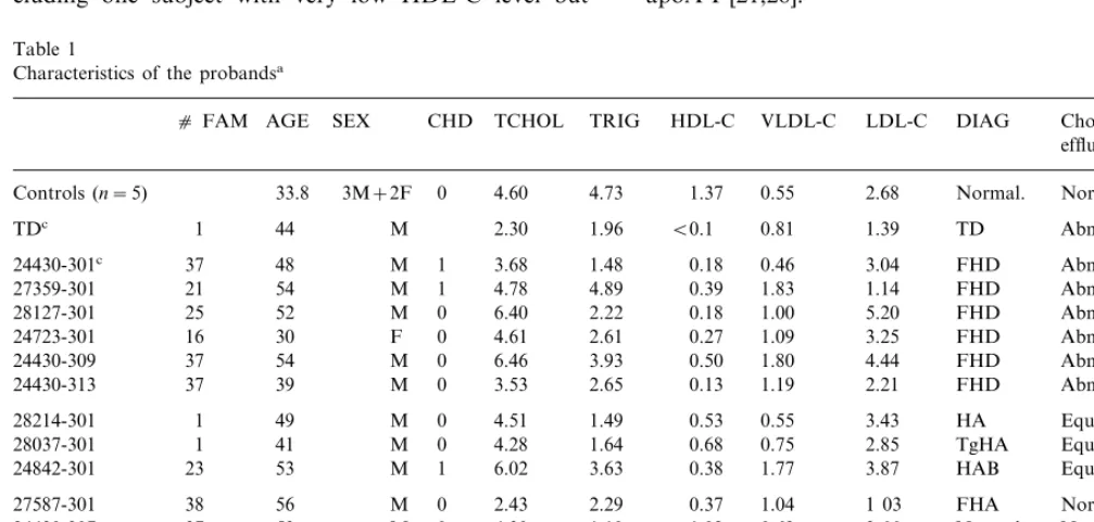
cm2 flasks. After subsequent passages, cells were incu-bated with DMEM containing 10% FBS (DMEM-FBS) in 75 cm2stock flasks for 5 – 15 passages. After the cells were cultured under defined experimental conditions; trypsin (0.05%) in 0.53 mM. EDTA-4Na (Gibco BRL) was used to separate the cells from the flask. Depending on the experiments, 5×10⁴or 5×10⁵cells were seeded in 35 mm petri dishes containing 2 ml of DMEM-FBS. Fibroblasts from 23 subjects were used for these exper-iments: five normolipidemic controls, one subject with TD (cell line TDI as previously described [19]), and 16 subjects with hypoalphalipoproteinemia (Table 1), in-cluding one subject with very low HDL-C level but
with associated mild hypertriglyceridemia, mild hyper-glycemia and elevated apoB level (24292-301), and three subjects from the same kindred (24430-301, 309 and 313). A fourth subject from kindred 24430, subject 24430-307, is normolipidemic and was also included in the analysis.
2.4. Cellular cholesterol labeling and loading
The protocol for cellular cholesterol efflux experi-ments was described in detail elsewhere [28]. The cells
were 3
H-cholesterol labeled during growth and free cholesterol loaded in growth arrest. We seeded 5×10⁴ cells in 35 mm cell culture dishes; at :50% confluence,
0.2 mCi/ml 3H-cholesterol was added. When the cells reached confluence, they were washed in PBS
contain-ing 1 mg/ml BSA and the medium was replaced by
DMEM without serum (growth arrest) containing 2 mg/ml BSA and 20 mg/ml free cholesterol for 24 h. Cellular cholesterol pools were allowed to equilibrate
for another 24 h in DMEM containing 1 mg /ml BSA
and efflux studies (0 – 24 h) were then carried out using 10 mg/ml purified apoA-I. We used apoA-I rather than HDL3 because differences in cholesterol efflux between control and FHD cells were more pronounced with apoA-I [21,28].
Table 1
Characteristics of the probandsa
cFAM AGE SEX CHD TCHOL TRIG HDL-C VLDL-C LDL-C DIAG Cholesterol effluxb
33.8 3M+2F 0 4.60
Controls (n=5) 4.73 1.37 0.55 2.68 Normal. Normal
1 44 M 2.30 1.96 B0.1 0.81 1.39 TD Abnormal
TDc
Abnormal
48 M
24430-301c 37 1 3.68 1.48 0.18 0.46 3.04 FHD
Abnormal
54 M
27359-301 21 1 4.78 4.89 0.39 1.83 1.14 FHD
Abnormal
24430-309 3.93 0.50 1.80 4.44 FHD Abnormal
24430-313 37 39 M 0 3.53 2.65 0.13 1.19 2.21 FHD Abnormal
1 49 M 0 4.51
28214-301 1.49 0.53 0.55 3.43 HA Equivocal
28037-301 1 41 M 0 4.28 1.64 0.68 0.75 2.85 TgHA Equivocal
Equivocal 53
24842-301 23 M 1 6.02 3.63 0.38 1.77 3.87 HAB
38 56 M 0 2.43
27587-301 2.29 0.37 1.04 1 03 FHA Normal
37 53 M 0 4.30
24430-307 1.10 1.02 0.62 2.66 Normal Normal
1 51 M 0 4.68
27413-301 2.76 0.65 1.26 2.76 HA Normal
1.85 4.72
1 M 42
71 0.70 0.84 3.18 FHA Normal
24292-301
1 40 M 1 5.85
27730-301 2.04 0.28 0.93 4.68 HA Normal
27558-301 1 62 F 1 3.83 2.18 0.34 1.05 2.44 HA Normal
45 1
23855-301 M 1 3.50 1.12 0.68 0.51 2.31 HA Normal
aCHD refers to coronary heart disease, TCHOL to total cholesterol, TRIG to triglyceride levels, expressed in mmol/L, as are levels of HDL-C,
VLDL-C and LDL-C;cFAM refers to the number of subjects examined in each family. DIAG refers to the lipoprotein phenotype identified in the proband. FHD: familial HDL deficiency; TgHA: hypertriglyceridermia with low HDL; TD: Tangier disease; HAB: hyperapob; HA: hypoalphalipoproteinemia.
b 3H-cholesterol efflux at 24 h.
2.5. Preparation of HDL3 and apoA-I
HDL3 was freshly prepared from a plasma pool of
normolipidemic donors. Lipoproteins were isolated by standard sequential ultracentrifugation with density (d) adjusted with KBr (HDL3 d=l.125 – 1.210 g/ml). The preparation was extensively dialyzed in PBS (NaCl, 138
mM; KCl, 2.7 mM; NaOH, 51.7 mM; KH2PO4, 0.575
mM; EDTA, 0.385 mM; pH 7.4) and stored at 4°C for up to 1 month. Protein concentration was determined by the method of Lowry. ApoA-I was isolated by gel permeation chromatography as described [15] after iso-lation from whole blood of total HDL particles by ultracentrifugation. The HDL preparation was delipi-dated in acetone:ethanol (1:1) and diethyl ether. HDL proteins were then evaporated to dryness under a
stream of N2and resuspended in 50 mM glycine, 4 mM
NaOH, 0.5 M NaCl and 6 M urea (pH 8.8) at the
concentration of 20 – 30 mg/ml. HDL proteins were
fractionated at 4°C on two Sephacryl S-200 (Pharma-cia) column (2.6×100 cm) equilibrated and eluted with
the same buffer (45 ml/h). Fractions containing the
apoA-1 peak were extensively dialyzed in 0.01 M NH4HCO3, then lyophilized and resuspended in PBS at
concentration of 1 mg/ml. Protein purity on each
apoA-I fraction was assayed on PAGGE and appropri-ate fractions pooled, dialyzed in PBS and Iyophilized
before being stored at −70°C.
2.6. Cholesterol efflux studies
Efflux studies were carried out from 0 to 24 h in the presence of purified apoA-I (10mg protein/ml medium). Efflux was determined as percent of 3H cholesterol in the medium after the cells were incubated for specified periods of time. All experiments were performed in triplicate, in the presence of cells from one control subject and the cells from the study subjects to be examined; some experiments included cells from one subject with TD. All results were confirmed at least twice. We performed experiments on the kinetics of cholesterol efflux in the presence of apoA-I at times 0, 4, 6, 12 and 24 h (data not shown). The most significant difference in efflux was observed at the 24-h time point. Because of the biological variability in efflux in control
subjects, we considered a \40% reduction from the
mean of all controls (set at 100%) as a significant reduced efflux.
2.7. Cell fusion studies
The 3H-cholesterol labeled cells were treated with
trypsin (0.05% trypsin, 0.53 mM EDTA-4Na) to detach cells from the flasks. The cells were counted and 200 000 cells of each cell type (400 000 cells in total) were plated together in 35 mm Petri dishes and grown
overnight until confluence in 2 ml DMEM-FBS. The cells were washed once with PBS and then incubated for exactly 57 s with 0.7 ml of a 40% polyethylene glycol 1000 (J.T. Baker) solution in PBS. After exten-sive washes with freshly prepared DMEM, the cells were incubated in 1 mL of DMEM-FBS and this medium was changed after 1 h. The loading with free cholesterol (20mg/ml) was performed 8 h after the cell fusion. The efflux protocol was then followed as de-scribed above. All fusion experiments were carried out at times 0 and 24 h. As a control, all experiments were carried out in the absence or the presence of PEG treated cells. As a control, we also included a fusion of FHD with FHD and TD with TD cells. Cellular proteins were determined in order to ascertain that cell loss did not occur when incubated in the presence of PEG. However, as PEG is highly cytotoxic, a certain amount of cell loss is inevitable. In such experiments, a large percentage of cells will not become fused; further-more, cells from one kind could fuse with other cells of
the same kind. Thus, the percentage of TD+FHD
fused cells was expected to be relatively small. In the
TD+FHD cells without PEG, the cholesterol efflux is
expected to be the mean level of efflux seen with the FHD cells and TD cells individually. In the presence of
PEG, this TD+FHD fusion would be expected to
increase efflux above this level, if the cells complement each other. However, a lack of increase in cellular cholesterol efflux would suggest that complementation did not occur.
2.8. Sequencing of the ABCA1 gene
All the exons of the ABCA1 gene were sequenced as previously described [22,25].
3. Results
The clinical and biochemical characteristics of the probands are listed in Table 1. We studied 14 probands with unexplained hypoalphalipoproteinemia, one pa-tient with classical TD (previously described in refer-ence [19]) and completed families on eight kindred. In one kindred (24430), we report data on the affected proband (301), two affected brothers (309 and 313) and one normolipidemic, non-affected brother (307) (Fig. 1). Thus, we present data on 15 separate probands and three additional family members from one kindred. Subjects were selected on the basis of an unexplained decrease in HDL-C the absence of diabetes and severe hypertriglyceridemia, a normal pattern of migration of apoA-I on IEF and the lack of clinical features seen in TD or LCAT deficiency [15]; specifically, clinical exam-ination of the probands did not reveal corneal
S
.
Mott
et
al
.
/
Atherosclerosis
152
(2000)
457
–
468
461
Fig.
1.
(
Continued
S.Mott et al./Atherosclerosis152 (2000) 457 – 468 463
Fig.
1.
(
Continued
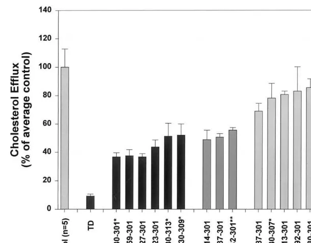
Fig. 2. Cellular cholesterol efflux in control subjects (n=5, normalized to 100%) one subject with TD, six subjects with FHD, three subjects with equivocal results (the lack of family data does not allow unambiguous assignment to FHD), seven subjects with unexplained hypoalphalipo-proteinemia and one subject (24430-307) who is the normolipidemic brother of affected sibs 24430-301, 24430-309 and 24430-313 (from Table 1). Cholesterol efflux was measured at 24 h, normalized to a percentage of controls. In a typical experiment, the level of cholesterol efflux in control subjects at 24-h was 8 – 12% of total cholesterol (medium plus cell3H-cholesterol); that of FHD cells was 4 – 6% and TD cells 2% of total
cholesterol.
hepatomegaly or enlarged spleen or evidence of a pe-ripheral demyelinating neuropathy. Colonoscopy was not performed on any subject to test for the presence of hystiocytic infiltration of the mucosa, a finding consid-ered by some to be pathognomonic of TD [14]. We have previously reported that plasma triglyceride levels were mildly to moderately elevated in FHD; therefore only patients with triglyceride B95th percentile were
included. The mean plasma lipid and lipoprotein cholesterol values for the study subjects are shown in Table 1. Reference values for HDL-C were taken from the Lipid Research Clinical data tables [29]. As previ-ously noted, many cases of reduced HDL-C are sec-ondary to elevated triglycerides and (or) increased apoB-containing lipoprotein secretion by the liver. Cel-lular cholesterol efflux was examined on fibroblasts obtained from each patient. We have previously re-ported a decrease in cholesterol efflux in cells from subjects 24430-301 and his brother 24430-313, and sub-ject 24723-301 [28]. These results were confirmed in the present study and three new probands and one addi-tional family member were identified: subjects 27359-301, 28127-301 24842-27359-301, and 24330-309 were also found to have decreased cellular cholesterol efflux (Fig.
2 and Table 1). Thus, 7/14 (50%) of the probands
studied had a reduced cellular cholesterol efflux defect. Mutations in these affected subjects have been previ-ously reported [25]: subject 24430-301 had a C6370T
mutation, leading to a nonsense mutation
Arg2084STOP; patient 24723-301 had a D2017-19
dele-tion, leading to a DLeu631 deletion; patient 28127-301 had a C2665T mutation, leading to a nonsense muta-tion Arg849STOP and patient 27359-301 had a 6 bp
deletion D5618-23, leading to a DGlu1833 and DAsp
1834 deletion. The patient with TD was heterozygote for two mutations: a T4369C (Cys1417Arg) mutation and a mutation leading to a truncated mRNA species [22]. In the remaining seven probands, no cholesterol efflux defect was identified. These cases, therefore, are still considered to be FHD but possibly caused by a different genetic disorder than mutations at the ABCA1 gene locus. In the other cases, the patients were consid-ered to have primary hypoalphalipoproteinemia (HA) or, on the basis of family studies, as having familial hypoalphalipoproteinemia (FHA) of yet unknown causes. In other families, a pattern of familial hyper-triglyceridemia with hypoalphalipoproteinemia (FT-gHA) was identified, as we previously reported [13]. We pooled the results from five control subjects and arbi-trarily set this value at 100%. Because of variability in the assay, we consider values that fall within 75 – 125% of controls as normal cholesterol efflux.
S.Mott et al./Atherosclerosis152 (2000) 457 – 468 465
We had previously classified subject 24842-301 as hav-ing familial combined hyperlipidemial hyperapoB on the basis of elevated cholesterol, triglyceride and apoB levels [15]. However, on repeated testing, cells from this patient had a lesser degree of defective efflux and the cholesteryl ester mobilization was reduced, compared with normal [15]. Using DNA markers spanning the ABC1-CERP genomic region, we were unable to show co-segregation of the low HDL-C trait with the chro-mosome 9q31 region in this subject. Extensive haplo-type analysis of the region at 9q31 in these families fail to show segregation with the low HDL-C trait, al-though family 28037 is limited in size and, in family
28214, no other subject had and HDL-C B5th
percentile.
There is a significant correlation (r=0.545, P=
0.029) between the HDL-C level and cholesterol efflux in our 17 (14 probands plus three additional family members) subjects tested (the TD subject and controls were excluded from this analysis). This suggests that
30% of the variability in HDL-C levels in our
sub-jects with low HDL-C may be due to cellular choles-terol efflux.
We performed family studies in eight kindred. Three of these families have been reported elsewhere [22 – 25]. In six subjects from four kindred (indicated in Table 1), a cellular cholesterol efflux defect was identified. The pedigree of the four kindred with a cholesterol efflux defect are shown in Fig. 1a – d. The most consistent pattern of inheritance is Mendelian autosomal co-domi-nant, homozygous FHD subjects have TD. Subjects with heterozygous TD disease have been reported as
being clinically normal but present a low half-normal HDL-C compared with age-matched controls [30].
We next investigated whether these familial forms of decreased HDL-C were associated with atherosclerotic vascular disease. In 8/18 subjects, there was clinically evident CAD) (as evidenced by strict angiographic criteria, myocardial infarction, or myocardial revascu-larization) [27]. The association of the low HDL-C trait with CAD must, however, be interpreted with caution. Because of the referral bias of the clinic, many patients have pre-established cardiovascular disease [27]. The prevalence of a reduced HDL-C level in our patients with premature CAD is 50% [27,31] according to the National Cholesterol Education Program (NCEP)
crite-ria (HDL-C B0.9 mmol/l). The presence of vascular
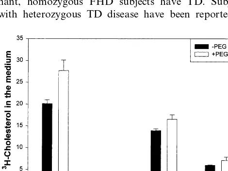
disease in the family was established through direct interview of all family members. The presence of vascu-lar disease in the coronary, aorto-iliac and carotid vascular beds was sought on clinical grounds. In sub-jects with FHD, no segregation with the presence of a low HDL-C and vascular disease was identified [15]. Cell fusion studies were carried out on fibroblasts from a control subject, a subject with FHD and a subject with TD. We determined the optimal time for PEG fusion to be exactly 57 s; longer incubation times resulted in increased cell death due to the toxicity of PEG, as documented by cell protein content, and shorter incubation times resulted in a lack of fusion (data not shown). Approximately 40% of the cells were found to be diploid (or more) by microscopic examina-tion and flow cytometric analysis of the cells. After staining for DNA, cells revealed a marked increase in polyploidy after PEG fusion. Cellular protein mass after PEG incubation and extensive washes was not significantly different than when the cells were incu-bated without PEG. The results of one cell fusion experiment are shown in Fig. 3. Results from four separate fusion experiments yielded similar findings. As a control for PEG fusion, cellular cholesterol efflux is not significantly affected by the presence of PEG in
control, FHD or TD cells. The TD+FHD fused cells
showed a 24-h efflux level intermediate between that observed in individual FHD and TD cells. The fused
TD+FHD cells did not exhibit increased efflux,
sug-gesting that TD cells do not complement the genetic defect in FHD. On the basis of these results, we confirm that FHD and TD represent different manifes-tations of the same genetic defect.
4. Discussion
In the present study, we examined 14 probands (and three family members, 17 subjects) with a marked re-duction in plasma HDL-C levels. In two of these
sub-jects, a syndrome of familial combined
hyperlipidemia/hyperapoB was identified. In 7/14 (50%) probands (from four kindred), a cellular cholesterol efflux defect was identified, an entity referred to as FHD. We have previously postulated that a reduced cholesterol efflux onto apoA-I containing nascent parti-cles (either pre-b migrating or small HDL3 particles) would lead to small HDL particles that are rapidly catabolized. This was documented in two subjects with FHD from the same family (24430-301 and 24430-313, Table 1) in whom stable isotope kinetic studies were performed. In these subjects, apoA-I catabolism was increased 4 – 5-fold compared to control subjects [17]. Similar findings have been reported by Emmerich et al. [32] in a subject with unexplained low HDL-C levels. This supports the concept that an intracellular choles-terol transport and efflux defect can lead to hyper-catabolism of HDL particles. However, in half of the
cases examined (7/14, of which five have a primary
form of hypoalphalipoproteinemia and two had a com-bination of elevated apoB and increased triglyceride levels), no efflux defect was identified with our proto-col. These subjects have a primary form of HDL defi-ciency that remains unexplained.
Patients exhibiting moderate to severe HDL-C defi-ciency comprise therefore a heterogeneous group. The search for a genetic etiology in such patients has been thwarted by a lack of precise phenotypes. The identifi-cation of subjects with FHD (defined here as a defect in cellular cholesterol transport and efflux) has allowed a molecular diagnosis, with mutations at the ABCA1 gene having been identified, segregating with the low HDL trait [22,25].
Investigations of fibroblasts obtained from subjects with Tangier disease have shown that these cells have a
profound defect in apoA-I or HDL3-mediated cellular
cholesterol efflux [18 – 20]. Similarly, one report on sub-jects with severe HDL deficiency but without clinical evidence of TD has also shown a defect in cellular cholesterol efflux [33]. We have reported similar find-ings on our patients.
The results obtained with the cell fusion experiment confirm that FHD and TD represent different manifes-tations of a similar genetic defect. Tangier disease re-sults from a homozygous (or compound heterozygous) genetic defect at the ABCA1 gene locus while FHD (defined as a defect in cellular cholesterol efflux) is caused by heterozygous mutations at the ABCA1 gene. Because FHD is transmitted as an autosomal co-domi-nant trait, the phenotypic defect in FHD is less severe than TD, but far more frequent. Cell fusion experi-ments suffer severe limitations in interpretation. First, only a percentage of cells are fused with PEG; second, there are no cell markers that allow distinction between fused cells (i.e. fusion between FHD and TD cells) and third, cell fusion methodology is best applied in reces-sive disorders.
Tangier disease is considered a very rare lipoprotein disorder [14,30]. But a defect in cellular cholesterol transport and efflux is identified in half of the patients we have examined in the present study. It is possible, therefore, that anomalies in cellular processing of cholesterol represent a hitherto unrecognized etiology for moderately severe hypoalphalipoproteinemia. The TD/FHD cellular phenotype therefore might be a rela-tively common cause of low HDL-C states.
The treatment of subjects with FHD presents a clini-cal challenge. Reports from Serfaty – Lacrossnie`re on one subject with Tangier disease show that these pa-tients show little or no response to conventional treat-ments (despite an increase in apoA-I levels by a factor of two, the clinical significance of raising apoA-I from 2 to 4 mg% is doubtful) [30]. Subject 24430-301 has been treated sequentially for three-month periods with lovastatin 20 mg/d, niacin 3000 mg/d and fenofibrate 300 mg/d with little effect on HDL-C levels (data not shown). Although anecdotal, these results suggest that
the cellular defect in TD/FHD might be difficult to
overcome with currently available lipid modulating agents.
Cholesterol efflux from the cell follows two processes: first, diffusion of membrane cholesterol onto an accept-ing particle, and second, the active transport of intra-cellular cholesterol to the plasma membrane. Efflux of cholesterol from the membrane appears to be mediated by passive desorption of cholesterol from the cell mem-brane to apoA-I-phospholipid complexes, following a concentration gradient of free cholesterol and acceptor particles [34 – 36]. The shape, composition and confor-mation of HDL particles may also influence cholesterol efflux [37,38]. Intracellular cholesterol translocation to the plasma membrane may be mediated through apoA-I-cell interaction via yet uncharacterized specific mem-brane proteins, the nature of which continue to stir controversy [39]. Subsequent work has suggested that a HDL receptor may act as a signal-transducing protein, which promotes intracellular cholesterol transport to the cell membrane. In this model, the binding of HDL stimulates the activation and movement to the plasma membrane of protein kinase C (PKC), most likely via the phosphoinositol pathway [40]. The PKC isoforms identified in fibroblasts are PKC a, d, oand z (theb1,
b2, g and h are not seen in fibroblasts or expressed in very small quantities) [41,42]. HDL3 binding to cells stimulates the phosphorylation of several proteins, the roles of which are not fully known but include ERK-1 and ERK-2, members of the mitogen-activated protein kinases (MAPK) [42,43]. The current concept is that this mechanism translocates cellular cholesterol from various pools to the cell membrane where it will be
available for efflux by desorption. HDL3 induces the
hydrolysis of phosphatidylcholine by phospholipases C
S.Mott et al./Atherosclerosis152 (2000) 457 – 468 467
apoA-I-mediated activity of phospholipase C and D (as assessed by the formation of DAG and phosphatidic acid) was found to be markedly impaired [44]. A similar pattern of abnormal phospholipase C and D activation in TD cells can be reproduced by incubation with pertussis toxin, suggesting the involvement of a G protein in TD [44]. Cellular cholesterol content is regu-lated by: a) cholesterol synthesis in the smooth endo-plasmic reticulum (via HMG CoA reductase); b) receptor-mediated endocytosis of LDL or selective free cholesterol uptake from LDL; c) cholesterol efflux from plasma membrane to cholesterol acceptor particles (pre-dominantly HDL), and d) intracellular cholesterol
es-terification via the enzyme acyl-CoA:cholesteryl
acyltransferase (ACAT) [34,45]. The first two pathways are coordinately regulated at the level of gene transcrip-tion. Cells obtain cholesterol from two predominant sources: thede no6osynthesis involving the HMG CoA
reductase pathway or, preferentially, the endocytosis of LDL particles [46]. Surprisingly, relatively little is known about the intracellular trafficking of cholesterol and its transport to the plasma membrane from the various pools of origin [34]. There is accumulating evidence that the trans-Golgi network is a common pathway for transport of cellular cholesterol to the plasma membrane [47].
In the study by Rogler et al. in fibroblasts from a patient with Tangier disease, the efflux defect was re-versible by the selective activation of PKC with
1,2-dioctanoylglycerol (DOG) (10−5 mol
/l) [20]. This strongly suggests that translocation of cellular choles-terol is mediated, at least in part, by a PKC-mediated pathway and can be overcome by activation of the signal transduction pathway.
In conclusion, moderately severe HDL-C states are frequently caused by a defect in cellular cholesterol efflux. Half such patients identified, including those with Tangier disease, have mutations at the ABCA1 gene [22 – 25]. It remains to be determined whether the cholesterol efflux pathway can be modulated by hor-monal mediators or from selective activation of the signaling transduction pathway.
Acknowledgements
Supported by an Operating Grant from the Medical Research Council of Canada (MT 15432) and the Heart and Stroke Foundation of Canada and by a salary award from the Fonds de la recherche en sante´ du Que´bec (JG Jr). The authors wish to express their gratitude to the clinical staff of the Clinical Research Institute of Montre´al for their support for this project and to Paule Marchand for editorial assistance. The advice of David Watkins for help in establishing the cell fusion protocol is gratefully acknowledged.
References
[1] Eisenberg S. High density lipoprotein metabolism. J Lipid Res 1984;25:1017 – 58.
[2] Barter PJ, Rye K-A. High-density lipoproteins and coronary heart disease. Atherosclerosis 1996;121:1 – 12.
[3] Genest J Jr, Marcil M, Denis M, Yu L. High density lipo-proteins in health and in disease. J Invest Med 1999;47:31 – 42. [4] Castelli WP, Garrison RJ, Wilson PW, Abbott RD, Kalousdian
S, Kannel WB. Incidence of coronary heart disease and lipo-protein cholesterol levels: the Framingham study. J Am Med Acad 1986;256:2835 – 8.
[5] Gordon T, Castelli WP, Hjortland MC, Kannel WB, Dawber TR. High density lipoprotein as a protective factor against coronary heart disease: the Framingham study. Am J Med 1977;62:707 – 14.
[6] Genest J Jr, McNamara JR, Ordovas JM, et al. Lipoprotein cholesterol apolipoprotein A-1 and B and lipoprotein (a) abnor-malities in men with premature coronary artery disease. J Am Coll Cardiol 1992;1979:27 – 8.
[7] Assmann G, Schulte H, von Eckardstein A, Huang Y. High-den-sity lipoprotein cholesterol as a predictor of coronary heart disease risk: the PROCAM experience and pathophysiological implications for reverse cholesterol transport. Atherosclerosis 1996;124:1 – 20.
[8] Goldbourt U, Medalie JH. High-density lipoprotein cholesterol and incidence of coronary heart disease-the Israeli Ischemic heart disease study. Am J Epidemiol 1979;109:296 – 308. [9] Roma P, Gregg RE, Meng MS, et al. In vivo metabolism of a
mutant form of apolipoprotein A-I, apo A-IMilano, associated
with hypoalphalipoproteinemia. J Clin Invest 1993;91:1445 – 52. [10] Calabresi L, Franceschini G. High density lipoprotein and coro-nary heart disease: insights from mutations leading to low high density lipoprotein. Curr Opin Lipidol 1997;8:219 – 24. [11] Genest J Jr, Cohn J. Plasma triglyceride-rich lipoproteins and
high density lipoprotein disorders associated with atherosclero-sis: biomedicine’98 vascular biology symposium. J Invest Med 1998;46:351 – 8.
[12] Gaziano JM, Hennekens CH, O’Donnell CJ, Breslow JL. Fast-ing triglycerides, high density lipoprotein, and risk of myocardial infarction. Circulation 1997;96:2520 – 5.
[13] Genest J Jr, Bard J-M, Fruchart J-C, Ordovas JM, Schaefer EJ. Familial hypoalphalipoproteinemia in premature coronary artery disease. Arterioscler Thromb 1993;13:1728 – 37.
[14] Assmann G, vonEckardstein A, Brewer HB. Familial high den-sity lipoprotein deficiency: Tangier disease. In: Scriver CR, Beaudet AL, Sly WS, Valle D, editors. The metabolic and molecular basis of inherited disease, 7th. USA: McGraw-Hill, 1995:2053 – 72.
[15] Marcil M, Boucher B, Frohlich J, Davignon J, Solymoss BC, Genest J Jr. Severe familial HDL deficiency (FHD) in French Canadian kindred: clinical, biochemical and molecular character-ization. Arterioscler Thromb Vasc Biol 1995;15:1015 – 24. [16] Schaefer EJ, Ordovas JM. Metabolism of apolipoproteins A-1,
A-II, and A-IV. Meth Enzymol 1986;129:420 – 43.
[17] Batal R, Tremblay M, Krimbou L, Mamer O, Davignon J, Genest J Jr, Cohn JS. Familial deficiency characterized by hypercatabolism of mature apoA-1 but not proapoAI. Atheroscler Thromb Vasc Biol 1998;18:655 – 64.
[18] Walter M, Gerdes U, Seedorf U, Assmann G. The high density lipoprotein- and apolipoprotein AI-induced mobilization of cel-lular cholesterol is impaired in fibroblasts from Tangier disease subjects. Biochem Biopyhs Res Commun 1994;205:850 – 6. [19] Francis GA, Knopp RH, Oram JF. Defective removal of cellular
[20] Rogler G, Trumbach B, Klima B, Lackner KJ, Schmitz G. HDL-mediated efflux of intracellular cholesterol is impared in fibroblasts from Tangier disease patients. Arterioscler Thromb Vasc Biol 1995;15:683 – 90.
[21] Remaley AT, Schumacher JA, Farsi JA, Nazih BD, Brewer HB Jr. Decreased reverse cholesterol transport from Tangier disease fibroblasts: acceptor specificity and effects of brefeldin on lipid efflux. Atheroscler Thromb Vasc Biol 1997;17:1813 – 21. [22] Brooks-Wilson A, Marcil M, Clee S, et al. Mutations in the ATP
binding cassette (ABC1) transporter genein Tangier disease and familial HDL deficiency (FHA). Nat Genet 1999;22:336 – 45. [23] Bodzioch M, Orso E, Klucken J, et al. Nat Genet 1999;22:347 –
51.
[24] Rust S, Rosier M, Funke H, et al. Tangier disease is caused by mutations in the gene encoding ATP binding cassette transporter 1. Nat Genet 1999;22:352 – 5.
[25] Marcil M, Brooks-Wilson A, Clee S, et al. Mutations in the ABC1 gene in familial HDL deficiency with defective cholesterol efflux. Lancet 1999;354:1341 – 6.
[26] Genest J Jr, Martin-Munley SS, McNamara JR, et al. Familial lipoprotein disorders in patients with premature coronary artery disease. Circulation 1992;85:2025 – 33.
[27] Weber M, McNicoll S, Marcil M, et al. Metabolic factors clustering lipoprotein cholesterol, apolipoprotein B, lipoprotein (a) and apolipoprotein E phenotypes in premature coronary artery desease in French Canadians. Can J Cardiol 1997;13:253 – 60.
[28] Marcil M, Lu Y, Krimbou L, Boucher B, Oram J, Cohn JS, Genest J Jr. Cellular cholesterol transport and efflux from fibroblasts are abnormal in subjects with familial HDL defi-ciency. Arterioscler Thromb Vasc Biol 1999;19:159 – 69. [29] The Lipid Research Clinics Population Studies Data Book, vol.
1. The prevalence study. Washington, D.C.: Department of Health and Human Services, Public Health Service, July 1980 p.28 – 81. (NIH publication number 80-1527).
[30] Serfaty-Lacrosniere C, Civeira F, Lanzberg A, et al. Ho-mozygous Tangier disease and cardiovascular disease. Atherosclerosis 1994;107:85 – 98.
[31] Genest J Jr, McNamara JR, Salem DN, Schaefer EL. Prevalence of risk factors in men with premature coronary artery disease. Am J Cardiol 1991;67:1185 – 9.
[32] Emmerich J, Verges B, Tauveron I, et al. Familial HDL defi-ciency due to marked hypercatabolism of normalapoA-I. Arte-rioscler Thromb 1993;13:1299 – 306.
[33] Eberhart GP, Mendez AJ, Freeman MW. Decreased cholesterol efflux from fibroblasts of a patient without Tangier disease, but
with markedly reduced high density lipoprotein cholesterol lev-els. J Clin Endocrinol Metab 1998;83:836 – 46.
[34] Fielding G, Fielding PE. Intracellular cholesterol transport. J Lipid Res 1997;38:1503 – 21.
[35] Liscum L, Munn NJ. Intracellular cholesterol transport. Bioch-ern Biophys Acta 1999;1438:19 – 37.
[36] Phillips MC, Gillotte KL, Haynes MP, Johnson WJ, Lund-Katz S, Rothblat GH. Mechanisms of high density lipoprotein-medi-ated efflux of cholesterol from cell plasma membranes. Atherosclerosis 1998;137:13 – 7.
[37] De la Llera Moya M, Atger V, Paul JL, et al. A cell culture system for screening human serum for ability to promote cellular cholesterol efflux: relations between serum components and efflux, esterification, and transfer. Arterio Thromb 1994;14:1056 – 65.
[38] Davidson WS, Gillotte KL, Lund-Katz S, et al. J Biol Chem 1995;270:5882 – 90.
[39] Fidge N. High density lipoprotein receptors, binding proteins, and ligands. J Lipid Res 1999;40:187 – 201.
[40] Mendez AJ, Oram JF, Bierman EL. Protein kinase C as a mediator of high density lipoprotein receptor-dependent efflux of intracellular cholesterol. J Biol Chem 1991;266:10104 – 11. [41] Choi SW, Park HY, Rubeiz NG, Sachs D, Gilchrest BA. Protein
kinase C-alpha levels are inversely associated with growth rate in cultured human dermal fibroblasts. J Dermatol Sci 1998;18:54 – 63.
[42] Deeg MA, Garver WS, Bierman EL, Oram JF. HDL stimulates phosphorylation of 18 and 80 kDa proteins in cholesterol-loaded human skin fibroblasts. Circulation 1993;88:1215 Abstracts. [43] Deeg MA, Bowen RF, Oram JF, Bierman EL. High density
lipoproteins stimulate mitogen activated protein kinases in hu-man skin fibroblasts. Arterioscler Thromb Vasc Biol 1997;17:1667 – 74.
[44] Walter M, Reinecke H, Gerdes U, et al. Defective regulation of phosphatidylcholine-specific phospholipases C and D in a kin-dred with Tangier disease: evidence for the involvement of phosphatidylcholine breakdown in HDL- mediatedcholesterol efflux mechanisms. J Clin Invest 1996;98:2315 – 23.
[45] Liscum L, Underwood KW. Intracellular cholesterol transport and compartmentation. J Biol Chem 1995;270:15443 – 6. [46] Goldstein JL, Brown MS. Regulation of the mevalonate
path-way. Nature 1990;343:425 – 30.
[47] Mendez AJ, Uint L. Apolipoprotein-mediated cellular choles-terol and phospholipid efflux depend on a functional Golgi apparatus. J Lipid Res 1996;37:2510 – 24.