117 THE MICROSCOPE • Vol 57:3, pp 117-120 (2009)
What’s in the Pot? An Investigation Into the Use
of a Byzantine Ceramic Vessel
1Meggan King
McCrone Research Institute*
1 Presented at Inter/Micro 2008.
* 2820 S. Michigan Avenue, Chicago, IL 60616
KEYWORDS
Ceramic, pottery, water vessel, Amorium, Turkey, Byzantine, archaeology, polarized light microscopy (PLM), scanning electron microscopy (SEM), micro-chemistry
ABSTRACT
During the 2007 excavation season at the Byzan-tine city of Amorium, a shard of ceramic with an as-sociated residue, which was believed to have been in the interior of this piece of ceramic vessel, was col-lected and submitted to the author for analysis. The study was divided into two parts: analysis of the resi-due and analysis of the ceramic shard. Identification of the residue revealed that the ceramic may have been used as a water vessel in the past. Characterization of the ceramic shard could yield information regarding the source material and manufacturing process of the vessel.
INTRODUCTION
Amorium is a medieval city of the Byzantine Em-pire located in central western Turkey. A brief history of Amorium has been published previously in The
Mi-croscope (1), and additional information about the
exca-vation is available from the Amorium Excaexca-vation Project (2).
In this study, a ceramic shard and its associated lining (residue) was collected from area AM-07, A-20,
Area 13 during the 2007 excavation season at the Amorium excavation site and submitted to the author for analysis (Figures 1 and 2).
MATERIALS AND METHODS
Analysis was performed on objects after they were cleaned to eliminate interference. The supernatant liq-uid that was used for washing, along with any debris that was removed from the pieces were retained.
Residue
The residue was first examined using a stereomi-croscope. A small piece of the residue was submerged in distilled water and observed under the polarized light microscope (PLM) to determine whether the due was water soluble. A small fragment of the resi-due (approximately 0.5 cm x 0.5 cm) was washed by placing it into a microcenterfuge tube with distilled water and a small amount of ethyl alcohol.
The tube was then held in an ultrasonic bath for several minutes and any debris was allowed to settle. The fragment was then removed and allowed to dry. All of the supernatant liquid was retained. After dry-ing, a piece of the clean fragment was prepared for examination using PLM by crushing and mounting the resulting particles in n=1.66 refractive index liq-uid.
118 THE MICROSCOPE 57 (2009)
Ceramic Shard
The ceramic shard was first examined macroscopi-cally and then by stereomicroscopy. A small fragment (approximately 1 cm x 0.5 cm) was removed from the original ceramic shard. This piece was then washed and allowed to dry using the same technique that was used for the residue. The fresh fractured surfaces were examined in order to assess the ceramic fabric and at-tempt to determine the forming method of the vessel. A portion of the fragment was also crushed, using a mortar and pestle, to examine the mineral composi-tion by PLM and reflected light microscopy. To pre-pare the crushed material for microscopical analysis,
it was first separated by size fraction. The crushed material was placed in a 50 mL beaker along with a few drops of ethanol and 5-7 mL of distilled water. The beaker was swirled and then placed in an ultrasonic bath for 1 minute. The supernatant liquid containing the fine silt and clay fraction was decanted into a new clean beaker and retained.
This process was repeated multiple times until the supernatant liquid remained clear after sonication, leaving behind the coarse mineral fraction. The coarse mineral fraction was allowed to dry and was then separated by density using bromoform (density = 2.9 g/mL). Bromoform separations allow “light” minerals
Figure 1. Concave surface of Byzantine ceramic shard (left) and associated residue (right).
Figure 2. Convex surface of Byzantine ceramic shard (left) and associated residue (right).
Figure 3. Plane-polarized light photomicrograph of a suspected calcium carbonate particle mounted in n=1.66, polarizer oriented N-S.
119 MEGGAN KING
to be separated from “heavy” minerals. Light miner-als will float on the surface of the bromoform and can be removed with a pipette while heavy minerals will sink to the bottom of the microcenterfuge tube. The light mineral fraction was prepared by mounting par-ticles in n=1.540 refractive index liquid and the heavy mineral fraction was prepared by mounting particles in n=1.660 refractive index liquid. Optical properties determined by PLM were used to identify the miner-als present.
RESULTS AND DISCUSSION
Residue
Stereomicroscopy was used for initial observa-tions and it was noted that the material is
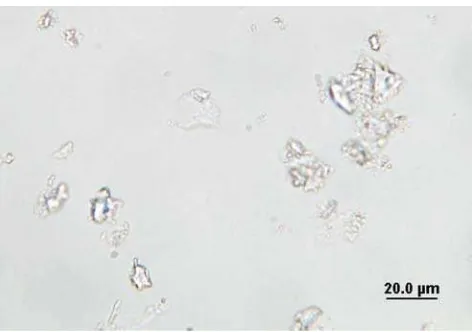
approxi-mately 300 μm thick, pitted, porous and brittle. It was also determined that this residue is not readily water soluble. Under the PLM, the residue appears to be composed almost entirely of calcium carbonate due to the high birefringence and large contrast change observed in the n=1.660 liquid mounting medium when the polarizing filter or stage is rotated (Figures 3 and 4).
Carbonates were also detected by submersing a portion of crushed residue in dilute hydrochloric acid and observing the effervescence of CO2. The resulting
spectra from SEM/EDS analysis are shown in Figures 5 and 6. The major elements present in the residue are calcium, carbon and oxygen. Minor amounts of mag-nesium, aluminum, silicon, phosphorus, sulfur and po-tassium were also present.
Figure 5. EDS spectrum of the concave surface of the residue. Major elements detected include calcium, carbon and oxygen.
Figure 6. EDS spectrum of the convex surface of the residue. Major elements detected include calcium, carbon and oxygen.
Figure 8. Tourmaline (shown with arrow) in light fraction mounted in n=1.66, plane polarized light.
120 THE MICROSCOPE 57 (2009)
Ceramic shard
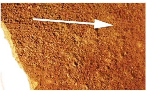
The ceramic shard has a thickness ranging from 3.4 mm to 4.9 mm. The concave surface of the ceramic piece has a red-orange color (Figure 1); the convex sur-face of the ceramic piece has a dark brown-black col-ored surface (Figure 2). The interior surface (Figure 7) exhibits “rilling” — undulating ridges and striations on the wall surface (3).
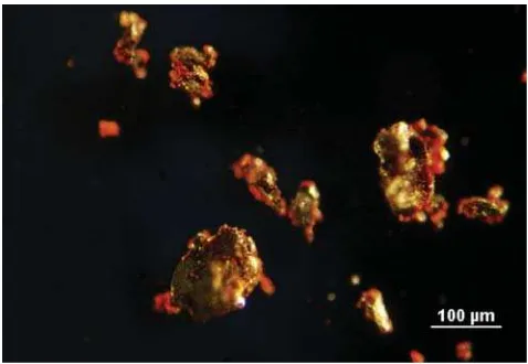
The light fraction is composed mainly of quartz and calcite with small amounts of alkali feldspar and tourmaline (Figure 8). The quartz and calcite are of-ten intimately associated (Figure 9). Many quartz and feldspar grains are cemented together with diagenetic calcite (4).
The heavy fraction is mainly composed of iron ox-ides many of which are magnetic and likely magne-tite; the non-magnetic iron oxides are likely hematite (Figure 10). Some carbonates and biotite mica are also present.
CONCLUSIONS
Due to the relative uniform thickness of the shard, presence of parallel striations (rilling) on the interior surface, and the cultural context of the piece, it is most likely that the vessel was formed using a wheel-throw-ing technique.
The residue is composed almost entirely of calcium carbonate, with small amounts of other trace elements as seen by SEM/EDS.
The ceramic piece has a mineral composition con-sisting primarily of calcite, quartz and iron-oxide based minerals. The intimate association of the quartz and calcite found in the light mineral fraction indi-cates that some of the ceramic source material is most likely from sedimentary rocks.
Assuming that the ceramic piece was once part of a vessel and that the residue was at one time attached to the ceramic piece, the analysis performed by PLM and SEM/EDS support the conclusion that the residue is a calcium carbonate scale. This would be expected, if the vessel was used to store or boil water.
ACKNOWLEDGEMENTS
Thanks to Dr. Gary Laughlin of McCrone Research Institute (McRI), Skip Palenik of Microtrace, LLC, An-drew Bowen of Stoney Forensic, and all the staff at McRI for their support and assistance. I am also grate-ful for the help of Jane Foley, Assistant Director/Head of Conservation of the Amorium Excavation Project and Dr. Christopher Lightfoot, the project’s director.
REFERENCES
1. King, Meggan, Bowen, Andrew, Brinsko, Kelly and Sparenga, Sebastian. “Skeleton Crystals,” The
Mi-croscope, 56 (3), pp 125-130,2008.
2. The Amorium Excavations Project, www.amoriumexcavations.org (accessed October 30, 2009).
3. Rice, Prudence M. Pottery Analysis a Sourcebook.
The University of Chicago Press, Chicago, 1987. 4. Palenik, S.J., Microtrace, LLC, Elgin, IL. Personal communication, June 2008.
Figure 9. Quartz grain (black arrow) with diagenetic calcite (white arrow), crossed polars with Red I plate.