Detection of Dermatophyte between Toes of Medical Students Wearing
and not Wearing Socks in Universitas Padjadjaran using Direct
Microscopic Examination
Venna Magarita,1 Ramlan Sadeli,2 Hendra Gunawan3
1Faculty of Medicine Universitas Padjadjaran 2Department of Microbiology Faculty of Medicine
Universitas Padjadjaran, 3Department of Dermatology and Venereology Faculty of Medicine
Universitas Padjadjaran/Dr. Hasan Sadikin General Hospital Bandung
Abstract
Background: Dermatophytes thrive in warm, humid, and moist areas. Wearing the same socks more than a day is one predisposing factor. The aim of this study is to detect the dermatophyte between the fourth and fifth toes of medical students of Universitas Padjadjaran, who wear and do not wear socks.
Methods: This was a cross-sectional descriptive study performed in Microbiology Laboratory of Faculty of Medicine Universitas Padjadjaran from September to October 2013. Direct microscopic examination using 10% Potassium Hydroxide (KOH) was performed on 50 medical students, 25 students who wear socks and 25 students who do not wear socks. The samples were taken from scraped skin between the fourth and fifth toes using the cellophane tape method. The sample size is obtained via stratified random sampling.
Results: From all participants, two students (4%) showed positive result of dermatophyte hyphae and 1 student (2%) showed positive result of dermatophyte hyphae and arthrospore. Of these three students, two students wear socks and one student does not wear socks. The two students who wear socks changed their socks more than a day.
Conclusions: Dermatophyte was detected in only a few medical students of Universitas Padjadjaran, who wear and do not wear socks. [AMJ.2015;2(4):480–4]
Keywords: Dermatophyte, direct microscopic examination, medical students
Correspondence: Venna Magarita, Faculty of Medicine, Universitas Padjadjaran, Jalan Raya Bandung-Sumedang Km.21, Jatinangor, Sumedang, Indonesia, Phone: +6287827590863 Email: [email protected]
Introduction
Dermatophytosis is a superficial fungal
infection involving stratum corneum of the skin, hair, or nails.1 Tinea pedis is one contagious
dermatophyte infection of the toe webs and soles of the feet, mostly found between the
fourth and fifth toes.2,3 Dermatophytosis is
common infection and occurs worldwide. It is estimated that over eight million people visit their primary health care physician for tinea related symptoms each year.4
Dermatophytes thrive on warm, humid, and moist areas.5 Some predisposing factors
of getting infected include hot and humid weather, high perspiration, wearing occlusive footwear, and wearing the same socks for more than a day. Transmission of dermatophytes like Trichophyton rubrum, Trichophyton
interdigitale and Epidemophyton floccosum is
usually via feet. In this site, infections are often
chronic and may remain subclinical for many years.4 Based on the observation, there are
medical students of Universitas Padjadjaran who wear and do not wear socks. Few of them who wear socks do not replace it at least once a day, predisposing them to get dermatophyte infection. Therefore, the aim of this study is to detect the dermatophyte between the
fourth and fifth toes of medical students of
Universitas Padjadjaran who wear and do not wear socks using 10% Potasium Hydroxide (KOH) under direct microscopic examination.
Methods
This was a cross-sectional descriptive study conducted during September to October 2013 in Microbiology Laboratory, Faculty of Medicine, Universitas Padjadjaran.
With a stratified random sampling, a total
of Universitas Padjadjaran class of 2010 participated in this study; 25 students wearing socks, and the other 25 students not wearing socks. From this number, 15 of them were males and 35 females. The samples of students’ informed consent were obtained for the study.
Samples were obtained using cellophane tape to collect the skin samples that had already been scraped between the fourth
and fifth toes. The tape was then placed onto
microscope slide that had a drop of 10% KOH. The slide was labeled into two; those who wore socks and those who did not wear socks, using a label pen.
The microscope slides were examined under a microscope (400×). Here, the hyphae (long,
branching, rod-shaped filaments of uniformed
width with separation lines) or arthrospore was visualized. Data was presented in a table in the form of frequency and percentage.
Results
All 25 students who wore socks used cotton socks. As for the students who did not wear socks, all 25 of them wore closed shoes.
The result of the direct microscopic examination using 10% KOH showed positive
findings in 3 out of the total 50 students (6%).
From 25 students wearing socks, 2 students were positive for dermatophyte and from 25 students who did not wear socks only 1 student was positive for dermatophyte (Table 3).
Out of all 50 students, 2 male students were
Table 1 Respondent Characteristic
Characteristic Wear socks Do not wear socks
n=25 n=25
Sex
Male 15 0
Female 10 25
Age (year old)
20 0 1
21 13 10
22 6 7
23 5 5
24 0 1
25 0 0
26 0 0
27 1 1
positive for dermatophyte (4%). One male student showed positive for dermatophyte hyphae and 1 male student showed positive for dermatophyte hyphae and arthrospore. In females, only 1 participant out of 35 females
Table 2 Routine of Participants wearing Socks based on the Frequency of their changing Socks
Wearing socks n=25
Changing socks
everyday 11
more than a day 14
Table 3 Percentage of Dermatophyte Presence
Presence of Dermatophyte n=3
Wear socks (+) 2
Hyphae 1
Hyphae and arthrospore 1
arthrospore 0
Do not wear socks (+) 1
Hyphae 1
Hyphae and arthrospore 0
arthrospore 0
showed positive for dermatophyte hyphae. Of all 50 students, one female student was positive for dematophyte (2%). Both students who wore socks and were positive for dermatophyte changed their socks more than a day (Table 4).
Discussion
Out of 50 students who participated in this study, only 3 students were positive for dermatophyte (6%). This might be because the sensitivity for KOH is only 73.3% and
the specificity is 77.7%. Therefore, KOH is
used only for complementary diagnosis of tinea pedis. The perfect gold standard for the diagnosis of the disease are positive KOH smear, positive culture, and also clinical exam showing tinea pedis.6 Furthermore, during
microscopic view of KOH smear, textile fibers
from socks may obfuscate proper discernment of dermatophyte.7 However, KOH smear is a
good screening test for knowing the presence of the infection.6
In this study, hyphae itself was positive in 2 participants while arthrospore was not found in any participants under direct microscopic examination. Arthrospores are the asexual spores formed by the hyphae in the pathologic stage.8 Occasionally, hyphae are broken up
into arthrospores that are first arranged as a
string of pearls and later become detached. This phenomenon can be seen regularly in hair follicles, hair shafts, and nails, but less often in stratum corneum. In addition, hyphae and spores are transparent, and thus they are hard to detect.9
Based on this study, dermatophyte were positive in only one female and two males. The result of this study was similar to a study performed in 2012 discussing the comparison of dermatophytes and other agents of human dermatitis between males and females in Jos, Plateau State, Nigeria10. The study showed
that 63.3% males had positive isolation for dermatophytes compared to 56.5% females. The reason underlying males to be more prone to dermatophyte infection than females is that male skin has larger pores, has more blood supply, and has more sebaceous glands that are active. Thus, males will be more prone to sweating causing more oily skin than females. Additionally, there is a difference between sex hormones, which make males more prone to be infected. Other than that, females are known to mount more forceful immune responses especially humor responses. In addition, the age of the onset of tinea pedis is in late childhood or young adult.4 In this study,
all of the students were between the ages of 20 until 27 years old. Thus, it explains why in this age group positive result can be found.
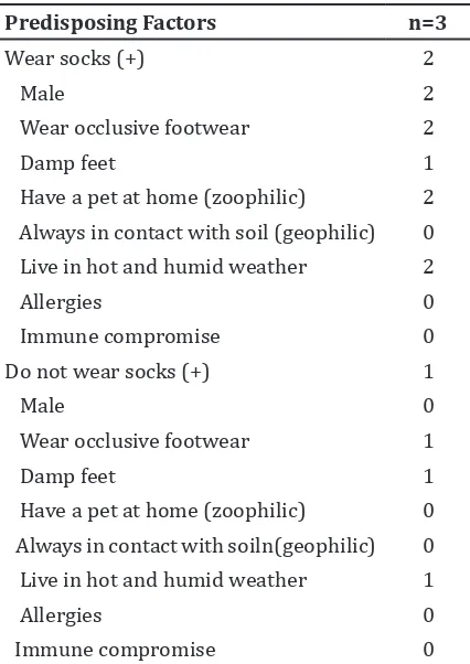
Out of the three students who were positive for dermatophyte, two of the students wore
Table 4 Percentage of Routine of Changing Socks in Students who were Positive
Table 5 Percentage of Predisposing Factors Present in students who were
Wear occlusive footwear 2
Damp feet 1
Have a pet at home (zoophilic) 2
Always in contact with soil (geophilic) 0
Live in hot and humid weather 2
Allergies 0
Immune compromise 0
Do not wear socks (+) 1
Male 0
Wear occlusive footwear 1
Damp feet 1
Have a pet at home (zoophilic) 0
Always in contact with soiln(geophilic) 0
Live in hot and humid weather 1
Allergies 0
Immune compromise 0
socks (66.7%) (Table 3). Those who did not wear socks were positive in one individual (33.3%). Occlusion of the feet with socks can predispose one to a dermatophyte infection.11
Furthermore, socks are a way in which dermatophytes can spread.12 However, the
type of socks can influence the amount of
dermatophytes presence. Nylon socks shows larger number of dermatophyte isolated than the cotton and wool socks. Fibers of socks made from nylon and cotton are loose enough for dermatophytes to go through and thus, both are not good enough in preventing the adhesion of dermatophytes.13 However, in this
study the type of socks worn by both students wearing socks and showing positive result for dermatophyte were the cotton socks. Socks worn should be replaced at least once a day.14
In this study, however, the students who wear socks and who were positive for dermatophyte changed their socks more than a day.
Dermatophytes grows predominantly in warm and humid areas. This is because warm and damp conditions help to promote the growth of dermatophytes.15 In this study, two
students have damp feet, which consist of one student wearing socks and one student not wearing socks.
Tinea pedis is so contagious and is transmitted via direct or indirect contact with skin lesions of the infected people or contaminated environments.14,16 Based on
their ecology, dermatophytes can be classified
into anthropophilic. Dermatophytes spread from person to person either by direct contact or via fomites, zoophilic. Dermatophytes spread to humans from animals or via fomites and geophilic. Dermatophytes are found in the environment and spread when in contact with soil.4 Zoophilic organisms of dermatophyte
are chiefly found in animals and cause marked inflammatory reactions in humans who have
contact with infected cats, dogs, cattle, horses, birds or other animals.10 The students in this
study spent most of their time in the faculty of medicine together and therefore are in close contact almost every weekday. However, most students here do not open and share their shoes and socks in the faculty and thus, lesser chance for a transmission event to occur. This explains why the result was only positive for dermatophyte in 3 out of 50 participants (6%). Those three students might have contracted dermatophyte from anthropophilic, zoophilic or geophilic sources.
There are several limitations in this study. The detection of dermatophyte was done only using direct microscopic examination and
not culture because culture of dermatophyte takes a very long time.17 Therefore, culture is
not performed in this study. Other than that, the type of shoe material worn by the students is different. Wearing non-porous material can increase the moisture and temperature of the feet unlike wearing the open shoes.12,17
All the students in this study wore closed shoes, however the type of material worn is
different. Thus, it might influence the presence
of dermatophyte. Furthermore, some students might wear socks and might not wear socks alternately but they informed the researcher regarding the one most frequently worn. Some students might also forget if they were using any antifungal medication or having any underlying foot disease. Therefore, they were excluded in the study. In this study, it can be concluded that dermatophyte was detected in only a few medical students of Universitas Padjadjaran, who wore and did not wear socks. It is recommended that proper and hygiene behavior should be sought in order to reduce and eliminate the number of people infected with dermatophyte.
References
1. Newman WA. Dorlands pocket medical dictionary. 28th ed.Philadelphia: Elsevier Saunders; 2009. p. 232.
2. Tanaka K, Katoh T, Irimajiri J, Taniguchi H, Yokozeki H. Preventive effects of various types of footwear and cleaning methods on dermatophyte adhesion. J Dermatol.
2006;33(8):528−36.
3. Fauci A, Braunwald A, Kasper D, Hauser S, Longo D, Jameson J, Loscalzo J. Harrison`s principle of internal medicine. 17th ed. New
York: Mc Graw Hill Medical; 2008. p.1264 4. Wolff K, Johnson RA. Fitzpatricks color
atlas and synopsis of clinical dermatology. 6th ed. New York: Mc Graw Hill Medical; 2009. p. 693, 4, 700, 1114.
5. PubMed Health. Athlete`s foot (Tinea Pedis). 2013; [cited 2013 May 14] Available from: http://www.ncbi.nlm.nih. gov/pubmedhealth/PMH0001878/. 6. Levitt JO, Levitt BH, Akhavan A, Yanofsky H.
The sensitivity and specificity of potassium
hydroxide smear and fungal culture relative to clinical assessment in the evaluation of tinea pedis: a polled analysis. Dermatol Res Pract. 2010; 2010:764843. 7. Karan A, Alikhan A, Feldman SR.
Microscopically differentiating
8. Cervantas O. Dermatophytosis. The center for Food Security and Public health,Iowa State University,Institude for International Cooperation in Animal Biologics, Iowa State University College of Veterinary Medicine; 2005.
9. Andersson EB. Dermatopathology. Berlin: Springer Verlag Berlin Heidelberg; 2006. p. 78.
10. Aleruchi C, Adesoji M, Louisa T, James D, Samson S, Emenike I.Comparison of dermatophytes and other agents of human dermatitis between males and females in Jos, Plateau State, Nigeria. Afr J Cln Exper
Microbiol. 2012;13(2):84−9.
11. Gordon CC, Alimuddin Z. Manson`s tropical diseases. 22nd ed. Philadelphia: Elsevier Saunders. 2009.
12. Carlo CJ, MacWilliams BP. Tinea Pedis (Athlete’s Foot); [cited 2013 May 13] Available from: http://www.bhchp.org/
BHCHP%20Manual/pdf_files/Part1_PDF/
TineaPedis.pdf
13. Watanabe K, Taniguchi H, Nishioka K, Maruyama R, Katoh T. Preventive effects of various socks against adhesion of dermatophytes to healthy feet. Nihon
Ishinkin Gakkai Zasshi. 2000;41(3):183−6.
14. Justad J. Athlete`s foot Best Practice Guideline; 2012; [Cited 2013 May 14] Available from: http://www. dphhs.mt .gov/dsd/ddp/documents/ AthletesFoot.pdf.
15. Kumar V, Tilak R, Prakash P, Nigam C, Gupta R. Tinea pedis- an update. Asian J Medical
Sciences. 2011; 2(2):134−8
16. Macit I, Ferdi T, Salih H, Turker G, Muhsin A. Epidemiology of tinea pedis and toenail tinea ungium in worshippers in the mosques in Adana, Turkey. J Dermatol.
2005;32(9):698−704.
17. Hainer BL. Dermatophyte infections. Am