The combination of
Acalypha indica
–Centella asiatica
extracts decreases the neuronal
damage in hypoxia-induced hippocampal injury animal model
pISSN: 0853-1773 • eISSN: 2252-8083 • https://doi.org/10.13181/mji.v27i3.1697 • Med J Indones. 2018;27:137–44
• Received 29 Nov 2016 • Accepted 29 May 2018
Copyright @ 2018 Authors. This is an open access article distributed under the terms of the Creative Commons Attribution-NonCommercial 4.0 International License (http://creativecommons.org/licenses/by-nc/4.0/), which permits unrestricted non-commercial use, distribution, and reproduction in any medium, provided the original author and source are properly cited.
Siti Farida,1 Desak G.B. Krisnamurti,1 Lisnawati,2 Ninik Mudjihartini,3 Imelda M. Sianipar,4 Erni H.
Purwaningsih1
1 Department of Medical Pharmacy, Faculty of Medicine, Universitas Indonesia, Jakarta, Indonesia 2 Department of Anatomical Pathology, Faculty of Medicine, Universitas Indonesia, Jakarta, Indonesia
3 Departmen of Biochemistry and Molecular Biology, Faculty of Medicine, Universitas Indonesia, Jakarta, Indonesia 4 Department of Physiology, Faculty of Medicine, Universitas Indonesia, Jakarta, Indonesia
ABSTRACT
Background: Approximately 80–85% of strokes are ischemic and lead to alterations in neuronal cell morphology and cell death.
There is a lack of studies on the effect of the combination of Acalypha indica L. (AI) and Centella asiatica L. (CA) in terms of its neurotherapy property. This study was conducted to investigate the neurotherapeutic effect of the combination of AI–CA extracts
in improving rat’s hippocampal neuron injury post-hypoxia.
Methods: A total of 36 Sprague-Dawley rats were categorized into six groups and placed in a hypoxia chamber for 7 consecutive
days. Then, they were moved to normoxia cages and treated for 7 consecutive days as follows: control group without treatment as a negative control; treatment groups were administered citicoline 50 mg/kgBW as a positive control; three different dose combinations of AI150–CA150, AI200–CA150, and AI250–CA150 mg/kgBW, respectively. Histological analyses were performed
to assess the improvement in nerve cell damage in the hippocampus.
Results: Treatment with citicoline significantly decreased the damage of nerve cells (30.8%); the combination of the AI–CA
extracts of AI150–CA150, AI200–CA150, and AI250–CA150 also significantly decreased the damage of nerve cells (36%, 36.4%,
and 30.4%, respectively) compared to the control rats (15.4%).
Conclusion: The combination of AI–CA extracts decreased the neuronal damage in the hypoxia-induced hippocampal injury
animal model. The improvement effect of the combination of AI–CA extracts was not significantly different to citicoline.
Corresponding author: Erni H. Purwaningsih
The Basic Health Research (Riset
Kesehatan Dasar) data published in 2013
indicated that stroke is the major cause of death in Indonesia.1 Cerebral hypoxia caused by ischemia
leads to the alteration of nerve cell morphology and cell death, in which the nerve cells become pyknotic. The most vulnerable areas of the brain to hypoxia is the hippocampus, contains stem cells of the nervous system that lay on the dentate gyrus.2 Neuronal ischemic cells resulting from stroke undergo morphological changes. In the early stages, the nerve cells exhibit shrinking, vacuolization, and condensation, eventually become pyknotic and necrotic.2 In an emergency situation, there are three management objectives of stroke: preventing acute brain injury by restoring perfusion to the ischemic non-infarct area, reversing nerve injury as early as possible, and preventing further neurologic injury by protecting cells in the ischemic penumbra area from further damage by increasing the glutamate levels.3
Citicoline is a neuroprotective agent that has been reported to improve nerve membrane damage caused by cerebral ischemia through several mechanisms, such as increasing the synthesis of phosphatidylcholine, decreasing the levels of free fatty acids, and restoring the levels of post-ischemic sphingomyelin.4,5 Although citicoline is an effective drug for stroke, it has several limitations in terms of its usage and affordability. These limitations have led researchers to identify an alternative drug from herbs, which has more effective, safe, and affordable compounds. Previous studies have demonstrated that the root extract of Acalypha
indica L. (AI) exhibited neuroprotective and
neurotherapeutic effects on the neuromuscular junction and was able to inhibit the activity of neurotoxin on isolated frog tissues ex vivo6 and in
vivo.7 The extract was also found to be effective in
improving the rat hippocampal nerve cells at doses of 400 and 500 mg/kg BB.8 On the other hand,
Centella asiatica L. (CA) extract has been reported
to be a peripheral vasodilator in neurological disorders and also improves circulation.9 CA is also
known that able to influence the central nervous system, enhance the excitatory brain neurons, and improve the learning ability.9 Moreover, the
leaf extract of CA at the dose of 500 mg/kg BW showed a neuroprotective effect against stress-induced neuronal atrophy of hippocampus in
mice.10 Therefore, the administration of the
combination of water extracts of AI and CA could improve hippocampus neuron injury caused by cerebral hypoxia. Thus, the objective of this study is to investigate the neurotherapeutic effect of AI and CA extracts combination in improving rat’s hippocampal neuron injury post-hypoxia in comparison with citicoline.
METHODS
Animal model of hypoxia
All procedures in this study were approved by the Ethics Committee of the Faculty of Medicine Universitas Indonesia (Number: 464/H2.F1/ETIK/2013). This investigation was a single-blind experimental study conducted in male Sprague-Dawley (SD) rats. Male rats were chosen because this study did not evaluate the reproductive function of rats. A total of 36 healthy male SD rats, aged 12 weeks and weighed 250– 275 g, obtained from Balai Penelitian Veteriner, Bogor. All SD rats were caged, each cage containing three rats, and provided access to food and water ad libitum. The animals were exposed to a 12-h light–dark cycle and maintained at a temperature of 22°C±2°C. The 36 rats were randomly divided into six groups (n=6 in each group).
After 1 week of acclimatization period, the rats were placed in a hypoxia chamber containing 10% O2 and 90% N2, supplied at a rate of 3 L/ min, for 7 consecutive days. The hypoxia status was assigned by the parameter values of pH=7.09,
pCO2=82.5, pO2=15.8, and HCO3=24.7 (hypoxia was
reached if PO2<60%). After the hypoxia procedure, all rats were replaced in a normoxia chamber under normal atmospheric conditions. To ensure that the induction has achieved the hypoxic status in this study, one rat from each post-induction hypoxia group was euthanized for analyzing blood gas and histopathology examination of the brain. All rats were then treated under normoxia for 7 consecutive days as follows: the control group was administered only aquadest (without treatment); the treatment groups were administered citicoline 50 mg/kg BW; and the combinations of AI150–CA150, AI200– CA150, and AI250–CA150 mg/kg BW, respectively.
Extraction procedure
A. indica L. was collected from Depok
were weighed, dried, and powdered. The powder was macerated with 70% ethanol for 24 h. Subsequently, the residue was remacerated three times with the same solvent, and the extract was mixed into the previous extract. The collected extract was then concentrated by a rotary vacuum evaporator until it had dried. The leaf extract of
C. asiatica L. was obtained from Balai Penelitian
Tanaman Rempah dan Obat (Balittro) in a dry
powder form.
Drug dosage
The doses of AI used in this study were 150, 200, and 250 mg/kg BW, and the dose of CA was 150 mg/kg BW. Based on a previous study by Suswati et al11 who used 400 and 500 mg/kg BW,
a lower AI dose was used in this study. Similarly, the dose of CA was also lower in this study than the dose of 500 mg/kg BW used by Halim and Ibrahim.8 The combination of the lower doses
of AI+CA was expected to have a beneficial neurotherapeutic effect. All treatments were administered orally using a nasogastric tube for 7 consecutive days.
Oral administration of drugs
The final condensed extract of AI–CA was dissolved in aquadest to obtain an appropriate suspension with 1% carboxymethyl cellulose (CMC) Na just before administration every day. The required dose of the extract was taken in a syringe attached with a nasogastric tube containing 2 ml of aquadest per rat. The control group was administered only aquadest of the same volume.
Hippocampus sample preparation
One day after the last treatment, the rats were euthanized by an anesthetic treatment using ether. The brain was extirpated and then immersed in 10% formalin buffer before histological preparation with HE staining. The hippocampus was cut at the dentate gyrus that divided into an outer and an inner layer. The nerve cell damage on the outer layer of the dentate gyrus of the hippocampus was analyzed by measuring the percentage of pyknotic cells, chromatin condensed, and nuclear fragmented under a light microscope, at 400× magnification, connected to the Optilab program. The nerve cell damage on the inner layer of the dentate gyrus was not examined as this part was less sensitive to hypoxia.10 The number of pyknotic
cells, chromatin condensed, and nuclear fragmented was counted from 500 nerve cells on the outer layer of the dentate gyrus of the hippocampus.
Statistical analysis
All data were analyzed by the Statistical Package for the Social Sciences for Windows (SPSS Inc., Chicago, IL, USA) and expressed as mean±SD. The distributions of continuous variables were assessed for normality using the Shapiro–Wilk test. One-way analysis of variance was used to analyze the number of nerve cells damaged (pyknotic cell, condensated chromatin, and nuclear fragmented). The analyses were continued using the post-hoc test. Statistical significance was accepted at p<0.05, with an α value of 0.05 and 95% confidence interval.
RESULTS
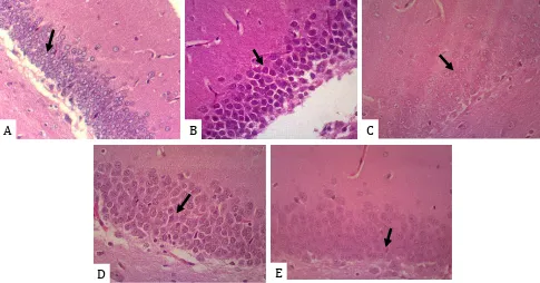
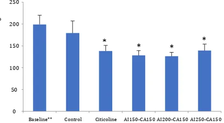
The difference of the histopathological alterations of the nerve cells damage (pyknotic, condensated chromatin, nuclear fragmented) on the outer layer of hippocampal dentate gyrus after treatment for 7 consecutive days among groups were showed in the Figure 1A, 1B, 1C, 1D, and 1E. Treatment with citicoline significantly decreased the damage of nerve cells (30.8%); the combination of the AI–CA extracts of AI150– CA150, AI200–CA150, and AI250–CA150 also significantly decreased the damage of nerve cells (36%, 36.4%, and 30.4%, respectively) compared to that in the control rats (10.4%).
Figure 2 shows the number of nerve cells damage (pyknotic, condensated chromatin, and nuclear fragmented) in the control group (without treatment) was not significant decrease (15.4%) compared to baseline (post-induction hypoxia). The treatment group with citicoline 50 mg/kg BW showed the percentage of total nerve cell damage significantly decrease (-30.8%) compared to baseline (before treatment) (p=0.027). Similar result has also showed the percentage of nerve cell damage in all treatment groups of combination of AI150–CA150, AI200–CA150, AI250–CA150 extracts were significantly decrease (-36%, -36.4%, and -30.4%, respectively) compared to baseline (p=0.003, p=0.002, and p=0.029, respectively). Post-hoc analyze showed significantly difference between control and treatment groups, but no statistical differences were found between citicoline and three different doses of combination AI–CA extracts. The lowest percentage of nerve cells damage was achieved in AI150–CA150 and AI200– CA150 groups given A. indica L. 150, 200 mg/kg
BW and C. asiatica L. 150 mg/kg BW.
DISCUSSION
During hypoxia, intracellular hypoxia-inducible factor (HIF) 1α rapidly accumulates in the nucleus of the cell.12,13 Increased HIF1α expression contributes to mitochondrial activities14 and increases the formation of reactive
oxygen species (ROS).15,16 When such high ROS concentrations in the brain are not overcome by increasing the levels of endogenous antioxidants, it leads to oxidative stress. This condition results in nerve cell damage as characterized by the pyknotic cell, condensated chromatin, and nuclear fragmented appearance. This study showed demonstrated nerve cell damage caused due to brain hypoxia based on the high number of pyknotic cells, condensated chromatin, and nuclear fragmented on the outer layer of the dentate gyrus of the hippocampus after placing the rats in a hypoxia chamber for 7 consecutive days (Figure 1B).
When the decrease in pO2 is not too severe and oxygenation is still restored, the injury could
A
B
C
D
E
Figure 1. Representative photomicrographs of hippocampal dentate gyrus post-hypoxia before and after treatment. (A) Normal
be reversed.16 However, the control (C) group
without treatment still showed high damage in the nerve cells (pyknotic nerve cells, condensated chromatin, and nuclear fragmented) and low density of nerve cells after removing the rats from under the normoxia condition to normal atmosphere for 7 consecutive days (Figure 1C). This finding suggests that physiological improvement alone under the normoxia condition for 7 days post-hypoxia is not effective. This condition may be caused by the duration of hypoxia, i.e., 7 consecutive days, which affects a greater area of the hypoxic brain.16 Inversely, a
highly obvious improvement was detected in almost all the therapeutic groups treated with the combination of the extract of AI at doses of 150, 200, and 250 mg/kg BW and CA at the dose of 150 mg/kg BW (Figure 1E) and also in the group treated with citicoline at the dose of 50 mg/ kg BW (Figure 1D). Significant differences were observed between the control and all treatment groups, but there was no significant difference among the treatment groups. The results of this study indicated that the combination of AI+CA extracts could improve nerve cell damage or protect against the progression of the hippocampal nerve cells post-hypoxia damage. The decrease in nerve cell density in the treatment
groups was similar to that in the citicoline group, indicating that the maturation of nerve cells could be stimulated by treatment with both citicoline and the combination the AI–CA extracts. These findings could also demonstrate that the damage occurring in the nerve cells after 7 days’ post-hypoxia (10% O2, 90% N2) was still reversible through an appropriate treatment for 7 days.
Regarding the mechanism of action, citicoline could improve nerve cell damage due to the cerebral ischemia by increasing phosphatidylcholine synthesis, decreasing free fatty acid levels, and restoring post-ischemic sphingomyelin levels.4 While, the combination
of AI–CA extracts has a lot of polyphenol and flavonoid compounds which have rich on hydroxyl groups (OH-) that react to free radicals
as a scavenger.17 This effect could improve the
neuronal cell damage caused by hypoxia.17–20 Moreover, the combination of AI–CA extracts have an anti-inflammatory activity21 and cell proliferation stimulation.22 Medicinal plants have been known that contain several compounds besides polyphenol and flavonoids that support each other to provide a beneficial effect.17 The
root extract of AI has been used in several studies6–8,11 and its compounds have been proven 0
50 100 150 200 250
Baseline** Control Citicoline AI150-CA150 AI200-CA150 AI250-CA150
The
num
ber
of
ner
ve
cel
l dam
age
*
*
*
*
250
200
150
100
50
0
Baseline** Control Citicoline AI150–CA150 AI200–CA150 AI250–CA150
The number of nerv
e cell damage
Figure 2. The number of nerve cell damage among groups. *Statistically significant difference compared to baseline (p<0.05);
to possess higher antioxidant activities than those of the leaves.18 The phytochemical screening of AI
showed the presence of alkaloids, phenols, tannins, glycosides, saponins, and steroids that possess an antioxidant effect.17 The antioxidant effect of AI
reduces the higher ROS concentrations induced by hypoxia through the radical-scavenging mechanism. Besides this mechanism, AI has also been demonstrated to prevent the chain reaction of oxidation radicals, delay or inhibit the oxidation process, and increase the shelf life by slowing the lipid peroxidation process,16,17 leading to an improvement in nerve cell damage due to cerebral hypoxia. Furthermore, the anti-inflammatory effect of AI in Wistar albino rats has been reported by Dutta et al21 Moreover, the anti-inflammatory and the proliferation stimulation activities of AI were supported by Yolanda et al,22 who reported that acalyphin present in AI extract decreased the activity of the phospholipase A2 (PLA2) enzyme, leading to reduction in the inflammatory response and promotion of the proliferation of nerve cells. AI also possesses neuroprotective and neurotherapeutic effects on the neuromuscular junction and inhibits the activity of neurotoxin on isolated frog tissues ex vivo and in vivo.6
In addition to the finding of AI, Yolanda et al22 demonstrated that a single AI root extract, which contains acalyphin, increased the viability of post-hypoxic rat hippocampal tissue culture cells through the inhibition of PLA2.22 Besides these studies on AI, Rahman et al19 demonstrated that
CA, which contains active compounds such as polyphenol, flavonoid, β-carotene, and vitamin C, also exhibits antioxidant activities. The results of the present study are also supported by Mohamed and Abdou20 who reported that CA enhanced the activity of endogenous antioxidants such as superoxide dismutase (SOD), catalase, reduced glutathione (GSH), glutathione peroxidase (GPX), and reduced lipid peroxidase in rats with diabetes mellitus (DM) induced by streptozotocin. CA has also been reported to possess peripheral vasodilator activity in neurological disorders, improves the blood circulation,9 and has
anti-inflammatory properties.23 In addition, Yulianti et al24 demonstrated that nanoparticles of CA increased the proliferation of fibroblasts and keratinocyte cells of the skin. Moreover, this finding was supported by the result of an in vivo study conducted by Sunanda et al25 who showed that a single CA leaf extract at a dose of 300 mg/kg BW can improve the spatial memory performance
and help improve cognitive impairment caused due to oxidative stress.
To date, no studies have supported the effect of the combination of AI–CA extract that has been assumed to have a beneficial neurotherapeutic effect. Based on several studies mentioned above for AI and CA, this reason was also supported by Suswati et al11 proved that AI
extract at the doses of 400 mg/kgBW and 500 mg/kgBW has a neurotherapeutic effect and that a single CA leaf extract can improve the spatial memory performance impaired due to oxidative stress at the dose of 300 mg/kg BW. However, this study has demonstrated that the neurotherapeutic effect was already seen at the dose of A. indica L. 150, 200, and 250 mg/kg
BW which combined with C. asiatica L. 150 mg/
kg BW at each combination dose. These results were presumably due to the addition of CA, which may work together with AI to their same activities as an antioxidant17–20 or by exhibiting anti-inflammatory activity.21 Unfortunately, we could not determine whether the combination effect of AI–CA extracts is synergistic because we were not able to use the quantitative isobole methods developed by Loewe et al26 to evaluate it due to several active compounds contained in each medicinal plant and complex. Still, the isobole method is used to evaluate the synergistic effect of two or more single compound that have similar effect.26,27 Based on these results, the clinical effect of the combination of AI and CA extracts can be applied to patients with stroke or brain injury as an adjuvant therapy, particularly as a neurotherapeutic agent.
The limitation of this study is that the quantitative isobole method developed by Loewe et al26 could not be applied because of the several active compounds contained in each medicinal plant and complex. Therefore, further studies are required to determine whether the combination of AI–CA and citicoline has the same receptor target using immunohistochemical examinations.
be due to the antioxidant, anti-inflammatory, and cell proliferation stimulation activities.
Conflict of interest
None of the authors have any conflicts of interest to declare in relation to this manuscript.
Acknowledgments
This study was funded by a research grant from Universitas Indonesia. We would like to thank the Department of Biochemistry and Molecular Biology and the Department of Anatomical Pathology at the Faculty of Medicine Universitas Indonesia for providing the laboratory facilities. We also thank Prof. Dr. Frans Ferdinal, M. Biomed, who allowed us to use the hypoxia chamber, and Dr. Ninik Mujihartini, M. Biomed, who taught us how to use the hypoxia chamber. We also thank all our colleagues at the Department of Medical Pharmacy, Faculty of Medicine Universitas Indonesia, and our students Ermono, Hatim, Raymond, Setyo, Jody, Eka, Wynne, Yashinta, Farah, Monica, Ireska, and Kevin who contributed to this research. neuroprotection and neurorepair in ischemic stroke. Brain Sci. 2013;3(3):1395–414.
5. Martindale (ed). Citicoline. In: Martindale`s the Extra Pharmacopea. 31st ed.1995.p1742.
6. Purwaningsih EH, Ibrahim N, Zain H, Tedjo A. Neuro-protection and neuro-therapy effects of Acalypha indica Linn. water extract ex vivo on musculus gastrocnemius Frog. Makara Journal of Health Research. 2008;12(2):70–5.
7. Purwaningsih EP, Ibrahim N, Zain H. The nerve protection and in vivo therapeutic effect of Acalypha indica extract in frogs. Med J Indones. 2010;19(2):96–102.
8. Halim HDP, Ibrahim N. Efek neuroprotektif ekstrak akar Acalypha indica 500 mg/kgBB terhadap perubahan inti sel saraf hipokampus pascahipoksia serebri. eJKI. 2013;1(2):113–7.
9. Veerendra Kumar MH, Gupta YK. Effect of different extracts of Centella asiatica on cognition and markers of oxidative stress in rats. J Ethnopharmacol. 2002;79(2):253–60.
10. Anissa RF. Pengaruh pemberian ekstrak air daun pegagan (Centella asiatica Linn.) terhadap kemampuan kognitif dan kadar neurotrasmiter monoamin pada hipokampus tikus galur Wistar jantan dewasa [Unpublished thesis]. Institut Teknologi Bandung. 2006. Indonesian.
11. Suswati L. Perbaikan neuron hipokampus pascahipoksia serebri dengan penggunaan ekstrak air akar tanaman akar kucing (Acalypha indica L.). Tesis. Universitas Indonesia Library. 2010. [Cited 2014 Feb 8] Available from http://lib.ui.ac.id/abstrakpdf. jsp?id=20339828&lokasi=lokal
12. Powell FL. Functional genomics and the comparative physiology of hypoxia. Annu Rev Physiol. 2003;65(1):203–30.
13. Semenza GL. Hydroxylation of HIF-1: oxygen sensing at the molecular level. Physiology. 2004;19(4):176–82. 14. Agani FH, Pichiule P, Chavez JC, LaManna JC. The role
of mitochondria in the regulation of hypoxia-inducible factor 1 expression during hypoxia. J Biol Chem. 2000;275(46):35863–7.
15. Zepeda AB, Pessoa Jr A, Castillo RL, Figueroa CA, Pulgar VM, Farías JG. Cellular and molecular mechanisms in the hypoxic tissue: Role of HIF-1 and ROS. Cell Biochem Funct. 2013;31(6):451–9.
16. Michiels C. Physiological and pathological responses to hypoxia. Am J Pathol. 2004;164(6):1875–82.
17. Kumar, Nirmalababurao, Rani AR. Antimicrobial, antioxidant activity and phytochemical screening of Acalypha indica crude leaf extract. I J Pharm Clin Res. 2016;8(6):583–8.
18. Shanmugapriya R, Ramanathan T, Thirunavukkarasu P. Evaluation of antioxidant potential and antibacterial activity of Acalypha indica Linn. using in vitro model. Asian J Biomed Pharm Sci. 2011;1(1):18–22.
19. Rahman M, Hossain S, Rahaman A, Fatima N, Nahar T, Uddin B, et al. Antioxidant activity of Centella asiatica (Linn.) urban: impact of extraction solvent polarity. J Pharmacogn Phytochem. 2013;1(6):27–32.
20. Mohamed NA, Abdou HM. The hypoglicemic and antioxidative effects of Centella asiatica againts STZ-induced diabetic disorders in rats. Int J Pharma Bio Sci. 2015;6(2):621–33.
21. Dutta S, Mariappan G, Dipankar S. Anti-inflammatory effect of chloroform and aqueous extract of Acalypha indica Linn. against carrageenan induced paw edema in Wistar albino rats. J Herb Med Toxicol. 2010;4(1):153–6. 22. Yolanda S, Andraini T, Kusuma I. Acalypha indica root
extract increases post-hypoxic rat hippocampal tissue culture cell viability via phospholipase A2 inhibition. Med J Indones. 2013:22(3);136–40.
23. Somchit MN, Sulaiman MR, Zuraini A, Samsuddin L, Somchit N, Israf DA, et al. Antinociceptive and antiinflammatory effects of Centella asiatica. Indian J Pharmacol. 2004;36(6):377–80.
24. Yulianti L, Bramono K, Mardliyati E, Freisleben H-J. Effects of Centella asiatica ethanolic extract encapsulated in chitosan nanoparticles on proliferation activity of skin fibroblasts and keratinocytes, type I and III collagen synthesis and aquaporin 3 expression in vitro. J Pharm Biomed Sci. 2016;06(05):315–27. 25. Sunanda BP, Latha K, Rammohan B, Uma MS,
effects of Centella asiatica against scopolamine induced cognitive impairment in mice. Indian J Pharm Educ. 2014;48(4):31–4.
26. Loewe S. (1953). The problem of synergism and
antagonism of combined drugs. Arzneimittelforschung 3. p.285–90.