URCHIN EGGS
Muh. Akbar Bahar, Gemini Alam, Marianti A. Manggau, Mufidah, Suparman
Faculty of Pharmacy, Hasanuddin University email: [email protected]
ABSTRACT
Ongkea cortex, the wood bark of Mezzettia Parviflora Becc, is a traditional medicine originated from Southeastern Sulawesi (Indonesia). It has been empirically known to have antitumor property. In this study, we examined the antiproliferative activity and obtained the antimitotic compound of ongkea cortex. Antimitotic activity was ultimately determined by the inhibition of cleavage-stage of newly fertilized sea urchin (Lytechinus variegatus) eggs. A bioassay-guided fractination was performed in order to find the bioactive substance of ongkea cortex. The IC50 values of methanolic extract, ethyl acetate-soluble part of metanolic extract and ethil acetat insoluble part of metanolic extract were 1221.68 µg/mL, 2.69 µg/mL, and 15.15 µg/mL, respectively. Ethyl acetate-soluble part of metanolic extract was further investigated. It was partitionated using vacuum liquid column chromatoghraphy with different solvent system by increasing their polarities. There were three different fractions obtained. Fraction III exerted the highest inhibition activity with IC50 value of 1.33 µg/mL. It was separated subsequently to result four groups of compounds. III-C group presented the most potent inhibition activity with IC50 value of 0.7147 µg/mL. It was then subjected to preparative TLC and yieldedsix groups of subfractions. III-C-3 subfraction was indicated as the most potent compound with IC50 value of 0.3378 µg/mL. It was ten times weaker compared with antimitotic activity of Vincristine with IC50 of 0.0351 µg/mL. As a conclusian, ongkea cortex might have antimitotic property with the highest rate inhibition activity exhibited by III-C-3 compound.
Keywords: ongkea cortex, Mezzettia Parviflora Becc, sea urchin eggs, antimitotic compound, antiproliverative activity
INTRODUCTION
Ongkea cortex (Mezzettia
Parviflora Becc.), belongs to annonaceae family, is a traditional substance that has been empirically used in Southeast Sulawesi (Indonesia) to treat some diseases such as hipercholesterolemia, asthma, diabetes mellitus and tumor [1].
The familiy of annonacea has a broad range of pharmacological activities
such as antimicrobial, antifungal,
antitumor, and insecticidal properties. It
such as benzylisoquinoline, acetogenin,
c-benzil-flavonoid and diterpenoid
alkaloids [2]. Based on the concept of chemotaxonomy, chemical compounds of plants from the same familial groups are mostly similar.
Previous studies have reported
that ongkea cortex has
the first study trying to evaluate the possibility of ongkea cortex to be developed as an anticancer agent.
To analyse theantiproliferative activity of ongkea cortex, we used an inhibition of sea urchin (Lytechinus variegates) eggs development assay. It is a proper bioassay model to study biological activities such as antimitotic, teratogenic and antineoplastic activities [6].
In this study, the antitumor activity of ongkea cortex was evaluated to provide scientific based information as a traditional medicine. A bioassay-guided fractination was performed in order to find the bioactive compound of ongkea cortex.
METODE PENELITIAN Samples Collection
Ongkea cortex was obtained from Buton regency, Southeast Sulawesi. Samples were cleaned, shattered, dried and powdered.
Samples Extraction
Dried powdered portions of 1.6 Kg of ongkea cortex were extracted by maceration method with 12 Litres of methanol for 3x3x24 h. The results were collected and rotoevaporated to yield viscous methanol extract.
Partition of methanol extract
Methanol extract was
partitionated with ethil acetate using liquid-solid partition method. It produced ethyl acetate-soluble part of metanolic extract and ethil acetat insoluble part of
metanolic extract. The chemical
compound profiles of extracts were monitored by thin layer chromatography. Antimitotic activities of extracts were investigated by an inhibition of sea
Fractination of active extract
The most antimitotically active extract was fractionated using vacuum liquid column chromatoghraphy with diffrent solvent with increasing polarity; hexane, hexane:ethyl acetate (15:1; 10:1, 5:1), methanol. Fractions with similar chemical profiles based on thin layer chromatography were grouped and tested
against sea urchin (Lytechinus
variegates) eggs.
Determination of Antiproliferative Activity
RESULTS AND DISCUSSSION
The antiproliferative assay using sea urchin eggs has been well known to have a good sensitivity against toxic substance. It can help to assess the ability of natural compounds to be developed as an anticancer. The inhibition of growth cycle of sea urchin eggs can be associated with several steps such as DNA and RNA synthesis, protein synthesis, and mitotic spindle assembly [8].
A first screening showed that the IC50 values of methanolic extract, ethyl acetate-soluble part of metanolic extract
and ethil acetat insoluble part of metanolic extract were 1221.68 µg/mL,
2.69 µg/mL, and 15.15 µg/mL,
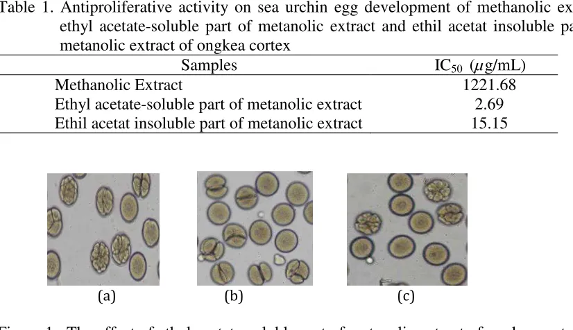
respectively (Table 1). Ethyl acetate-soluble part of metanolic extract possesed the highest inhibition rate. Figure 1 showed its effect on the initial development of sea urchin embryos. In order to find the active principles possibly responsible for antimitotic activity of ongkea cortex, ethyl acetate-soluble part of metanolic extract was further investigated by bioassay-guided fractination.
Table 1. Antiproliferative activity on sea urchin egg development of methanolic extract, ethyl acetate-soluble part of metanolic extract and ethil acetat insoluble part of metanolic extract of ongkea cortex
Samples IC50 (µg/mL)
Methanolic Extract 1221.68
Ethyl acetate-soluble part of metanolic extract 2.69
Ethil acetat insoluble part of metanolic extract 15.15
(a) (b) (c)
Figure 1. The effect of ethyl acetate-soluble part of metanolic extract of ongkea cortex on the initial development of sea urchin (Lytechinusvariegatus) embryos. (a) 1 µg/ml (44.34 % inhibition); (b) 10 µg/ml (54.4 % inhibition); (c) 100 µg/ml(76.28 % inhibition).
Table 2. Antiproliferative activity on sea urchin egg development of Fraction 1,II and III.
Samples IC50 (µg/mL)
Fraction I 7.39
Fraction II 7.48
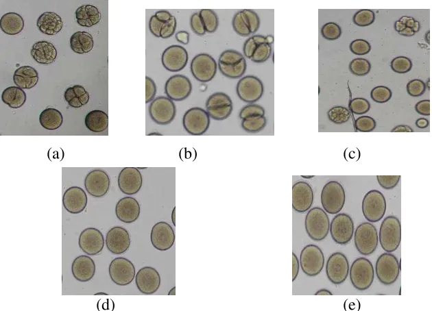
(a) (b) (c)
(d) (e)
Figure 2. The inhibition effect of fraction III against sea urchin development. (a) 0.2 µg/mL (3.37 % inhibition); (b) 5 µg/mL (55.95 % inhibition); (c) 25 µg/mL (84.5 % inhibition); (d) 25 µg/mL (100 % inhibition); and (e) 125 µg/mL (100 % inhibition)
Table 3. Antiproliferative activity on sea urchin egg development of Fraction III-A,III-B, III-C and III-D.
Samples IC50 (µg/mL)
Fraction III-A 1.2971
Fraction III-B 1.9480
Fraction III-C 0.7147
Fraction III-D 3.2569
It was partitionated using vacuum liquid column chromatoghraphy with different solvent system by increasing their polarities yielded three different group of bioactive fractions. Fraction III exerted the highest inhibition activity with IC50 value of 1.33 µg/mL while Fraction I and II had IC50 of 7.39 µg/ml and 7.48 µg/ml, respectively (Table 2). The inhibition effect of fraction III on sea urchin eggs was demonstrated by figure 2.
Fraction III was separated
0.7147 µg/mL; and 3.2569 µg/mL,
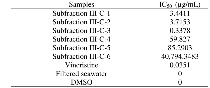
consecutively (Table 3). The most bioactive substance was seemingly in Fraction III-C. Furthermore, it was subjected to preparative TLC and yielded six groups of subfractions. Their IC50
were 3.4411 µg/mL; 3.7153 µg/mL;
0.3378 µg/mL; 59.827 µg/mL; 85.2903
Table 4. Antiproliferative activity on sea urchin egg development of subfraction III-C-1,III-C-2, III-C-3, III-C-4, III-C-5 and III-C-6.
Samples IC50 (µg/mL) compounds presenting 100% inhibition in sea urchin eggs assay at a concentration 16 µg/mL or less, they should be very potential to be investigated as anticancer agents in in vivo experiment [9]. Based on this information, it can be stated that
subfraction III-C-3 has a strong
antiproliferative activity. Therefore, the potency of subfraction III-C-3 of ongkea
antitumoral property with the highest rate inhibition activity presented by III-C-3 compound. We suggest to evaluate the cytotoxicity effect of III-C-3 compound using human cell lines and in vivo experiment with animal model.
REFERENCES
1. Fernandes-Silva CC, Freitas JC,
Salatino A, Salatino MLF. 2013. Cytotoxic Activity of Six Samples of Brazilian Propolis on Sea Urchin (Lytechinusvariegatus) Eggs.
Evidence-Based Complementary and Alternative Medicine. Article ID 619361.
2. Hakim, E.H., Achmad, S.A.,
Makmur, L., Mujahidin, D., dan Syah, Y.M. 2001. Profil Kimia
Annonaceae. Bull Soc. Nat. Prod.
Chem. (Indonesia). VolI : 1. Januari-Juni 2001.
3. Jacobs RS, White S, Wilson L. 1981. Selective compounds derived from marine organisms: effects on cell division in fertilized sea urchin eggs.
Fed Proc. 40 (1). 26-29.
4. Jimenez PC, Fortier SC, Lotufo
TMC, Pessoa C, Moraes MEA, Moraes MO, Costa-Lotufo LV. 2003. Biological activity in extracts of ascidians (Tunicata, Ascidiacea) from the Northeastern Brazilian coast. J. Exp. Mar. Biol. Ecol. 287, 93D101.
5. Militao GCG, Pinheiro SM, Dantas
INF, Pessoa C, De Moraes MO,
Costa-Lotufo LV, Lima MAS,
Silveira ER. 2007. Bioassay-guided fractionation of pterocarpans from roots of HarpalycebrasilianaBenth.
Bioorganic & Medicinal Chemistry.
15: 6687–6691.
6. Mufidah, Manggau MA, Tayeb R,
Alam G. 2007. Free radical
scavenging activity of ongkea
(Mezzetia parviflova becc.)
Woodbark. POKJANAS TOI.
Airlangga University.
7. Mufidah. 2011. Aktivitas
antiaterosklerosis ekstrak terstandar
klikaongkea (mezzetia parviflora
endotel. Disertasi. Program Studi
Ilmu Kedokteran. Universitas
Hasanuddin.
8. Mufidah, Manggau MA, Alam G,
Bahar MA, Kasim S, Rusdi M. 2012. Efek Antiagregasi Platelet Fraksi Klik
a Ongkea (Mezzettia Parviflora
Becc.). Majalah Farmasi dan
Farmakologi, 16. (1). 51 – 54.
9. Mufidah M, Wahyudin E, Lawrence
GS, Subehan, Manggau MA, Alam G. 2012. Phytochemical analysis and
antioxidant activity of Mezzetia
parviflora Becc. Woodbark extract.