PRESTORAGE LEUKOREDUCED FILTRATION (PLF) DECREASE SOLUBLE HUMAN LEUKOCYTE ANTIGEN-I (sHLA-I) LEVEL IN
THROMBOCYTE CONCENTRATE STORED FOR FIVE DAYS
1 Herawati, S.,
1 Department of Clinical Pathology Faculty of Medicine Udayana University,
Bali-Indonesia
ABSTRACT
Backgroud: Thrombocyte concentrate is one of the important blood component to improve patient's clinical condition. In order to provide thrombocyte concentrate with good therapeutic effect, the preparation process and storage condition should be maintained properly. One attempt to maintain good quality of thrombocyte concentrates is by doing Prestorage Leukoreduced Filtration (PLF) method during preparation of thrombocyte concentrates.
Aims: The purpose of this study was to determine the effect of PLF on sHLA-I level in thrombocyte concentrates stored for 1, 3 and 5 days. Methods: This is an experimental study with posttest only control group design, enrolling 34 thrombocyte concentrates and randomly assigned into PLF group and control group.
Conclusion: It can be concluded that PLF decrease sHLA-I level in thrombocyte concentrates stored for five days compared with control groups.
INTRODUCTION
The use of blood components has been increased in the last decade because of the progress in the field of bone marrow transplantation and the increased use of chemotherapy in hematologic malignancy. Thrombocyte concentrate is one of the important blood component to improve patient's clinical condition. Thrombocyte concentrate can be obtained from the pooled random donor platelet concentrate and from single donor platelet apheresis. Thrombocyte concentrate from pooled random donor platelet concentrate can be prepared by Platelet Rich Plasma (PRP) method which is well known in United States and Indonesia and Buffy Coat (BC) method which is mostly used in Europe 1,2,3.
In order to provide thrombocyte concentrate with good therapeutic effect, the preparation process and storage condition should be maintained properly for good viability of thrombocyte concentrates.Viability of thrombocyte concentrate is the ability of thrombocyte concentrate which is transfused to the recipients to circulate without experiencing early destruction in the recipient's body 2,4.
granule release of platelets, changes in the cytoskeleton of platelet, phosphatidyl serine exposure on the outer surface membrane of platelets and microvesiculation 5,6.
Many research related to platelet storage lesion have been carried out, aiming to maintain the viability of platelets in vitro and in vivo. One attempt to maintain thrombocyte concentrates quality is by doing preparation of thrombocyte concentrates with Prestorage Leukoreduced Filtration (PLF). PLF aims to reduce residual leukocytes contained in thrombocyte concentrates. Several studies have shown that residual leukocytes, especially monocytes are a major source of proinflammatory cytokines such as interleukin-1 (IL-1),interleukin-6 (IL-6),interleukin-8 (IL-8) and tumor necrosis factor- (TNF-) that play a role in transfusion reactions 4,5,7,8,9.
Residual leukocytes in thrombocyte concentrates associated with Human Leukocyte Antigen (HLA). HLA is a heterodimer and highly polymorphic glycoprotein. HLA class I antigens expressed on all nucleated cells, while expression of HLA class II antigen is limited to Antigen Presenting Cells (APC) and activated T lymphocytes. The presence of HLA-I antigen in thrombocyte concentrates is considered as a marker of immunological reactivity and cellular fragmentation therefore it can be used as an indicator of the performance of PLF in thrombocyte concentrates. The purpose of this study was to determine the effect of PLF on sHLA-I level in thrombocyte concentrates stored for 1, 3 and 5 days 5,10.
MATERIAL AND METHODS
Data and specimen collection
randomly assigned into PLF group and control group. PLF group was thrombocyte concentrate with PLF treatment and control group without PLF treatment.
Preparation of thrombocyte concentrates
Whole blood (350 ml) was collected into triple blood bags containing CPDA-1 anticoagulant. Criteria for inclusion include preparation of thrombocyte concentrate by PRP method with screening test result of Hepatitis B, Hepatitis C, HIV and VDRL were negative. Criteria for exclusion include hemolysis in thrombocyte concentrate, low platelet count (< 150 x 103/l) and use of any drug known to affect platelet function in the
72 hours prior to blood donation.
After collection of whole blood, within 4 hours was done centrifugation 375 x g for 15 minutes at 22C to form platelet rich plasma (PRP). The PRP was expressed into second satellite bag, then centrifuged again at 1.500 x g for 15 minutes at 22C to obtain platelet poor plasma (PPP). Plasma was expressed into third satellite bag and leaving 40 – 60 ml in the prepared thrombocyte concentrates unit which was left undisturbed for 1 – 2 hours 2,3.
PLF Group were 17 units of thrombocyte concentrates undergo PLF with AcrodoseTM Plus System (Haemonetics), and control group were
17 units of thrombocyte concentrates that did not undergo PLF. All the thrombocyte concentrates were then placed on a horizontal shaker at 22C for 5 days.
Samples from all thrombocyte concentrates were taken on day 1, day 3 and day 5 to determine the concentration of sHLA-I using an Enzyme Linked Immunosorbent Assay (ELISA) (Bioassay Technology Laboratory).
Data was expressed as mean SD. Independent Samples T-test procedure was used to compare means of PLF group and control group. Repeated measure one way anova was used to compare the difference of sHLA-I level in serial measure in both groups. Statistical significance for these tests using a value of 95% ( CI = 95 % ) with a p-value less than or equal to 0.05 as the limit of significance.
Ethical approval
The study protocol was approved by Institutional Ethics Committee of Sanglah Hospital / Faculty of Medicine Udayana University.
RESULTS
[image:5.612.127.513.460.582.2]Laboratory and basic characteristic of thrombocyte concentrate were listed in table 1.
Table 1
Laboratory and basic characteristic of thrombocyte concentrate
Parameter PLF group Control group
Mean thrombocyte count/unit 2,58 x 10¹⁰ 2,47 x 10¹⁰
Mean leucocyte count/unit 0,66 x 10⁶ 1,99 x 10⁶
Mean volume/unit 44,06 ml 42,29 ml
Swirling phenomena +2 +2
Data of mean sHLA-I level in control group and PLF group were listed in table 2.
Table 2
The difference of mean sHLA-I level in platelet concentrate stored on Day 1, 3, 5 for control group (without PLF) and treatment group (with PLF)
Storage
sHLA-I level (µg/ml) Mean difference of
sHLA-I
p value Control
(without PLF)
Treatment (with PLF)
Day 1 3,66±0,87 2,75±0,82 0,91 0,004*
Day 3 4,05±1,29 3,07±0,75 0,98 0,011*
Day 5 5,61±3,26 3,81±0,97 1,80 0,036*
* significant p<0,05
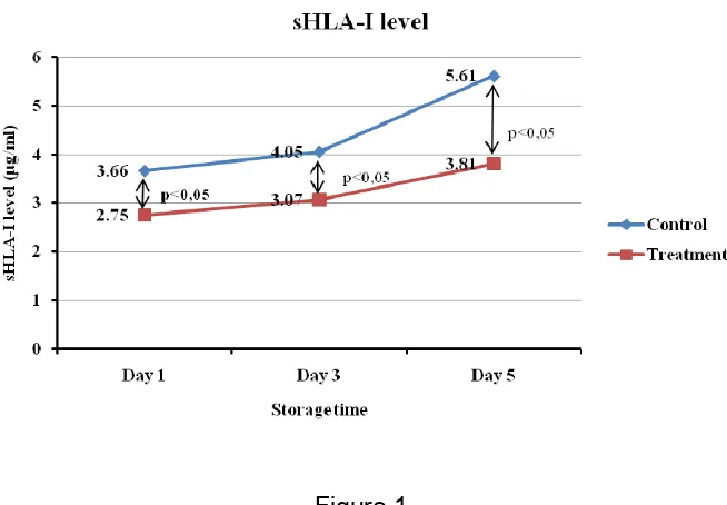
Mean sHLA-I level in PLF group at day 1 is 2,75±0,82 µg/ml and control group is 3,66±0,87 µg/ml, which is statistically significant (p < 0,05). Mean sHLA-I level in PLF group at day 3 is 3,07±0,75 µg/ml and control group is 4,05±1,29 µg/ml, which is statistically significant (p < 0,05). Mean sHLA-I level in PLF group at day 5 is 3,81±0,97 µg/ml and control group is 5,61±3,26 µg/ml, which is statistically significant (p < 0,05). It indicates that PLF group has lower mean sHLA-I level than control group and statistically significant.
Table 3
The difference of mean sHLA-I level between storage time in control group (without PLF) and treatment group (with PLF)
Group Mean
difference of sHLA-I level
p value
Treatment (with PLF)
Storage at first day vs third day 0,32 0,028* Storage at third day vs fifth day 0,74 0,001* Storage at first day vs fifth day 1,06 0,000* Control (without PLF)
Storage at first day vs third day 0,39 0,046* Storage at third day vs fifth day 1,56 0,038* Storage at first day vs fifth day 1,95 0,011* * significant p<0,05
Figure 1
The difference of mean sHLA-I level in treatment group (with PLF) and control (without PLF) in platelet concentrate stored on day 1, 3 and 5
DISCUSSION
The mean (± SD) sHLA-I level in PLF group were statistically lower than the control group both on the first day of storage ( 2,75 ± 0,82 µg/ml vs 3,66 ± 0,87 µg/ml, p = 0.004 ), storage on the third day ( 3,07 ± 0,75 µg/ml vs 4,05 ± 1,29 µg/ml, p = 0,011 ) , and the fifth day of storage ( 3,81 ± 0,97 µg/ml vs 5,61 ± 3,26 µg/ml, p = 0,036 ). sHLA-I level in PLF group was statistically lower than the control group indicating the role of PLF in immunological reactivity. PLF is a procedure to reduce the number of residual leukocytes contained in thrombocyte concentrates prior to storage using a filtration method. PLF eliminate residual leukocytes before undergoing apoptosis and necrosis and before release cytokines 6,7,11,12.
signal that increases the expression of IL-2 receptor. The second signal is required to induce a variety of cytokines which then resulted in the proliferation and differentiation of specific T- lymphocytes. Immunogenicity of HLA antigens on blood components depends on the ability of APC to stimulate recipient T cell by sending 2 different signals 6,10.
In vitro studies revealed that the HLA antigen-I plays a role in modulating the function of immunocompetent cells, so the presence of sHLA-I in thrombocyte concentrates is a marker of immunological reactivity. HLA-I antigen inhibit lymphocyte responses and cytotoxic T cell activity. HLA-I antigen modulate the function of immunocompetent cells through two ways: (1) HLA-I molecule binds to its physiological ligand and inhibits the T cell function through blockade of receptors and/or induce apoptosis, and (2) through indirect presentation in which HLA-I phagocytosis by APC, and then degraded into peptides and presented to CD4 T-cells that will cause immune tolerance or immune activation which depends on the activation capacity or tolerance capacity of HLA-I peptide
13,14.
Our findings are in agreement with Ahmed et al (2010) which reported a significant difference between sHLA-I level in thrombocyte concentrate with leukoreduction and thrombocyte concentrates without leukoreduction, where the sHLA-I level was lower in leukoreduction platelet concentrate (p < 0,05). Also reported the accumulation of proinflammatory cytokines (IL-1, TNF-, IL-6 and IL-8) and chemokines in platelet concentrates without leukoreduction 5,15.
elevated of sHLA-I level during storage of platelet concentrates, which can suppress platelet destruction and immunological reactivity in platelets. In platelet concentrates with PLF can still measured sHLA-I level, because associated with residual leukocyte which is found in platelet concentrates (< 1 x 10⁶), but much lower than the amount of residual leukocytes
contained in platelet concentrates without PLF. During storage, the release of antigen HLA-I from residual leukocytes due to membrane damage or cell death. In addition, increement of sHLA-I level during storage can be caused by in vitro aging which platelet loss its biological reactivity and viability, associated with platelet apoptosis. Platelet apoptosis related because platelets contain enough mitochondria and give an overview of apoptosis-like morphologic changes. So, the increement sHLA-I level during storage associated with residual leukocyte apoptosis and platelet apoptosis 6,11,12,16.
CONCLUSION
The present study conclude that PLF decrease sHLA-I level of thrombocyte concentrates stored for five days compared with control groups.
REFERENCES
1. Fisk,J.M., Pisciotto,P.T., Synder,E.L., Perrotta,P.L. 2007. Platelets and Related Products. In: Hillyer,C.D., Silberstein,L.E., Ness,P.M., Anderson,K.C., Roback,J.D. editors. Blood Banking and
Transfusion Medicine Basic Principles & Practice. Second Edition.
Molecular Immunology. Updated 6th Edition. Philadelphia: Saunders
Elsevier Inc. p.97 – 111.
2. Harris,S.B., Hillyer,C.D. 2007. Blood Manufacturing: Component Preparation, Storage, and Transportation. In: Hillyer,C.D., Silberstein,L.E., Ness,P.M., Anderson,K.C., Roback,J.D. editors.
Blood Banking and Transfusion Medicine. Second Edition.
Philadelphia : Elsevier Inc. p. 183 – 204.
3. Brecher,M.E. 2005. Technical Manual. Fifteenth Edition. USA : American Association of Blood Banks (AABB). p. 483-520.
4. Musuraca,V., Santilli,E., Leonardo,P., D’Ettoris,AR, Vecchio,S., Geremicca,W. 2005. Quality control of leucodepleted products, a comparative analysis through interleukin assays and residual leucocyte count. Blood Transfus;3:144-56.
5. Ahmed,A.S., Leheta,O., Younes,S. 2010. In vitro assessment of platelet storage lesion in leucoreduced random donor platelet concentrates. Blood Transfusion;8:28 – 35.
6. Dzik,W.H., Szczepiorkowski,Z.M. 2007. Leukocyten Reduced Products. In: Hillyer,C.D., Silberstein,L.E., Ness,P.M., Anderson,K.C., Roback,J.D. editors. Blood Banking and
Transfusion Medicine. Second Edition. Philadelphia : Elsevier Inc.
p. 359 – 382.
8. Singh,H., Chaudhary,R., Ray,V. 2003. Evaluation of platelet storage lesions in platelet concentrates stored for seven days. Indian J Med Res;118:243-246.
9. Ferrer,F., Rivera,J., Corral,J., Gonzalez-Conejero,R., Lozano,M.L., Vicente, V. 2000a. Evaluation of pooled platelet concentrates using prestorage versus poststorage WBC reduction: impact of filtration timing. Transfusion;40:781-788.
10.Abbas,A.K., Lichtman,A.H., Pillai,S. 2010. Cellular and Molecular
Immunology. Updated 6th Edition. Philadelphia: Saunders Elsevier
Inc. p.97 – 111.
11.Conte,R. 2002. Apoptosis: New Insight in Transfusion Immunobiology. LaTrasfusione Del Sangue;47(3):363-77.
12.Frabetti,F., Tazzari,PL., Musiani,D., Bontadini,A., Mattreini,C., Roseti,L., Tassi,C., Viggiani,M., Marini,M., Conte,R. 2000. White cell apoptosis in platelet concentrates. Transfusion;40:160-8.
13.Rabson,A., Roitt,I.M., Delves,P.J. 2005. Really Essential Medical
Immunology. Second Edition. USA: Blackwell Publishing Ltd. p:
45-9.
14.Klein,H.G., Anstee,D.J. 2005. Mollison’s Blood Transfusion in
Clinical Medicine. Eleventh Edition. USA : Blackwell Publishing.
p.48 – 114.


