MOLECULAR MECHANISMS OF RADICULAR CYST FORMATION
Shanty Chairani
Dentistry Study Program, Faculty of Medicine, University of Sriwijaya, Palembang, Indonesia Zone A Drg. M Isa Building, UNSRI Inderalaya, Palembang, Indonesia
Email : [email protected]
Abstract
Background : Radicular cysts are the most common cystic lesions affecting the jaws. They are most commonly found at the apices of teeth with necrotic pulps or defective root canal filling. It presupposes that physical, chemical or bacteria injury resulting in death of pulp followed by stimulation of epithelial cell rests of Malassez, which normally can be found in periodontal ligament. But it still unclear how are the epithelial cell rests stimulated to proliferate and how does the bone destruction occurs throughout cyst growth.
Aims : The purpose of this study is to give a short overview of the molecular mechanisms of radicular cyst formation.
Content : It was reported that the inflamed cells in periapical of necrotic teeth can induce the production of proinflammatory cytokines and growth factors, which those factors mediate proliferation of epithelial cell rests. As the cysts develop, the surrounding bone will get resorbed, The expression of receptor activator NFĸB-ligand (RANKL) and matrix metalloproteinases (MMP) were found in cyst cavity, which suggest the significance of these molecules in mediate bone resorption in inflammatory periapical lesions.
Conclusion : Radicular cysts are most likely induced by the initiation of an inflammatory focus of necrotic teeth which trigger proliferation of epithelial and bone resorption.
INTRODUCTION
A cyst is a pathologic cavity which is lined by epithelium.1 Radicular cysts
are most frequently seen in all kinds of all jaw cysts. They are referred to as odontogenic cysts, because they are lined by nonkeratinized stratified squamous epithelium, which is derived from odontogenic epithelium.1,2
Radicular cysts are usually found at the apices of necrotic teeth or defective root canal filling.3 However they may also be found on the lateral aspects of the
roots in relation to lateral accessory root canals.
Physical, chemical or bacteria stimuli can influence tooth and make it become necrotic. The death of dental pulp is followed by inflammatory reactions around the apical area.2,3,4 Until now, a large number of cells, cytokines, and
enzymes involved in these reactions have been described; however, the molecular mechanism underlying the pathogenesis of radicular cysts are not fully understood. The aim of this study is to give a short overview of the molecular mechanisms on how radicular cysts can be formed and developed.
Epithelial proliferation
The radicular cysts arise from the proliferation of the epithelial remnants of Malassez in response to stimulation of chronic inflammatory processes in the apical region of the necrotic teeth.1-4 The main factor in the pathogenesis of the
radicular cysts is bacterial endotoxin, which can be found in high amounts in the necrotic tooth. Endotoxin has mitogenic effect on epithelial cells.3 They were
reported to stimulate keratinocyte proliferation directly and indirectly by the stimulation of cytokines synthesis. The expression of cytokines and chemokines has been shown to be enhanced by bacterial endotoxins.4,5
factor-alpha (TGF-α).2,3-9 It is suggested that those molecules may modulate the
biochemical activity of the epidermal growth factor (EGF) receptors or upregulate the EGF receptor genes expression by influencing the transcription factors.2 Therefore, it will enhance ligand-receptor binding affinity and stimulate
proliferation of epithelial cell rests. Proinflammatory cytokines indirectly may also stimulate the proliferation and growth of the epithelial cell rests by inducing expression of keratinocyte growth factor (KGF) in the stromal fibroblasts.2
Chemokines and their receptor molecules can also be identified in radicular cysts.3,5,8,10 Silva et al,10 reported increasing expression of chemokine
receptors: CCR1, CCR2, CCR3, CCR5, and CXCR3 in radicular cysts. Compared to granulomas, cysts expressed the number of chemokines (RANTES, IP-10, and MCP-1) and chemokine receptors (CCR3, CCR5, CXCR1, and CXCR3) higher than granuloma.3,5,10 Thus, it suggested that
chemokines play a critical role in the development of granuloma to be cyst.10
The epithelial cell rests of Malassez are quiescent or stable cells, which are generally standby in the G0 phase of the cell cycle.2 For these cells, to
divide and proliferate, they have enter the cell cycle and undergo synthesis of RNAs and proteins (G1 phase) and synthesis of DNA and chromosome replication (S phase) as well as mitosis (M phase). Appropriate extracellular signals (mitogens) are required to stimulate cells in the G0 phase to enter the G1 phase of the cell cycle.
Several studies have evaluated the expression of cell proliferation markers in the epithelial lining of cystic lesions, such as PCNA and Ki-67.11-14
The proliferative capacity of cells, represented by mitosis, can be identified by the Ki-67 antigen, which is expressed in all active phases of the cell cycle, except in G0.13 PCNA is a nuclear nonhistone protein necessary for DNA
synthesis, which is elevated during the G1/S phase of the cell cycle. Quiescent and senescent cells have a very low level of PCNA mRNA.11 The expression of
PCNA and Ki-67 have been identified in the epithelial lining of radicular cysts.11-14 Those evidences prove that there are proliferative activity in radicular
One of extracellular signals that can stimulate quiescent cells to enter cell cycle is growth factor.2 Growth factors are multifunctional, because they are
involved in cell growth, cell differentiation, cell activation, secretion and chemotaxis. Binding of growth factors to their specific receptor proteins on the surface of cell membrane activates a series of intracellular target enzymes, protein kinases, which influence transcription and cell cycle control.2,3 Several
growth factors, such as EFG and KGF released by stromal fibroblast, TGF-α released by macrophages and lymphocytes, and insulin like growth factor (IGF) released by stromal fibroblasts have been identified in radicular cysts.2,8 These
growth factors will induce epithelial cell rests to divide and proliferate, then it might develop into an apical cysts.
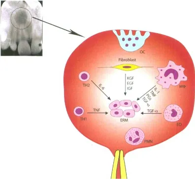
A schematic illustration of the mechanism of epithelial rest cells proliferation by pro-inflammatory cytokines and growth factors produced by host cells around periapical tissue of non-vital teeth is shown in figure 1.
Figure 1. A schematic illustration of the mechanism of epithelial rest cells proliferation2
Cyst development and expansion
Once formed, the cyst will experience a slow enlargement. Cystic expansion is influenced by a number of factors, like mural growth, hydrostatic enlargement, and bone resorbing factor.3 Most radicular cysts grow slowly and
suggested that the internal pressure is a major factor in the development of cysts. Expansion of cysts is associated with the hydrostatic pressure within the cyst, which is higher than capillary blood pressure. In order to balance the osmotic pressure, fluids from outside enters the lumen. These process makes cyst gradually enlarged.1,2,3
The risen osmotic pressure inside cyst is resulted from the numbers of high molecular weight proteins in cyst fluid, which coming from inflammatory exudates.2,3 Other factor that contribute in increasing the osmotic pressure of
cyst fluid are mast cells.15 The number of mast cells were found higher in
subepithelial zone of radicular cysts. Mast cells may contribute to cyst enlargement in three ways 1) by direct release of heparin into the luminal fluid, 2) by release of hydrolytic enzymes which could degrade capsular extracellular matrix components, thereby facilitating their passage into the fluid, and 3) by the action of histamine on vascular permeability, thus leading the transudation of serum proteins.15
Increasing vascular permeability can also be induced by growth factor. Leonardi et al,16 reported that vascular endothelial growth factor (VEGF) was
expressed in the radicular cysts. A number of cell types, including keratinocytes, macrophages, fibroblasts, and lymphocytes are involved in VEGF secretion. VEGF has bioactivity to increase vascular permeability, resulting in accumulation of inflammatory cells and cyst fluid.16 Hepatocyte growth factor
(HGF) can also be found in radicular cysts. HGF might play a role in cyst expansion through inducing lining epithelial cell proliferation and its invasion into capsular connective tissue.8
Mechanism of bone resorption in radicular cyst
The most prominent destructive event connected with radicular cyst is the resorption of alveolar bone. The effector cells of this process are osteoclasts. Activated osteoclast will resorb the mineralized matrix and degrade organic components of bone.
the expression of both molecules in radicular cysts, with the number of RANKL was hgher than OPG (the ratio of RANKL / OPG: 1.40 ± 0, 04). Both RANKL and OPG are involved in osteoclasts signaling. RANKL is a molecule (ligand) required to stimulate differentiation of osteoclast precursor cells into mature osteoclasts by binding to its receptor, RANK expressed on the surface of osteoclast precursor cells. As a result, mature osteoclast become activated resulting in bone resorption.4,19 Whereas OPG is a molecule that acts as a
decoy receptor, inhibits RANKL-RANK interaction and thus bone resorption.4,19
Identification of RANKL and OPG in radicular cysts demonstrated that both molecules are contributing to osteoclast formation and bone resorption.
Inflammatory cytokines produced by host cells such as IL-1, TNF-α, and PGs can mediate bone resorption.2,4,7 Other interleukins that have been
implicated in alveolar bone loss are IL-6, IL-3 IL-11, IL-17, and IL-18. Those interleukins were reported to be expressed in radicular cyst.2,4,6,8 They can
stimulate proliferation of osteoclasts and induce the released of PGs by host cells.2 PGs are also a potent inductor for bone resorption by osteoclasts.
Other cytokines that play a role in the process of bone resorption was granulocyte-macrophage colony stimulating factor (GM-CSF) which has been identified in radicular cysts fluid.6 GM-CSF together with IL-3 can stimulate the
release of macrophage colony stimulating factor (M-CSF) which will then stimulate the differentiation of hematopoiesis stem cell into osteoclast.
Other molecule that contributing in bone resorption is matrix metallo proteinase (MMP). MMP is a protease enzyme that can degrade extracellular matrixs, including collagen, fibronectin, lamini, and proteoglycans. The expression of Matrix Metallo Proteinase (MMP) can be found in radicular cyst. Walhgren et al,20 reported that MMP 2, MMP 8 and MMP 9 are expressed in
DISCUSSION
The pathogenesis of radicular cyst involves the activation of epithelial cell rests of Malassez after physical, chemical, or bacterial injury. Three phases of cystic formation has been described : initiation, cyst formation, and cyst enlargement. It is established that the radicular cysts are a result of inflammatory process in the periapical tissues.2,4 Humoral and cellular immune
responses play a role in the pathogenesis of these lesions.
Constant irritation from necrotic pulps will result in an inflammatory response within the periapical area. There are upregulated the expression of proinflammatory cytokines (IL-1, IL-6, IL-8, and TNF-α), inflammatory mediators (PGs), chemokines, and growth factors (EGF, KGF, TGF-α, FGF, HGF) in radicular cysts, released by surrounding host cells.2,3,5,6,8 The high levels of
those molecules is most probably due to ongoing stimulation of the lesion by bacterial toxins released from the infected root canal. All of those molecules synergistically stimulate cell rests of Malassez to enter cell cycle and begin to proliferate. 3
As cyst grows, there are increasing vascular permeability induced by mast cell and formation of inflammation exudates originating from the lytic product of the dying cell in the cyst lumen.4 Therefore, the osmotic pressure of
the cyst fluid will increase, To equalize the osmotic pressure, fluid enters the lumen, resulting in expansion of the cyst.
The growth of radicular cysts will be accompanied by local bone destruction, resulted from activated osteoclasts. Proinflammatory cytokines, interleukins, prostaglandins, and TNF-α are known to stimulate bone resorption through the upregulation of RANKL.17 The actions of RANKL include promotion
of osteoclast differentiation, stimulation of osteoclast activation, survival, and adherence to bone surface.17,18 Therefore, the number of activated osteoclast
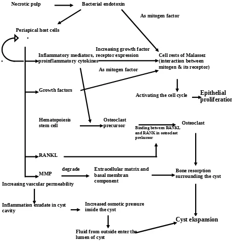
Schematic illustration of molecular mechanism of radicular cyst formation is shown in figure 2.
Fig 2. Schematic illustration of molecular mechanism of cyst formation
Understanding the molecular mechanism behind the pathogenesis of radicular cysts will provided the basis for the development of treatments for this lesions. Having eliminated successfully the initiating root canal infection by proper root canal treatment may prevail over tissue destructive events. The
Necrotic pulp Bacterial endotoxin
As mitogen factor
Cell rests of Malassez (interaction between mitogen & its receptor)
Epithelial proliferation Periapical host cells
Inflammatory mediators, proinflammatory cytokines
Growth factors
RANKL
As mitogen factor Increasing growth factor receptor expression
MMP Extracellular matrix andbasal membran component
degrade
Osteoclast precursor
Bone resorption surrounding the cyst
Cyst ekspansion Hematopoiesis
stem cell Osteoclast
Inflammation exudate in cyst cavity
Increased osmotic pressure inside the cyst
Fluid from outside enter the lumen of cyst
Activating the cell cycle
Binding between RANKL and RANK in osteoclast prekursor
lesion may heal completely by remineralization and the inflammatory characters will disappear. However, radicular cysts sometimes continuously expand after root canal therapy and eventually have to be removed by surgical procedure. This phenomenon suggests that processes other than those induced by infection contribute to the growth of these cysts. Hayashi et al,8 has identified
factors for angiogenesis, such as Ang, HGF, and IL-8, growth factors for epithelial cells (HGF), and bone resorbing factor (IL-6), which their releases were not induced by inflammatorty stimuli. Those finding could possibly explains the persistent growth of radicular cysts after root canal treatment.
The resorption of alveolar bone by osteoclasts is the most dominant destructive event from radicular cyst. The fact that RANKL is required for osteclast development suggest that agents which inbibit its activity may be theraupetic. Alternatively, soluble OPG, which inhibits osteoclast formation by blocking RANKL-RANK interaction, also have the potential to be developed for future theraupetic use for this lesion.
CONCLUSIONS
Radicular cysts are most likely induced by the initiation of an inflammatory stimulation of necrotic teeth. Humoral and cellular immune responses will get activated, thus trigger proliferation of epithelial cell rests of Malassez and stimulate bone resorption process.
REFERENCES
1. Cawson RA, Odell EW dan Porter S. Cawson’s Essential of Oral Pathology and Oral Medicine 7th Ed. Edinburgh: Churchill Livingstone, 2002: 103-7. 2. Lin LM, Huang GTJ dan Rosenberg PA. Proliferation of epithelial cell rests,
formation of apical cysts and regression of apical cysts after periapical wound healing. JOE 2007,33(8):908-16.
3. Kiss Csongor. Cell to cell interaction. Endodontic Topic 2004, 8:88-103. 4. Nair P, Sundqvist G, Sjogren U. Experimental evidence supports the
abscess theory of development of radicular cysts. Oral Surg Oral Med Oral Pathol Oral Radiol Endod 2008, 106:294-303.
6. Gervasio AM, Silva DAO, Taketomi EA, Souza CJA, Sung SSJ dan Loyola AM. Levels of GM-CSF, IL-3 and IL-6 in fluid and tissue from human radicular cysts. J Dent Res 2002, 81(1):64-68.
7. Jurisic V, Terzic T, Colic S, Jurisic M. The concentration of TNF-α correlate with number of inflammatory cells and degree of vascularization in radicular cysts. Oral Diseases 2008, 14:600-5.
8. Hayashi M, Ohshima T, Ohshima M, Yamaguchi Y, Miyata H, Takeichi O, et al. Profiling of radicular cyst and odontogenic keratocyst cytokine production suggests common growth mechanisms. JOE 2008, 34(1):14-21.
9. Teixeira-Salum TB, Rodrigues DBR, Gervasio AM, Souza CJA, Rodrigues Jr V, Loyola AM. Distinct Th1, Th2 and Treg cytokines balance in chronic periapical granuloma and radicular cysts. J Oral Pathol Med 2010, 39:250-256.
10. Silva TA, Garlet GP, Lara VS, Martins Jr W, Silva JS, Cunha FQ. Differential expression of chemokines and chemokine receptor in inflammatory periapical disease. Oral Microbiol Immunol 2005, 20:310-6.
11. Oliveira MG, Lauxen IS, Chaves ACM, Rados PV, Filho MSA. Immunohistochemical analysis of the pattern of p53 and PCNA expression in odontogonec cystic lesions. Med Oral Patol Oral Cir Bucal 2008, 13(5):E275-80.
12. Nadalin MR, Fregnani ER, Silva-Sousa YTC, Perez DEC. Syndecan-1 (CD138) and Ki-67 expression in odontogenic cystic lesions. Braz Dent J 2011, 22(3):223-9.
13. Gadbail AR, Chaudhary M, Patil S, Gawande M. Actual proliferating index and p53 protein expression in cyst. Oral Diseases 2009,15:490-8.
14. Ayoub MS, Baghdadi Hm, El-Kholy M. Immunohistochemical detection of laminin-1 and Ki-67 in radicular cysts and keratocystic odontogenic tumors. BMC Clinical Pathology 2011, 11:4.
15. Shylaja S. Mast cells in odontogenic cysts. Journal of Clinical and Diagnostic Research [serial online] 2010 April [cited: 2011 October 15]; 4:2226-2236. 16. Leonardi R, Caltabiano M, Pagano M, Pezzuto V, Loreto C, Palestro G.
Detection of vascular endothelial growth factor/vascular permeability factor in periapical lesions. JOE 2003, 29(3):180-3.
17. Menezes RM, Bramante CM, Silva Paiva KB, Letra A, Carneiro E, Zambuzzi WF dan Granjeiro JM. Receptor activator NFĸB-ligand and osteoprotegerin protein expression in human periapical cysts and granulomas. Oral Surg Oral Med Oral Pathol Oral Radiol Endod 2006, 102 : 404-9.
18. Tay JYY, Bay BH, Yeo JF, Harris M, Meghji S, Dheen ST. Identification of RANKL in osteolytic lesions of the facial skeleton. J Dent Res 2004, 83(4): 349-53.
19. Robling AG, Castillo AB, Turner CH. Biomechanical and molecular regulation of bone remodeling, Annu Rev Biomed Eng 2006, 8:455–98