Daftar Pustaka
1. Jensen TK, Toppari J, Keiding N and Skakkebaek NE. Do environmental estrogens contribute to the decline in male reproductive health? Clinical chemistry. 1995; 41: 1896-901.
2. Fernandez MF, Olmos B, Granada A, et al. Human exposure to endocrine-disrupting chemicals and prenatal risk factors for cryptorchidism and hypospadias: a nested case-control study. Environmental health perspectives. 2007; 115 Suppl 1: 8-14.
3. Atanassova N, McKinnell C, Walker M, et al. Permanent effects of neonatal estrogen exposure in rats on reproductive hormone levels, Sertoli cell number, and the efficiency of spermatogenesis in adulthood.
Endocrinology. 1999; 140: 5364-73.
4. De Flora S, Micale RT, La Maestra S, et al. Upregulation of clusterin in prostate and DNA damage in spermatozoa from bisphenol A-treated rats and formation of DNA adducts in cultured human prostatic cells.
Toxicological sciences : an official journal of the Society of Toxicology. 2011; 122: 45-51.
5. Soto AM, Justicia H, Wray JW and Sonnenschein C. p-Nonyl-phenol: an estrogenic xenobiotic released from "modified" polystyrene.
Environmental health perspectives. 1991; 92: 167-73.
6. Varayoud J, Ramos JG, Bosquiazzo VL, Lower M, Munoz-de-Toro M and Luque EH. Neonatal exposure to bisphenol A alters rat uterine implantation-associated gene expression and reduces the number of implantation sites. Endocrinology. 2011; 152: 1101-11.
7. Li YJ, Song TB, Cai YY, et al. Bisphenol A exposure induces apoptosis and upregulation of Fas/FasL and caspase-3 expression in the testes of mice. Toxicological sciences : an official journal of the Society of Toxicology. 2009; 108: 427-36.
8. Hulak M, Gazo I, Shaliutina A and Linhartova P. In vitro effects of bisphenol A on the quality parameters, oxidative stress, DNA integrity and adenosine triphosphate content in sterlet (Acipenser ruthenus) spermatozoa. Comparative biochemistry and physiology Toxicology & pharmacology : CBP. 2013; 158: 64-71.
9. Lee PC. Disruption of male reproductive tract development by administration of the xenoestrogen, nonylphenol, to male newborn rats.
Endocrine. 1998; 9: 105-11.
10. Fusani L, Della Seta D, Dessi-Fulgheri F and Farabollini F. Altered reproductive success in rat pairs after environmental-like exposure to xenoestrogen. Proceedings Biological sciences / The Royal Society. 2007; 274: 1631-6.
12. Winarni TI. Alteration of Rat Reproductive Organ In Adulthood Caused by The Exposure of Foreign Estrogenic Compounds (Mosquito Insecticides) During Early Life. Biomedical Science Diponegoro University. Semarang: Diponegoro, 2004.
13. Dang VH, Nguyen TH, Lee GS, Choi KC and Jeung EB. In vitro exposure to xenoestrogens induces growth hormone transcription and release via estrogen receptor-dependent pathways in rat pituitary GH3 cells. Steroids. 2009; 74: 707-14.
14. Edmunds JS, McCarthy RA and Ramsdell JS. Permanent and functional male-to-female sex reversal in d-rR strain medaka (Oryzias latipes) following egg microinjection of o,p'-DDT. Environmental health perspectives. 2000; 108: 219-24.
15. Furuya M, Adachi K, Kuwahara S, Ogawa K and Tsukamoto Y. Inhibition of male chick phenotypes and spermatogenesis by Bisphenol-A. Life sciences. 2006; 78: 1767-76.
16. Ganong WF. Buku Ajar Fisiologi Kedokteran. 17 ed. Jakarta: EGC, 1999. 17. Guyton H. Buku Ajar Fisiologi Kedokteran. 9 ed. Jakarta: EGC, 1996. 18. Francis S.G. JDB. Endokrinologi Dasar & Klinik. 4 ed. Jakarta: EGC,
1995.
19. Thomais Vlachogianni ea.
Chemical pollutants with endocrine disrupting properties: adverse health ef fects to humans and wildlife.
Department of Chemistry, University of Athens, University Campus Zogra fou, 15784 Athens, Greece2013.
20. RS M. Endocrine modulation. Center for Population Health Risk Assesment. Institute for Population Health2002.
21. D H. Enviromental effect on reproductive health : the endocrine disruption hypothesis. 1997: 1-11.
22. BJ D. The effect of enviromental hormones on reproduction. 1998: 1249-64.
23. RI DK. Pengenalan Pestisida, Direktorat Jenderal Pemberantasan Penyakit Menular dan Penyehatan Lingkungan. Jakarta2000.
24. Endah JdN. Menggendalikan Hama & Penyakit Tanaman. Tanggerang: AgroMedia Pustaka, 2005.
25. Tarumingkeng RC. Insektisida; Sifat, Mekanisme Kerja dan Dampak Penggunaannya. UKRIDA: Agro Media Pustaka, 1992.
26. Sudarmo S. Pestisida. Yogyakarta: Agro Media Pustaka, 1991.
27. Crinnion WJ. Environmental medicine, part 4: pesticides - biologically persistent and ubiquitous toxins. Alternative medicine review : a journal of clinical therapeutic. 2000; 5: 432-47.
28. W. M. Health effect of pesticides. Regional Awarness Workshop2002. 29. Guven M, F Bayran. Endocrine Change in Patient With Acute
Organophosphate Poisoning, Human and Experimental Toxicologi. 1999. 30. McClusky LM, de Jager C and Bornman MS. Stage-related increase in the
of male rats to p-nonylphenol. Toxicological sciences : an official journal of the Society of Toxicology. 2007; 95: 249-56.
31. Lubis HS. Deteksi Dini dan Penatalaksanaan Keracunan Pestisida Golongan OrganoFosfat Pada Tenaga Kerja. FKM USU2002.
32. JFX P. Kematian Karena Keracunan Baygon Suatu Tinjauan Kasus di Laboratorium Ilmu Kedokteran Karya Ilmiah Fakultas Kedokteran Universitas Diponegoro Tahun 1987-1991. 1993.
33. Sadler TW. Embriologi Kedokteran Langman. 7 ed. Jakarta: EGC, 1996. 34. R.F M. Young I.D.Emery's elements of medical genetics. London: Churcill
Livingstone2001.
35. P.L. W. Gray's anatomy. 13 ed. London: Churchill Livingstone1995. 36. H.M NEaB. Andrology: Male reproductive health and disfunction. Berlin:
Springer1997.
37. Richard M. Sharpe CM, Catrina Kivlin and Jane S. Fisher. Proliferation and functional maturation of Sertoli cells, and their relevance to disorders of testis function in adulthood. Reproduction. 2003.
38. Anna Hejmej ea. Antiandrogenic and Estrogenic Compounds: Effect on Development and Function of Male Reproductive System. Pubmed. 4: 53. 39. Pan YQ and Xu C. [Role of estrogen in male reproduction]. Zhonghua nan
ke xue = National journal of andrology. 2005; 11: 847-50.
40. Carreau S, Bois C, Zanatta L, Silva FR, Bouraima-Lelong H and Delalande C. Estrogen signaling in testicular cells. Life sciences. 2011; 89: 584-7.
41. Carreau S, Lambard S, Delalande C, Denis-Galeraud I, Bilinska B and Bourguiba S. Aromatase expression and role of estrogens in male gonad : a review. Reproductive biology and endocrinology : RB&E. 2003; 1: 35. 42. Fustini FM RV, and Carani C. Oestrogen deficiency in men: where are we
today ? Eur J Endocrinol. 1999.
43. O'Donnell L, Robertson KM, Jones ME and Simpson ER. Estrogen and spermatogenesis. Endocrine reviews. 2001; 22: 289-318.
44. Ando S, Carani C and Lombardi G. Insights into the role of estrogen in the male genital tract: a report on an estrogen and male reproduction workshop, Isola Capo Rizzuto, Italy, 23-24 September 1999. Trends in endocrinology and metabolism: TEM. 2000; 11: 248-50.
45. Sharpe RM, Atanassova N, McKinnell C, et al. Abnormalities in functional development of the Sertoli cells in rats treated neonatally with diethylstilbestrol: a possible role for estrogens in Sertoli cell development.
Biology of reproduction. 1998; 59: 1084-94.
46. Atanassova N, McKinnell C, Walker M, et al. Comparative effect of neonatal exposure of male rats to potent and weak (enviromental) estrogens on spermatogenesis at puberty and the relationship to adult testis size and fertility: evidence for stimulatory effect of low estrogen levels.
Endocrinology. 2000; 141: 3898-907.
47. Thyroid Hormone Stimulates the Proliferation of Sertoli Cells and Single Type A Spermatogonia in Adult Zebrafish (Danio rerio) Testis.
Lampiran 2. Cara kerja sediaan histopatologi
1) Menyiapkan wadah yang di isi dengan larutan formalin 10% bufer dengan volume minimal 5 kali volume jaringan
2) Testis yang telah diambil, segera di masukkan kedalam wadah tersebut
3) Memberi identitas pada semua wadah dengan identitas masing-masing kelompok perlakuan
4) Dikirim ke Sentra Diagnostik Patologi Anatomi disertai dengan formulir pengantar
5) Preparat kemudian dipotong dengan keteblan maksimal 3-4 cm
6) Setelah dipotong diletakkan di dalam kaset jaringan, dan dimasukkan ke wadah yang berisi formalin 10% bufer
7) Dilakukan proses pembuatan blok parafin, kemudian didinginkan di dalam lemari es
8) Blok parafin dipotong menjadi lebih tipis menggunakan microtome sesuai kebutuhan
9) Pita parafin dimekarkan dengan ditempelkan langsung pada kaca benda yang telah dibasahi dengan air
10) Dimulai proses pengecatan dengan Hematoxylin Eosin
11) Preparat diberi cat Hematoxylin
12) Kemudian di diferensiasi menggunakan air kran
13) Diberi cat Eosin
14) Kemudian di dehidrasi menggunakan alkohol 70%
15) Pada prosesl ‘clearing’ menggunakan larutan xylol
Lampiran 3. Hasil pengamatan sel Sertoli
Kelompok Lapang Pandang Jumlah Seluruh lapang pandang
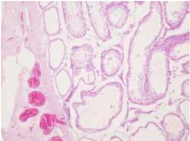
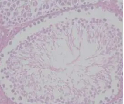
Lampiran 4. Gambar histopatologis testis/sel sertoli Tikus Sprague Dawley
Gambar histopatologis testis tikus kelompok kontrol dengan perbesaran 1000x. (1) Sel Sertoli. (2) Sel Leydig. (3) sel spermatogonia
Gambar histopatologis testis tikus kelompok perlakuan 3 dengan perbesaran 400x.
Gambar histopatologis testis tikus kelompok perlakuan 4 dengan perbesaran 400x.
Lampiran 5. Dokumentasi penelitian
Tempat pelaksanaan penelitian Laboratorium untuk hewan coba
Lampiran 6. Hasil perhitungan data dengan SPSS
kelompok perlakuan Statistic Std. Error
Variance 872,700
3. Tes Normalitas masing-masing kelompok
Tests of Normality
*. This is a lower bound of the true significance.
a. Lilliefors Significance Correction
4. Plots
5. Uji Oneway ANNOVA
Within Groups 53045,600 20 2652,280
Total 96909,040 24
6. Uji Post Hoc
Post Hoc Tests
Multiple Comparisons Dependent Variable: jumlah sel Sertoli
LSD
Std. Error Sig. 95% Confidence Interval
Lower Bound Upper Bound
1