Corresponding author: [email protected]
Staphylococcus epidermidis
: how to turn
from commensal to be a pathogen lifestyle
Titik NuryastutiDepartment of Microbiology, Faculty of Medicine, Universitas Gadjah Mada, Yogyakarta, Indonesia
DOI: http://dx.doi.org/10.19106/JMedSci005001201813
ABSTRACT
Staphylococcus epidermidis normally is a commensal inhabitant of healthy human skin and mucosa, but also a common nosocomial pathogen in immunocompromised patients, neonates, and patients with indwelling medical devices. To distinguish the pathogen and commensal strain is a big challenge when identifying this agent with its related infection. This mini-review aims to summarize recent research in this area with a special emphasis on the virulence factor of generating genotypic and phenotypic diversity in S. epidermidis. By living between a commensal and pathogen, S. epidermidis needed to establish many strategies to face different clinical environments, including the new ecological niche of biomaterials. In addition, the growing number of immunocompromised patients increased the risk for a very sensitive host. However, further exploration of the relationship between virulence factor and in vivo pathogenesis is still needed. According to the virulence factor of these bacteria, which are considered as a real pathogen, strict control measures should be taken for S. epidermidis infection.
ABSTRAK
Staphylococcus epidermidis merupakan bakteri komensal kulit dan mukosa pada manusia, tetapi akhir-akhir ini banyak ditemukan sebagai agen patogen infeksi nosokomial terutama pada pasien immunocompromised, neonatus, dan pasien dengan peralatan medis invasif. Saat ini, bagaimana membedakan S. epidermidis strain patogen dan komensal masih merupakan tantangan besar, baik di laboratorium maupun bagi klinisi. Tinjauan ini bertujuan untuk mendiskusikan peran faktor virulensi S. epidermidis dalam menyebabkan keragaman genotipik dan fenotipik serta keterkaitannya dengan perubahan karakterisitik S. epidermidis, sebagai bakteri komensal maupun patogen. Dengan hidup di antara pola komensal dan patogen, S. epidermidis perlu menyusun banyak strategi untuk menghadapi lingkungan klinis yang beragam, termasuk beradaptasi dengan permukaan biomaterial yang merupakan bahan dari peralatan medis invasif. Selain itu, meningkatnya jumlah penderita immunocompromised, menyebabkan peningkatan kepekaan host terhadap infeksi S. epidermidis. Namun, penelitian lebih lanjut tentang hubungan antara faktor virulensi dan patogenesis infeksi in vivo masih diperlukan. Dengan pertimbangan bisa berperan sebagai bakteri patogen, tindakan pengendalian yang ketat harus dilakukan untuk infeksi S. epidermidis.
Keywords: Staphylococcus epidermidis – commensal – pathogen - virulence factor -
INTRODUCTION
Staphylococcus epidermidis is a coagulase-negative Staphylococcus, considered as a part of the normal mucosa and skin microlora of humans and other mammals.1,2 It is considered as a member of the Staphylococci genus, which are gram-positive bacteria belonging to the family Staphylococcaceae. They are clustering, non-motile and non-spore forming cocci, facultative anaerobes and produce catalase. Currently, there are 35 known species of the genus Staphylococcus, from which 15 species are indigenous to humans, while the others are non-human pathogens.2,3 Coagulase-negative staphylococci (CNS) are grouped together as Staphylococcus saprophyticus (S. saprophyticus), Staphylococcus lugdunensis (S. lugdunensis), Staphylococcus schleiferi (S. schleiferi), Staphylococcus haemolyticus (S. haemolyticus), Staphylococcus caprae (S. caprae) or S. epidermidis based on their inability to clot blood plasma. Coagulase-negative staphylococci are widely distributed over the surface of the human body, where they constitute the majority of the commensal bacterial skin microlora.1
Culture analysis has revealed that Staphylococcus spp. are the most abundant organisms colonizing moist areas. These moist sites include the umbilicus, the axillary vault, the inguinal crease (side of the groin), the gluteal crease, the sole of the foot, the popliteal fossa (behind the knee), nares anterior, and the antecubital fossa (inner elbow).1 Staphylococci occupy an aerobic niche on the skin and probably use the urea present in sweat as a nitrogen source.3 In spite of being a saprophyte and opportunistic bacterium, this bacteria is involved in balancing the epithelial microlora and serves as a reservoir of resistance genes, which might be transferred to the closely related but more virulent
organisms, such as Staphylococcus aureus (S. aureus).4 Accordingly, S. epidermidis
maintains a commonly mutualism relationship with its host and serves as a shield, preventing colonization of potentially more harmful bacteria by producing lantibiotics, which are lanthionine-containing antibacterial peptides, also known as bacteriocins that may provide an added level of protection against certain common pathogens. Additionally, acting as skin microbiome, this bacteria promote the integrity of cutaneous defence through elicitation of host immune responses. 4,5 As an innocuous commensal microorganism, S. epidermidis was for a long time seen as an virulent species. However, today this bacterium is considered the most frequent cause of healthcare associated infections (HAIs), namely those related with indwelling medical devices. Overall, S. epidermidis is the most common species in HAIs, followed by S. haemolyticus, S. hominis, and S. capitis.6–8 it has not been established that adherence and bioilm formation are closely linked phenotypes for clinical isolates. In this study, the initial adhesion to different materials (acrylic and glass For example, S. epidermidis may be involved in prosthetic joint, vascular graft, surgical site, central nervous system shunt and cardiac device infections.5,9–11
but treatment failure is also associated with the ability of S. epidermidis to form bioilms on inert surfaces of medical devices from where these sticky, multilayered aggregates of bacteria are hard, if at all possible, to completely remove.13,14
Additionally, the increasing use of biomaterials in modern medicine has improved the quality of life of many patients. However, as a drawback, the occurrence of biomaterial-associated infections (BAI) is increasing and now becoming a serious health threat to
patients, as well as a inancial burden to the
society. S. epidermidis, generally regarded as an opportunist pathogen, is now recognised as a real “new” pathogen, since it is the major etiologic agent of BAI.10,11,15 Biomaterial-associated infections is generally related to
microbial bioilm formation, deined as a
microbial community encased in a matrix of self-produced extracellular polymeric substances (slime). Slime affects antimicrobial resistance as well as the effectiveness of the host immune system14,16. Currently, no effective non-invasive technique exists to
prevent or destroy bioilms associated with
BAI. Systemic antibiotics predominantly
attack a bioilm infection through the outermost layers of the bioilm, which are
usually ineffective as bacteria continue to grow from the inner layers combined with an increased production of extracellular polymeric substances. This virulence constitutes the main reason why biomaterial implants related to an infection nearly always have to be removed.17 In addition, the use of
antibiotics and disinfectants in hospitals puts a high selective pressure on bacteria to select for resistant and well-adapted variants.18,19 However, this unique pattern does not yet explain why just S. epidermidis and not any other bacteria, was able to conquer and occupy this novel ecological niche.
Notably, it has been shown that the genomic structure of S. epidermidis represents an amazingly versatile microorganism living in a grey area between commensalism and pathogenicity. S. epidermidis employs sophisticated regulatory networks to quickly adapt its metabolism to changing external conditions, to communicate with its neighbours in the same ecological niche, or to escape the host’s immune response.8,20 Genomic analyses demonstrated the presence of numerous mobile genetic elements in S. epidermidis genomes, including methicillin resistance-mediating SCCmec elements and insertion sequences (IS). IS elements seem to be important driving forces that keep the S. epidermidis genome extremely lexible and trigger heterogeneous gene expression.21It is suggested that well-adaptability properties both on the regulatory and genetic level might have contributed to the evolutionary success of S. epidermidis as a nosocomial pathogen.8 Meanwhile, due to the ubiquitous prevalence of S. epidermidis as a commensal bacterium, clinicians often face the challenge to decide whether an isolate represents the causative
agent of an infection or an unspeciic culture
contamination. Nowadays, our understanding of how S. epidermidis becomes a commensal or pathogen is far from complete and many questions still remain. This review addresses the questions concerning how the recent mechanism of commensal and infectious lifestyles of S. epidermidis takes place, which more focusing on literature about virulence properties of S. epidermidis i.e
bioilm formation, icaADBC presence and the mechanism of regulating gene expression, the role of small colony variant and methicillin resistance gene, as well as its genomic
DISCUSSION
Bioilm formation, major pathomechanism of HAIs infection
One of the main virulence characteristics of S. epidermidis is related with their adhesion
to substratum surfaces and subsequent bioilm
formation.5,10 A bioilm is a population of cells growing on a surface and enclosed in a self-produced matrix of extracellular polymeric
substance (EPS). Bioilms are notoriously
dificult to eradicate and are a source of
many recalcitrant infections.14 Bacterial
bioilm formation comprises a number of
physical, biological, and chemical processes. The relative contribution of each process
changes throughout bioilm development
and depends on prevailing environmental and hydrodynamic conditions.22 In general,
bioilm formation can be described in ive
phases 5,23,24 as shown in FIGURE 1.
FIGURE 1. The phases of bioilm formation in S. epidermidis. Graphs
were modiied from Vuong et al., Nuryastuti, and Bos et
Substratum surfaces will irst become covered with a conditioning ilm consisting
of proteins and glycoproteins, such as
ibronectin, vitronectin, ibrinogen, albumin,
and immuno-globulins, many of which serve as binding ligands to receptors on colonizing bacteria, although adhesion can also occur to
bare substratum surfaces. Bioilm formation
continues with the transport of bacteria to the substratum-liquid interface, which is governed by a combination of transport mechanisms, including Brownian motion, gravity, diffusion, convection, or the intrinsic motility of a microorganism.23,24 Subsequently, in the second phase, microbial adhesion may occur which is initially of a reversible nature. Factors involved in the initial adhesion to
a substratum surface include non-speciic
interactions originating from both the bacterial cell and substratum surfaces. These
non-speciic interactions are governed by
physicochemical properties such as surface charge, hydrophobicity, and chemical structure of both the bacteria and substratum surface. In the third phase, reversible adhesion of bacteria changes to irreversible, amongst others due to protein-protein interactions and the production
of EPS. The fourth phase in bioilm formation
is surface colonization. Adhering bacteria grow and divide, forming microcolonies that are considered to be the basic organizational
units of a bioilm. Entrapment of other
planktonic bacteria in the EPS also occurs, resulting in a multi-layered and mature
bioilm.5,23 The last step is detachment of individual bacteria or aggregates, which allows bacteria to disseminate into other areas for further surface colonization. In the clinical setting, this last step generally leads
to severe systemic infections.5 As a
pivotal structural component of microbial
bioilms, EPS has received much attention.
In general, EPS consists of polysaccharides,
eDNA and proteins in a hydrated environment. 26,27polysaccharide intercellular adhesin (PIA Recently eDNA
was found to be a major structural component of bacterial EPS where it plays a role in bacterium-surface and bacterium-bacterium interactions. The EPS produced by S. epidermidis consists mostly of polysaccharide intercellular adhesin (PIA).26
icaADBC and the mechanism of regulation of expression
Production of PIA, a key virulence factor of S. epidermidis, is subject to on-off switching, resulting in phenotypic variability (phase variants).11,15,28 Polysaccharide intercellular adhesion production is stimulated through the action of membrane bound sensory proteins within the bacterial cell wall. Polysaccharide intercellular adhesion synthesis is catalyzed by proteins encoded within the ica operon, a gene cluster consisting of icaADBC. The
icaA gene product is a transmembrane
protein with homology to N-acetyl-glucosaminyltransferases. The functions of
icaB and icaC are less well deined. However, icaB is likely to be secreted while icaC is predicted to be an integral membrane protein.
icaD might act as a link between icaA and icaC
and represent a novel enzyme combination. When icaA is co-expressed with icaD, the transferase activity increases 20 fold.11,29
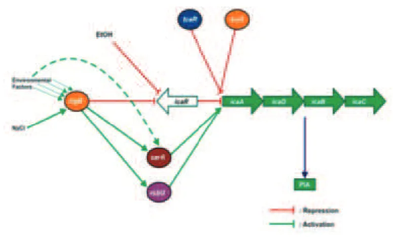
Extracellular polymeric substance production is vital, but metabolically expensive for S. epidermidis and therefore well-regulated (FIGURE 2). Regulation of ica
-expression and bioilm formation is negatively
controlled by the ica operon regulator, IcaR
and teiocoplanin-associated locus regulator,
TcaR. It is also inluenced by environmental
conditions that are potentially toxic for the bacterial cell. The exposure of S. epidermidis
sub-MIC (Minimal Inhibitory Concentration) concentrations of certain antibiotics, glucose, iron limitation and oxidative stress have all been shown to elevate ica-expression and
bioilm formation.30–32 Moreover, the global stress response factor σB, positively regulates
ica-expression by negatively regulating icaR
expression, while staphylococcal accessory regulator A (sarA) and regulators of sigmaB (rsbU) act similarly.33 In addition, the LuxS system involved in quorum sensing in S. epidermidis, recently emerged as another
negative regulator of bioilm formation.11
FIGURE 2. The schematic overview of regulatory network controlling expression of icaADBC in S. epidermidis. Graphs were modiied from Nuryastuti.23
The genetic and molecular basis of bioilm formation in S. epidermidis is multi-faceted. It has been reviewed that there are two distinct mechanisms of bioilm development; through an ica-dependent and an ica-independent mechanism of bioilm development.11 Bioilm production by ica operon-encoded enzymes is currently the best-understood bioilm mechanism in staphylococci,11 which is regulated by several regulatory genes such as icaR, ơB, rsbU and sarA, including the
Recent studies imply that the multicellular
organization of bacteria in a bioilm is a
crucial mechanism in resisting unfavorable conditions.35 Heterogeneous gene expression is typically observed in clinical S. epidermidis
strains, and it is assumed that this ability is an advantage for adaptation of staphylococci to changing environmental conditions.8 Phase variation involves both regulatory pathways, e.g. in response to environmental signals, as well as genetic variations, by local genomic re-arrangements, altered activity of regulatory proteins or modulation of transcription or translation of the appropriate gene through strand slip mechanisms.8,11,23
Polysaccharide intercellular adhesin is the most important component of the staphylococcal slime and its production is catalyzed by proteins encoded within the
icaADBC operon. Different S. epidermidis
strains vary widely in the degree of PIA
or slime, and bioilm they produce.5,36 The importance of the ica operon has been
conirmed in numerous epidemiological
studies, which found a higher prevalence of the ica genes in clinical than in control skin isolates.37,38 Clinical strains of S. epidermidis obtained from urinary tract infection,39 as well as from paediatric cancer patients receiving chemotherapy are reported to be related with
ica-presence.25
Epidemiological studies have shown that the icaADBC operon is a typical feature of nosocomial S. epidermidis strains obtained from device-associated infections.
10,33 It is shown that icaADBC operon is
mostly prevalent in strains associated with intravascular catheter-associated bacteraemia and septicaemia.10,39 A study of the occurrence of ica operon among S. epidermidis isolates obtained from various origins has indicated
that the genetic information for bioilm
formation is rarely found in isolates obtained
outside of hospital settings.18 Interestingly, many of these studies found that invasive S. epidermidis strains signiicantly more often
carried icaADBC than colonizing commensal
S. epidermidis strains. Therefore, icaADBC -negative S. epidermidis strains were regarded as non-virulent and it was proposed to use
icaADBC as a genetic marker to distinguish invasive and contaminating S. epidermidis in blood cultures.37,38
Phenotypic and genotypic instability of bioilm-forming ability
Phenotypic variation in ica-presence is commonly observed in S. epidermidis. 8,28,40 Ziebuhr and coworkers identiied an insertional element (IS256) that was capable of inserting itself into the ica-locus resulting in ica-negative phenotypes.8 This disruption was shown to be reversible as precise excision from the ica-locus which observed at low incidence resulting in ica-positive phenotypes.
We and others have shown that a
signiicant proportion (42-85%) of clinical
isolates are ica-negative during culturing in the laboratory.28,40 In contrast to studies showing a reversible switching (phenotypic switching) between ica-positive and ica-negative phenotypes, the ica-locus was permanently lost in these strains. The absence of IS256 and phenotypic variation in these clinical
S. epidermidis isolates and the inability to switch back to ica-positive suggested a new
mechanism of switching in terms of bioilm
formation involving genetic instability.28
We showed that the presence of the
ica-locus in clinical isolates represents a disadvantage for growth in laboratory conditions. In line with this, it was recently suggested that the presence of the icaADBC
Strains that have a high level of PIA
production have a signiicant growth
disadvantage under commensal conditions and are therefore outcompeted by strains with more moderate or absent PIA production. Whereas PIA production enables staphylococci to survive and grow under
hostile, infection related conditions (bioilms),
during commensal colonization (as well as during planktonic growth), PIA production can be considered a burden that can easily be subsided. It is important to conclude that the ability to express different slime-producing phenotypes could provide staphylococci with
a greater degree of lexibility for colonizing a
range of different environments.33 Too much or no PIA production is only favourable
under speciic conditions while the ability to
regulate PIA production allows the organism to adapt to all conditions, both commensal and infectious.23
Other studies have found, in S.
epidermidis, IS256 detection is attributed
to the epidemic bioilm-forming clonal
lineages, and the element has been shown
to trigger heterogeneous bioilm expression by reversible transposition into
bioilm-associated genes and regulators.21,41
Thus, IS256 was shown to cause phase variation of icaADBC operon expression by alternating insertion in and precise excision from the PIA synthesis-mediating gene locus.20 While switch-off of PIA production through IS256 insertions occurs with a frequency of approximately 10-6 per cell and generation, restoration of PIA-dependent
bioilm formation by precise IS256 excision
was found to be an extremely rare event (10−11 per cell and generation).42
The role of small colony variant (SCV)
Small colony variants are naturally occurring subpopulations of bacteria demonstrating distinctive phenotypic
characteristics and pathogenic traits.
Phenotypically, SCVs have a slow growth
rate, atypical colony morphology associated with the formation of pinpoint or ‘fried egg’ colonies and unusual biochemical features. It was most extensively studied for staphylococci, especially for S. aureus as well as S. epidermidis.43 SCVs were recorded as being <1 mm in size (less than 1/10 of the normal cell size), with reduced pigmentation and haemolytic activity as described in literature.43,44 The tiny size of SCVs on solid agar is often due to auxotrophy for haemin and/or menadione, two compounds involved in the biosynthesis of electron transport chain components, which is associated with defects in electron transport and, consequently, altered membrane potential. The abnormal membrane potential, in turn, may confer on these variants innate resistance to aminoglycosides, since the ability of these antibiotics to gain access to intracellular target sites depends on the proton motive force. More importantly, some
reports have linked bacterial SCVs to several
recurring infections that are intractable to conventional treatment antibiotic regimes.44–46
Small colony variants have been associated with long-lasting, chronic, and recurrent infections, and it was suggested that this property was linked to the ability
of SCVs to survive intracellularly, thereby
being protected from the host immune system
and the action of antibiotics. Both bioilm formation and the SCV phenotype may
contribute to the recurrence and persistence of
staphylococcal infections; bacteria are either embedded in large, adherent bioilms on the
resulted in the upregulation of alternative
sigma factor B, which plays a central role in the augmentation of icaADBC expression and PIA production.43,47
Methicillin resistance gene
In addition to bioilm formation,
nosocomial S. epidermidis isolates are characterized by their pronounced resistance against commonly used antibiotics including methicillin. Methicillin resistance is, similar with S. aureus, mediated by the mecA gene encoding a penicillin binding protein with
reduced afinity to β-lactam antibiotics.48 However, in contrast to methicillin-resistant S. aureus (MRSA), attention paid to methicillin resistant S. epidermidis (MRSE) in hospital settings is not adequate enough, meaning they are not dealt with by using intense hygienic measures as those for MRSA. As a result, methicillin resistance rates among nosocomial
S. epidermidis isolates and other CoNS are extremely high and regularly exceed those of MRSA.48–50 It has been reported approximately
80% of S. epidermidis isolates from device-associated infections are considered as
MRSE, and also found to be multiresistant;
whereas commensal strains obtained from the community are mostly methicillin-sensitive S. epidermidis.10
The mecA gene and its regulators are located on large DNA elements that are termed staphylococcal cassette chromosome mec (SCCmec). In addition to the methicillin resistance determinant, SCCmec carry a set of recombinases and a wide variety of mobile DNA elements such as transposons, insertion sequences or integrated plasmids.1,15
To date, ive major SCCmec types have been
identiied, all of them can be distributed over
the S. epidermidis genome. Interestingly, SCCmec have been shown to be transferable among staphylococcal species. These genes are now regarded as mobile elements in
which extensive recombination and gene
shufling takes place.15,51 Obviously, they do not only serve as shuttles for the transfer of methicillin resistance but can also carry
other staphylococcal genes. MRSE is
often associated with additional antibiotic resistance, such as erythromycin (encoded by erm genes), ciproloxacin, clindamycin, aminoglycosides (encoded in aacA/aphD gene) or trimethoprim-sulfamethoxazole.15
The recent indings of genomic research
strongly suggest that S. epidermidis and other coagulase-negative staphylococci represent the gene pool for the ongoing generation of novel SCC types from which methicillin resistance in S. aureus might originate.1,12 Accordingly, it would be meaningful and reasonable to control MRSE and MR-CoNS by appropriate hygiene measures in a similar manner for MRSA, in order to lower MRSA burden in medical facilities, due to their role as reservoirs for the spread of resistance genes within microbial communities.
Genomic lexibility
It was demonstrated that clonal
diversiication in S. epidermidis is mainly based on genetic recombination, which is in contrast to S. aureus, a species known to evolve preferentially by point mutations.21,41
Multilocus sequence typing (MLST) analysis of a representative collection of clinical S. epidermidis isolates revealed a high degree of genetic diversity within the species, but the most widespread clone was ST2 or ST27 (sequence types). Especially, clonal complex ST2 isolates were found to be highly
lexible with respect to methicillin resistance
and prone to take up these mobile genetic elements. 21 Possibly, the successful spread of ST2 may be due to the fact that all ST2 isolates contain IS256 insertion sequences and ica
invasiveness in S. epidermidis. In addition, most ST2 isolates show in vitro capacity to
form bioilms.15,21
Instability of genetic material is often an indication of mobility, and in this respect it is also conceivable that the ica operon represents mobile DNA that has been lost in the commensal strain. S. epidermidis
isolates ST 2(ST27) represent an ideal genetic
background for bioilm and resistance genes,
resulting in well-adapted strains which are then selected in the hospital environment and causes device-related infection and bacteraemia. The presence of multiple copies of IS256 in the ST27 genome might support this adaptation process by an ongoing generation of novel phenotypic and genotypic variants. Therefore, the combination of
bioilm formation, antibiotic resistance, and genetic lexibility may explain why ST2 has
become the dominant clonal variant within medical facilities.8,41
Clinical manifestation of related infection
Staphylococcus epidermidis and other CoNS have for a long time been dismissed as culture contaminations which is mainly due to the fact that CoNS are primarily ubiquitous commensals of the human skin and mucosa. It is still a great challenge for the clinical microbiology laboratory to distinguish infecting strains from contaminants. In suspected S. epidermidis infections, where the pathogen is also a skin commensal that could contaminate skin swab or blood specimen if aseptic techniques are not followed, the same indistinguishable microorganism must be cultured from at least two separate specimens in order to differentiate a relevant infection from skin contamination.52 In contrast, for virulent species such as S. aureus or gram negative bacteria, a single positive clinical
specimen may be suficient to determine the
presence of a recent infection.53,54 However,
some groups of the population are prone to be infected with this microorganism. These higher risk groups include preterm neonates, immunocompromised individuals and patients with indwelling medical devices.1,5,10
The most important clinical manifestation associated with CoNS, particularly S. epidermidis is biomaterial-associated infections (BAI), which include a unique, complex constellation of many factors that have to be considered for their successful management.33 The increasing use of foreign
materials in almost all ields of modern
medicine is associated with a risk of bacterial infection.10 Morbidity and mortality of biomaterial-associated infections may vary according to the underlying patient condition, the microbial strain(s) that are implicated, and the type of device. Biomaterial-associated
infections contribute signiicantly to the
increasing problem of nosocomial infections. While a variety of microbial strains have been involved as causative organisms in biomaterial-associated infections, staphylo-cocci, particularly S. epidermidis, account for the majority of infections related to both temporarily inserted and permanently implanted biomaterials.5,10
The presence of a biomaterial
signiicantly compromises the host’s ability to
cope with infectious microorganisms. These microorganisms can reach a biomaterial implant in several ways and at different times post-implantation. Airborne microorganisms, inevitably present in the operating theater, can reach a biomaterial implant surface as early as before the implantation. Also during insertion of a biomaterial implant, microorganisms
from the commensal microlora of the skin
can contaminate a biomaterial implant. Peri-operative contamination is believed to be the most common cause of biomaterial associated infection.10,18,33
devices or damaged tissue can encase themselves in a hydrated matrix of extra cellular polymeric substances, a slimy layer,
and start growing into a bioilm. Bacteria organized in bioilms are at least 10-1000
times more resistant to antibiotics 14,16 and can cope much better with unfavorable external conditions as the host immune system than their
planktonic counterparts. The bioilm mode of growth represents a beneit for staphylococcal
strains enabling them to colonize inert surfaces of medical devices.8 Antibiotic
resistance of bacteria in the bioilm mode
of growth contributes to the chronic nature of these infections, which are notoriously
dificult to resolve. The mechanisms of
bacterial resistance in bioilms are different
from the now familiar plasmids, transposons, and mutations that convey innate resistance
to individual bacterial cells. In bioilms,
resistance seems to depend on multicellular strategies resulting in an impaired penetration of antibiotics to the target organisms and a decreased immune response.14,55
Biomaterial-associated infections comprise local (e.g., exit site) and systemic
infections. Originating from bacteremia or other systemic spread of causative organisms and depending on the nature and localization of the biomaterial inserted, sepsis, endocarditis, meningitis, joint sepsis, vertebral
abscesses, and other local manifestations due to metastatic seeding may result.1,19 These comprise infections commonly associated with prosthetic vascular grafts, prosthetic heart valves, cardiac devices, and coronary stents.
Moreover, local inlammation signs include
erythema, warmth, swelling, tenderness, and purulent drainage, which characterize exit-site infections.
It has been shown that S. epidermidis was the most frequent agents of central venous
catheter (CVC) and umbilical
catheter-associated BSIs (Blood Stream Infection) in
neonatal ICUs.5,25the coagulase-negative staphylococci (CoNS Besides BSIs, the CoNS group may cause further invasive infections in preterm infants, such as infective endocarditis, meningitis, and necrotizing fasciitis.5,12 Additionally, S.
epidermidis is also considered as the main cause of septicemia in febrile patients who suffer from chemotherapy-induced neutropenia, which is accounting for approximately 20 to
40% of cases.1,25
CONCLUSION
So far it is still a great challenge for clinician to distinguish S. epidermidis strains that may cause infection from those that live on the skin. However, the virulence
properties identiied in this paper, such as the presence of bioilm formation phenotype
including icaADBC operon, IS256, mecA,
SCV properties, together with patient
characteristics, might be used to consider the pathogenesis of infection caused by S. epidermidis. Nevertheless, up to date, the clues to distinguish between infectious and commensal strains of S. epidermidis are not clear yet. It is well understood the adhesion to host tissue is considered crucial during both these lifestyles.
By living on the verge of commensalism and pathogenicity, S. epidermidis has elaborated many strategies to overcome different clinical environments, including the new ecological niche of biomaterials. In addition, the growing number of immunocompromised patients increases the risk for a very sensitive host.
The formation of bioilms, the acquisition of
resistance characteristics and the enormous
lexibility of the genome of staphylococci
are characteristics that help their survival
in speciic environments and are the main
taken more seriously with adequate prevention applications for future infection control and hygiene measures.
ACKNOWLEDGEMENTS
I would like to thank Prof Henny van der Mei from Department of Biomedical Engineering, University of Groningen, The Netherlands for giving insight and comments that greatly improved this manuscript.
REFERENCES
1. Becker K, Heilmann C, Peters G. Coagulase-Negative Staphylococci. Clin Microbiol Rev. 2014;27(4):870–926. http://doi:10.1128/ CMR.00109-13
2. Christensen GJM, Brüggemann H. Bacterial skin commensals and their role as host guardians. Benef Microbes. 2014;5(2):201– 15. http://doi:10.3920/BM2012.0062
3. Grice EA, Segre JA. The skin microbiome. Nat Rev Microbiol. 2013;9(4):244–53. http:// doi:10.1038/nrmicro2537
4. Otto M. Staphylococcus epidermidis -the’accidental’pathogen. Nat Rev Microbiol. 2009;7(8):555–67. http://doi:10.1038/ nrmicro2182
5. Vuong C, Otto M. Staphylococcus epidermidis
infections. Microbes Infect. 2002;4(4):481–9. http://doi:10.1016/S1286-4579(02)01563-0 6. Cerca N, Pier GB, Vilanova M, Oliveira R,
Azeredo J. Quantitative analysis of adhesion and bioilm formation on hydrophilic and hydrophobic surfaces of clinical isolates of
Staphylococcus epidermidis. Res Microbiol. 2005;156:506–514. http://doi:10.1016/ j.resmic.2005.01.007
7. Gomes F, Teixeira P, Oliveira R. Mini-review :
Staphylococcus epidermidis as the most frequent cause of nosocomial infections : old and new ighting strategies. Biofouling. 2014;30(2):131-41. http://doi:10.1080/08927 014.2013.848858
8. Ziebuhr W, Hennig S, Eckart M, Kr H, Batzilla C, Kozitskaya S. Nosocomial infections by Staphylococcus epidermidis: how a
commensal bacterium turns into a pathogen. Int J Antimicrob Agents. 2006;28S:S14–20. http://doi:10.1016/j.ijantimicag.2006.05.012 9. Garrett TR, Bhakoo M, Zhang Z. Bacterial
adhesion and bioilms on surfaces. Prog Nat Sci. 2008;18(9):1049–56. http:// doi:10.1016/j.pnsc.2008.04.001
10. Brescó MS, Harris LG, Thompson K, Stanic B, Morgenstern M, Mahony LO, et al.
Pathogenic Mechanisms and Host Interactions in Staphylococcus epidermidis device-related infection. Front Microbiol. 2017;8(1401). http://doi:10.3389/fmicb.2017.01401
11. O’Gara JP. ica and beyond: Bioilm mechanisms
and regulation in Staphylococcus epidermidis
and Staphylococcus aureus. FEMS Microbiol Lett. 2007;270(2):179–88. http://doi:10.1111 /j.1574-6968.2007.00688
12. Suja KRS, Sheela P, Jyothis S, Radhakrishnan EK. Virulence factors associated with coagulase negative Staphylococci isolated from human infections. 3 Biotech. 2017;7(140):1–10. http://doi:10.1007/ s13205-017-0753-2
13. Mack D, Davies AP, Harris LG, Jeeves R, Pascoe B, Knobloch JK, et al. Staphylococcus epidermidis in Biomaterial-Associated Infections. In: T.F. Moriarty et al.,
editors, Biomaterials Associated Infection: Immunological Aspects and Antimicrobial Strategies. New York: Springer Science Business Media; 2013. p 25-57. http:// doi:10.1007/978-1-4614-1031-7
14. Lewis K. Minireview riddle of bioilm resistance. Antimicrob Agents Chemother. 2001;45(4):999–1007. http://doi:10.1128/ AAC.45.4.999
epidermidis in implant related infections. Indian J Med Res. 2012;136:483–90.
16. de la Fuente-Nunez C, Reffuveille F, Fernandez L, Hancock REW. Bacterial bioilm development as a multicellular adaptation: antibiotic resistance and new therapeutic strategies. Curr Opin Microbiol. 2013;580–9. http://doi:10.1016/j.mib.2013.06.013
17. Wilkins M, Hall-stoodley L, Allan RN, Faust SN. New approaches to the treatment of bioilm-related infections. J Infect. 2014; 69:S47–52.http://doi:10.1016/ j.jinf.2014.07.014
18. Lindsay D, von Holy A. Bacterial bioilms within the clinical setting : what healthcare professionals should know. J Hosp Infect. 2006;64:313-25 http://doi:10.1016/ j.jhin.2006.06.028
19. Mack D, Davies AP, Harris LG, Jeeves R, Pascoe B, Knobloch JKM, et al. Staphylococcus epidermidis in biomaterial-associated infections. In: T.F. Moriarty et al.,
editors. Biomaterials Associated Infection: Immunological Aspects and Antimicrobial Strategies. New York: Springer Science Business Media; 2013. p 25-57 http:// doi:10.1007/978-1-4614-1031-7_2
20. Schoenfelder SMK, Lange C, Eckart M, Hennig S, Kozytska S, Ziebuhr W. International Journal of Medical Microbiology Success through diversity – How Staphylococcus epidermidis
establishes as a nosocomial pathogen. Int J Med Microbiol. 2010;300(6):380–6. http:// doi:10.1016/j.ijmm.2010.04.011
21. Kozitskaya S, Olson ME, Fey PD, Witte W, Ohlsen K, Ziebuhr W, et al. Clonal analysis of
Staphylococcus epidermidis isolates carrying or lacking bioilm-mediating genes by multilocus sequence typing. J Clin Microbiol. 2005;43(9):4751–7. http://doi:10.1128/ JCM.43.9.4751
22. Bryers JD. Medical bioilms. Biotechnol Bioeng. 2009;100(1): 1–18. http://
doi:10.1002/bit.21838
23. Nuryastuti T. Environmental signals affecting
ica-expression in Staphylococcus epidermidis
bioilms. [Dissertation]. The Netherlands: University of Groningen; 2010.
24. Bos R, Mei HC Van Der, Busscher HJ. Physico-chemistry of initial microbial adhesive interactions-its mechanisms and methods for study. FEMS Microbiol Rev. 1999;23:179-230.
25. von Eiff C, Peters G, Heilmann C. Pathogenesis of infections due to coagulase negative staphylococci. Lancet Infect Dis. 2002;2(11):677–85. http://doi:10.1016/ S1473-3099(02)00438-3
26. Rohde H, Frankenberger S, Zähringer U, Mack D. Structure, function and contribution of polysaccharide intercellular adhesin (PIA) to
Staphylococcus epidermidis bioilm formation
and pathogenesis of biomaterial-associated infections. Eur J Cell Biol. 2010;89(1):103– 11. http://doi:10.1016/j.ejcb.2009.10.005 27. Cramton SE, Gerke C, Schnell NF, Nichols
WW, Gotz F. The intercellular adhesion (ica) locus is present in Staphylococcus aureus
and is required for bioilm formation. Infect Immun. 1999;67(10):5427–33.
28. Nuryastuti T, van der Mei HC, Busscher HJ, Kuijer R, Aman AT, Krom BP. recA mediated spontaneous deletions of the icaADBC operon of clinical Staphylococcus epidermidis
isolates: A new mechanism of phenotypic variations. Antonie van Leeuwenhoek, Int J Gen Mol Microbiol. 2008;94(2):317–28. http://doi:10.1007/s10482-008-9249-8
29. Arciola CR, Campoccia D, Ravaioli S, Montanaro L. Polysaccharide intercellular adhesin in bioilm: structural and regulatory aspects. Front Cell Infect Microbiol. 2015;5(7):1-10. http://doi:10.3389/ fcimb.2015.00007
disinfectants increase bioilm expression of
Staphylococcus epidermidis. J Antimicrobial Chemother. 2002;49:683–7.
31. Cramton SE, Ulrich M, Gotz F, Doring G. Anaerobic conditions induce expression of polysaccharide intercellular adhesin in
Staphylococcus aureus and Staphylococcus epidermidis. Infect Immun. 2001;69(6):4079–
85. http://doi:10.1128/IAI.69.6.4079
32. Rachid S, Ohlsen K, Witte W, Hacker RG, Ziebuhr W. Effect of subinhibitory antibiotic concentrations on polysaccharide intercellular adhesin expression in bioilm-forming
Staphylococcus epidermidis. Antimicrob Agents Chemother. 2000;44(12):3357–63. 33. Fitzpatrick F, Humphreys H, O’Gara JP.
The genetics of staphylococcal bioilm formation-will a greater understanding of pathogenesis lead to better management of device-related infection? Eur Soc Clin Infect Dis. 2005;11(12):967–73. http://doi:10.1111/ j.1469-0691.2005.01274.x
34. Li H, Xu L, Wang J, Wen Y, Vuong C, Otto M, et al. Conversion of Staphylococcus epidermidis
strains from commensal to invasive by expression of the ica locus encoding production of bioilm exopolysaccharide. Infect Immun. 2005; 73(5):3188–91. http://
doi:10.1128/IAI.73.5.3188
35. O’Gara JP, Humphreys H. Staphylococcus epidermidis bioilms: importance and
implications. J Med Microbiol. 2001;50:582– 7. http://doi:10.1099/0022-1317-50-7-582 36. Rohde H, Frankenberger S, Zähringer U, Mack
D. Structure, function and contribution of polysaccharide intercellular adhesin (PIA) to
Staphylococcus epidermidis bioilm formation
and pathogenesis of biomaterial-associated infections. Eur J Cell Biol. 2010;89(1):103– 11. http://doi:10.1016/j.ejcb.2009.10.005 37. Vandecasteele SJ, Peetermans WE, Merckx RR,
Rijnders BJA, van Eldere J. Reliability of the
ica, aap and atlE genes in the discrimination
between invasive, colonizing and contaminant
Staphylococcus epidermidis isolates in the diagnosis of catheter-related infections. Eur Soc Clin Infect Dis. 2003; 9(2):114–9. http:// doi:10.1046/j.1469-0691.2003.00544
38. Chessa D, Ganau G, Spiga L, Bulla A, Mazzarello V. Staphylococcus aureus and
Staphylococcus epidermidis virulence strains as causative agents of persistent infections in breast implants. PLoS ONE. 2016; 11(1): e0146668. http://doi:10.1371/journal. pone.0146668
39. Caiso V, Bertuccio T, Santagati M, Campanile F, Amicosante G, Perilli MG, et al. Presence of the ica operon in clinical isolates of
Staphylococcus epidermidis and its role in bioilm production. Clin Microbiol Infect. 2004;10(12):1081–8. http://doi:10.1111 /j.1469-0691.2004.01024
40. Nuryastuti T, Mei HC Van Der, Busscher HJ, Kuijer R, Aman AT, Krom BP. High frequency spontaneous deletions within the icaADBC
operon of clinical Staphylococcus epidermidis
isolates. I J Biotech. 2012;17(2):144–53. 41. Kozitskaya S, Cho S, Dietrich K, Marre
R, Naber K, Ziebuhr W. The bacterial insertion sequence element IS 256 occurs preferentially in nosocomial Staphylococcus epidermidis Isolates : association with bioilm
formation and resistance to aminoglycosides. Infect Immun. 2004;72(2):1210–5. http:// doi:10.1128/IAI.72.2.1210
42. Hennig S, Ziebuhr W. A transposase-independent mechanism gives rise to precise excision of IS 256 from insertion sites in
Staphylococcus epidermidis. J Bacteriol. 2008;190(4):1488–90. http://doi:10.1128/ JB.01290-07
43. Bogut A, Niedz J, Kozioł-Montewka M, Strzelec-Nowak D, Blacha J, Mazurkiewicz T, et al. Characterization of Staphylococcus epidermidis and Staphyloccocus warneri
prosthetic-joint infections. J Med Microbiol. 2014;63:176–85. http://doi:10.1099/ jmm.0.066068-0
44. Eiff C Von, Vaudaux P, Kahl BC, Lew D, Emler S, Schmidt A, et al. Bloodstream Infections caused by small-colony variants of coagulase-negative staphylococci following pacemaker implantation. Clin Infect Dis. 1999;29:932–4. 45. Tande AJ, Osmon DR, Greenwood-quaintance
KE, Mabry TM, Hanssen AD. Clinical characteristics and outcomes of prosthetic joint infection caused by small colony variant staphylococci. mBio. 2014;5(5):e01910-14. http://doi:10.1128/mBio.01910-14
46. Laham N Al. Mini-review: Formation, antibiotic resistance and clinical outcome of infections associated with small colony variants of staphylococci. In: Méndez-Vilas A, editor, Microbial pathogens and strategies for combating them: science, technology and education. Formatex; 2013. p 497–507. 47. Laham N Al, Rohde H, Sander G, Fischer A,
Hussain M, Heilmann C, et al. Augmented expression of polysaccharide intercellular adhesin in a deined Staphylococcus
epidermidis mutant with the
small-colony-variant phenotype. J Bacteriol. 2007;189(12):4494–501. http://doi:10.1128/ JB.00160-07
48. von Eiff C, Reinert RR, Kresken M, Brauers J, Hafner D, Peters G, et al. Nationwide German multicenter study on prevalence of antibiotic resistance in staphylococcal bloodstream isolates and comparative in vitro activities of quinupristin-dalfopristin. J Clin Microbiol. 2000;38(8):2819–23.
49. Seng R, Leungtongkam U, Thummeepak R, Chatdumrong W, Sitthisak S. High prevalence of methicillin-resistant
coagulase-negative staphylococci isolated from a university environment in Thailand. Int Microbiol. 2017;20(2):65–73. http:// doi:10.2436/20.1501.01.286
50. Watanabe S, Ohnishi T, Yuasa A, Kiyota H, Shimizu T, Hatta N, et al. The irst nationwide
surveillance of antibacterial susceptibility patterns of pathogens isolated from skin and soft-tissue infections in dermatology departments in Japan. J Infect Chemother. 2017;23(8):503–11. http://doi:10.1016/j. jiac.2017.05.006
51. Li M, Wang X, Gao Q, Lu Y, Lu Y. Molecular characterization of Staphylococcus epidermidis strains isolated from a teaching hospital in Shanghai, China. J Med Microbiol. 2009;58:456–61. http://doi:10.1099/ jmm.0.007567-0
52. Kristóf K, Pongrácz J. Interpretation of blood microbiology results – function of the clinical microbiologist. J Int Fed Clin Chem Lab Med. 2016;27(2):147–55.
53. Osmon DR, Berbari EF, Berendt AR, Lew D, Zimmerli W, Steckelberg JM, et al. Diagnosis and management of prosthetic joint infection: clinical practice guidelines by the Infectious Diseases Society of America. Clin Infect Dis. 2013;56(1):e1–25. http://doi:10.1093/cid/ cis803
54. Baron EJ. The role of the clinical microbiology laboratory in the diagnosis of selected infectious processes. J Clin Microbiol. 2011;49(9Suppl):S25 http://doi:10.1128/ JCM.00842-11