doi:10.1100/2012/201352
Review Article
Microvascular Guidance: A Challenge to Support
the Development of Vascularised Tissue Engineering Construct
Irza Sukmana
1, 21Medical Implant Technology-MediTeg Research Group, Department of Biomechanics and Biomedical Materials,
Universiti Teknologi Malaysia, P23 UTM Skudai, Johore, 81310 Johor Bahru, Malaysia
2Department of Mechanical Engineering, University of Lampung, Gedung H lantai 2, Jl. Prof. Soemantri Brojonegoro No. 1,
Bandar Lampung 35143, Indonesia
Correspondence should be addressed to Irza Sukmana,[email protected]
Received 18 October 2011; Accepted 20 November 2011
Academic Editor: Thierry Darribere
Copyright © 2012 Irza Sukmana. This is an open access article distributed under the Creative Commons Attribution License, which permits unrestricted use, distribution, and reproduction in any medium, provided the original work is properly cited.
The guidance of endothelial cell organization into a capillary network has been a long-standing challenge in tissue engineering. Some research efforts have been made to develop methods to promote capillary networks inside engineered tissue constructs. Capillary and vascular networks that would mimic blood microvessel function can be used to subsequently facilitate oxygen and nutrient transfer as well as waste removal. Vascularization of engineering tissue construct is one of the most favorable strategies to overpass nutrient and oxygen supply limitation, which is often the major hurdle in developing thick and complex tissue and artificial organ. This paper addresses recent advances and future challenges in developing three-dimensional culture systems to promote tissue construct vascularization allowing mimicking blood microvessel development and function encounteredin vivo. Bioreactors systems that have been used to create fully vascularized functional tissue constructs will also be outlined.
1. Introduction
Tissue engineering is an emerging field. In 1987, the National Science Foundation defined tissue engineering as “an inter-disciplinary field that applies the principles of engineering and the life sciences towards the development of biological substitutes that restore, maintain or improve tissue function” [1]. Tissue engineering involve the repair and restoration of various tissue and organ functions, while limiting host rejec-tion and side effects for the patient by delivering cells and/or biomolecules through the use of 3D scaffolds [2].
Although there have been some successes to replace and restore some tissues using tissue engineering approaches, these have been mostly limited to thin or avascular tissues, such as cartilage, skin, or bladder [2,3]. However, for thick and vascular tissues and for most organs, the lack of a suffi -cient supply of nutrients and oxygen to growing tissues, as well as the waste removal, represent two important factors that limit the successful development and implantation of engineered tissue constructs [4]. These issues of poor mass
transport and mass transfer have often led to the failure of the culture process or even to that of implants.
One possible strategy for creating thick engineered tissue substitutesin vitrois to use a bioactive scaffold that allows the guidance of endothelial cells promoting microvessel develop-ment in a directional fashion in order to vascularize the tissue construct [5]. It is quite well accepted among the scientific community that prevascularization of tissue construct ap-pears to be one of the most favorable and efficient approaches to address the problem of tissue survival due to a lack of oxy-gen and nutrient supply [6]. The concept of prevasculariza-tion mainly involves the incorporaprevasculariza-tion of endothelial cells into a bioactive scaffold to form a capillary network inside the structure prior to its implantation [7]. This could accel-erate the formation of functional microvessels within the core of an implant.
prevascularization enhanced the benefits of cardiomyocyte transplantation [6,8]. More recently, Levenberg et al. (2005) demonstrated that prevascularization improved the perfor-mance of skeletal muscle tissue constructs when implanted in mice [9]. Furthermore, studies aiming to induce and control vascularization may also advance general knowledge on the development of therapeutics targeting angiogenesis. Numer-ous pathological conditions are associated with insufficient oxygen and blood supply [2,10]. Also, uncontrolled angio-genesis is associated with many diseases, including rheuma-toid arthritis, macular degeneration, and tumour growth [11].
Advances in tissue engineering have brought significant knowledge on the mechanisms and parameters related to vascularization and angiogenesis development and blood microvessel network formation [11,12]. This paper will re-port status of the current research and development related to cell culture systems designed to promote angiogenesis and vascularization. It is necessary to improve our understanding of the mechanisms behind angiogenesis and to apply that knowledge to guide microvessel growth in order to support the development of artificial organs. Other aspects including the challenges in angiogenesis guidance, assessment of angio-genesis and lumen formation, and the use of bioreactor sys-tem to culture vascularized tissue constructs will also be out-lined.
2. Cell Types and Markers
Cells can be isolated from a patient or donor and seeded into a scaffold to allow cell proliferation and support prevascu-larization process [6,13]. Subsequently, these constructs can be implanted in the same patient. After implantation, hope-fully endothelial cells would develop connections with the existing blood microvessels of the surrounding tissue, form-ing a microvascular network and allowform-ing adequate tissue perfusion [13].
When tissues become thicker, cells located more than a few hundred microns (about 200 to 300µm) from capillaries suffer from a lack of oxygen and nutrients, resulting into necrotic conditions [14]. Currently, the development of thick and complex tissues and organs such as the heart, muscles, kidneys, liver, and lung relies, in part, on the knowledge and ability to stimulate microvascular network formation within tissue constructs. Therefore, knowing how to carry out cell seeding within a scaffold and to perform successfulin vitro
cultures to prevascularize tissue constructs prior to their im-plantation is highly important. As such, researchers rely on the increasing knowledge related to neovascularization (i.e., vasculogenesis and angiogenesis) process to stimulate vascu-lar network formation within three-dimensional tissue con-structs [12,14].
Blood vessels are composed of several cell types and the main ones are smooth muscle cells (SMCs), pericytes, fibro-blasts, and endothelial cells. Smooth muscle cells are found in blood vessels, such as in middle layer (i.e., tunica media) of large and small blood vessels, in lymphatic vessels, uterus, and in the gastrointestinal and respiratory systems. Behind the basic function of vascular SMC in blood vessels, that is,
to maintain vessels integrity and support the endothelium, they are highly specialized cells for the regulation of blood vessel diameter, vessel contraction, blood pressure, and flow distribution [15]. Furthermore, SMCs synthesize the connec-tive tissue matrix of the vessel wall, which is composed of elastin, collagen, and proteoglycans. There are some markers to identify smooth muscle cells, including smooth muscle
α-actin (SMαA), smooth muscle myosin heavy chain (SM-MHC) [15], SM22α, and calponin [16]. To date, SMαA is the most commonly used marker of SMC, which represents up to 70% of the actin population in vascular SMC.
Pericytes are perivascular-specific cells that are associated with capillaries and blood microvessel development. Peri-cytes have the capacity to differentiate into other cell types, including SMC, fibroblasts, and osteoblasts [17]. During blood microvessel development, the coverage of capillary by pericytes is important for the maturation, remodeling, and maintenance of the vascular system via the secretion of growth factors and/or modulation of the ECM. The impor-tant role of pericytes in capillary development in vitrohas been studied by some researchers [17, 18]. For example, endothelial cells cocultured with pericytes, separated by a cellulose membrane, resulted in inhibition of capillary growth, while when pericytes were in contact or close to endothelial cells, endothelial cell growth and capillary devel-opment were observed [19].
Fibroblasts are a cell type mostly found in connective tis-sues, including blood vessels, cartilage, soft tissues (e.g., der-mis), and bone. A connective tissue can be defined as a tissue that wraps, connects, nourishes, and supports all other tis-sues and organs [20]. Fibroblasts originate from mesenchy-mal cells—mesenchymesenchy-mal cells are progenitor cells capable to form connective tissues and the lymphatic system [21]. Dur-ing normal development and wound-healDur-ing process of con-nective tissues, fibroblasts produce fibres and secrete factors to maintain the ECM and provide structural support for the tissues. To date, fibroblasts from human skin or skin fibro-blasts have been cocultured with endothelial cells in many angiogenesis studies [18,19,22,23]. Also, 3T3 cells that were originally obtained from Swiss mouse embryo tissues have become a standard fibroblast cell line [18]. In an in vitro
study, skin fibroblasts have been reported to have a signif-icant effect over microvascular formation and angiogenesis development from HUVEC [22,23].
In the entire circulatory system, endothelial cells are lining on the inner layer of the vessels (i.e., tunica intima) and consist of more than 1013 cells for approximately 1 kg of
vessel [24]. The first culture of endothelial cells was reported by Shibuya around 1930, and then various techniques and sources of endothelial cells were investigated [25]. Forin vitro
and due to the available sources of endothelial cells, HUVEC have been utilized by many research groups [24,26].
Heterogeneity between endothelial cells has been report-ed not only between large vessel-derivreport-ed cells and those of microvascular origin, but also between organs [24,25]. For example, aortic endothelial cells are thicker and cover a small area compared to those from human pulmonary artery [27]. Also, endothelial cells from microvascular (e.g., human der-mal microvascular endothelial cells (HDMECs)) are elon-gated, while those from human umbilical vein (HUVEC) are polygonal [27,28].
Furthermore, endothelial cells derived from arteries are different than those isolated from veins. In arteries, endothe-lial cells are long and narrow, aligned in the direction of blood flow, and form a continuous endothelium with many tight junctions. Endothelial cells from veins are shorter and wider and not aligned in the direction of blood flow [29]. These heterogeneities reflect their difference in functionality in term of the release of vasoactive substances and their inter-action with leucocytes during normal vessel development and wound healing [28,29].
Some unique molecular markers as well as genes are pre-ferentially expressed by endothelial cells. For example, the ex-pression of CD31 (also known as platelet endothelial cell ad-hesion molecule-1 or PECAM-1), CD34, and von Willebrand factor (vWF), as well as dil-acetylated LDL uptake has been used in many studies to distinguish endothelial cells from other cell types [29, 30]. Also, cell adhesion molecules (CAM), such as ICAM-1 (intercellular adhesion molecule-1), VCAM-1 (vascular cell adhesion molecule-molecule-1), and E- and P-selectins can be used to identify endothelial cells during wound healing and angiogenesis [30,31].
Other cell-cell adhesive proteins, such as ESAM (endo-thelial cell selective adhesion molecule), VE-cadherin (vas-cular endothelial cadherin), and N-cadherin, are known to have a significant in angiogenesis regulation [27,30]. ESAM is a tight junction molecule that is responsible for the regu-lation of cellular permeability and for maintaining the polar-ity of endothelial cells, while VE-cadherin is an adherens jun-ction molecule that plays an important role in regulating cell growth and in the organization of new vessels during angio-genesis [30–32]. Another member of the cadherin family, N-cadherin is localized on the basal side of endothelial cells. During vessel maturation, N-cadherin is in contact with per-icytes or smooth muscle cells [32].
Furthermore, the expression of some genes to assess if endothelial cells are undergoing angiogenesis has been sug-gested. Some genes that are preferentially expressed during endothelial cell sprouting, lumen formation, and capillary network establishment are HESR-1, notch 1, notch 4, and
delta 4[23,33].
3. Vasculogenesis and Angiogenesis
Recent studies on blood microvessels have provided essential information to develop strategies allowing neovasculariza-tion. Microvessels can develop through two processes: vascu-logenesis and angiogenesis. Both can be potentially applied
to vascularize tissue constructs. Although in some papers both terms are often used to mean the same thing and are simply referred to as angiogenesis; the specific role of the microenvironmental factors and the overall mechanisms are different [34].
Vasculogenesis refers to the process of differentiation of endothelial progenitor cells (i.e., mesodermal, mesenchymal, or bone marrow cells) to form new blood vessels. For exam-ple, in dermal tissues, vasculogenesis occurs in three main steps: (1) differentiation of mesodermal cells into angioblasts or hemangioblasts; (2) differentiation of angioblasts or hem-angioblasts into endothelial cells; (3) the organization of new endothelial cells into a primary capillary plexus [34,35].
Angiogenesis can occur in two different ways: (1) intus-susceptive, the longitudinal splitting of existing vessels, and (2) sprouting angiogenesis, the outgrowth of a new branch from a preexisting vessel [36]. Compared to the intussuscep-tive angiogenesis that mostly occurs during new organ for-mation as well as during tumour development, sprouting angiogenesis is relatively well characterized [36]. Sprouting angiogenesis refers to the formation of new capillaries from preexisting blood vessels, which is mainly initiated by a sprouting process [36,37].
Angiogenesis involves four different stages: (1) endothe-lial cells interact with their ECM and the underlying base-ment membrane; (2) they proliferate, migrate, and commu-nicate with each others; (3) they form cell-cell connections and tube-like structures; (4) tube sprouting and remodelling occur to form microvessels containing multicellular lumen [37].
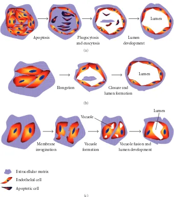
Angiogenesis as well as the availability of the numerous models now existing has been pioneered about 30 years ago by Folkman and Haudenschild (1980) when they demonstra-ted that new capillary blood vessels form in tumor progres-sion. They observed that capillary tube formation was ini-tiated from a vacuole structure within the endothelial cells that subsequently develop multicellular capillary lumen [38]. More recently, Kucera et al. (2007) have proposed three mod-els explaining how tube-like structures developed during the vascularization process: (1) cell death and phagocytosis, (2) Wrapping of the spaces around the extracellular matrix, and (3) vacuole formation from the coalescence of intracellular vacuoles. The models are presented onFigure 1, and below are the details [39].
3.1. Cell Death and Phagocytosis Model. Somein vitroas well
asin vivo studies of angiogenesis report that lumen
forma-tion was associated with apoptotic and cell death events. This model proposed some steps of the vascular lumen formation: endothelial cells organize themselves into a multicellular cord, and cells in the middle of the cord become apoptotic and die [40]. Finally, endothelial cells at the edge phagocytise the apoptotic cells, subsequently forming a multicellular ves-sel containing lumen [40, 41]. According to this model, dead cells define the lumen of the tube-like structures
(seeFigure 1(a)). However, this model fails to explain how
Apoptosis Phagocytosis and exocytosis
Lumen
Lumen
development
(a)
Elongation Closure and
lumen formation
Lumen
(b)
Vacuole
Lumen
Membrane invagination
Vacuole formation
Vacuole fusion and lumen development
Extracellular matrix
Endothelial cell
Apoptotic cell
(c)
Figure1: Models of the development of tube-like structures during vasculogenesis.
3.2. Wrapping Spaces around ECM Model. This model
main-ly suggests that most capillaries are initiated by the elonga-tion of endothelial cells to open multicellular structures. Sub-sequently, vascular tube can form when the elongated cell sheets are closed, even without vacuole formation (see
Figure 1(b)). This concept was initiated by Hirakow and
Hiruma (1983), when they studied the development of vas-cular lumen made of endothelial cells without the evidence of vacuole-like structures [42]. In a recent study, Parker et al. (2004) have shown that the vascular development was initiat-ed by the proliferation and migration of endothelial cells to form a cord-like structure, and vascular cords then undergo tubulogenesis to form vessels containing lumen [43]. How-ever, this model does not explain the polarity of endothelial cells during the development of capillary structures.
3.3. Vacuole Formation from the Coalescence of Intracellular
Vacuoles. The idea behind this model was proposed by
Folkman and Haudenschild (1980) when explaining angio-genesis and lumen formation within 3D constructs. This model involves several steps: formation either of single or
multiple vacuole(s) inside the endothelial cells and vacuoles subsequently fuse with each others. Finally, a continuous multicellular lumen will form when two or more cells with their vacuoles adhere to each others, therefore fusing (see
Figure 1(c)) [38].
More recent, Davis et al. (2000) have reported that endo-thelial cell lumens were forming in three-dimensional colla-gen and fibrin matrices. The authors reported that this pro-cess was controlled by the formation and coalescence of intracellular vacuoles and in the absence of cellular junctions [44]. Furthermore, the expression of cytoskeletal regulators controlling various cellular functions of endothelial cells, such as Cdc42 and Rac1 GTPases, were involved in morpho-genesis and vascularization processes [44,45].
observed during vascular tube development, it can thus be hypothesized that all these three models can be combined, in some way, to explain observations made in angiogenesis and vasculogenesis assays [46,47].
4. Promoting Angiogenesis in
Three-Dimensional Constructs
During vasculogenesis and angiogenesis development, cell-cell as well as cell-cell-ECM interactions are complex. While the roles of some individual factors during neovascularization have been investigated, the optimum combination between cell and their ECM can significantly vary from one material to another [48]. Therefore, some strategies have been pro-posed to promote and direct angiogenesis in 3D environ-ments.
Endothelial cells have been cultured in various matrices that have been precoated or not with adhesive matrix pro-teins or cell binding peptides. Surface chemistry and the im-mobilization mode can affect protein conformation, orienta-tion and anchorage strength, thus influencing cell behaviour [48]. Formation of capillary-like structures made of endothe-lial cells depends on the particular biological environment that will direct cell-cell as well as cell-biomaterial adhesion strength, which relies on biochemical and mechanical signals [49]. To date, there are three major strategies that have been used to study how biomaterials properties affect angiogenesis formation: (1) micropatterning, (2) surface modification, and (3) extracellular matrix modifications [48,49].
4.1. Micropatterning. Photolithography, micromoulding,
and microprinting are some examples of techniques used in micropatterning. Among them, laser microcontact printing is the most frequently studied [50]. It is a process in which biological molecules are printed directly onto a scaffold sur-face, and it has been broadly utilized by tissue engineering scientists. For example, a poly(ethylene glycol)-diacrylate (PEGDA) hydrogel, known to resist protein adsorption, has been micro-printed with the cell adhesive ligand, Arg-Gly-Asp-Ser (RGDS) in different concentrations and using diff e-rent patterns. Endothelial cells were cultured on these RGDS patterns and enhanced tube-like structure formation was ob-served on 50µm-wide stripes [51].
In another study, microcontact printing was used to form various patterns of fibronectin molecules on gold. Endothe-lial cells cultured on these fibronectin patterns formed tub-ular structures on 10µm-wide lines of fibronectin [52]. In a different study, when coated with gelatin, 20µm lines pro-moted endothelial cells to undergo capillary morphogenesis [53]. However, applying this technique to 3D scaffolds is pro-blematic.
4.2. Surface Modification. Proteins such as fibronectin,
gela-tin, collagens, and fibrinogen, only to name a few, have been used to coat synthetic polymers to increase their surface bio-activity. For example, collagen coating on the surface of elec-trospun poly(L-lactic acid)-co-poly(ε-caprolactone) scaffold increased endothelial cell adhesion and spreading [54,55].
Collagen type I has been known to increase the number of tube-like structures made of endothelial cells, thus support-ing angiogenesis and vasculogenesis inin vitroexperiments [56]. Furthermore, the proteolytic cleavage of collagen type IV provides an important binding site for endothelial cells undergoing angiogenesisin vitro[57].
Gelatin is used by many research groups to pre-coat tissue culture dishes. In tissue engineering, gelatin was used to coat poly(glycolic acid) (PGA) scaffold for the controlled release of some angiogenic growth factors [58]. When PGA scaffolds covered by bFGF- and VEGF-containing gelatin hydrogel were implanted in mice, significant angiogenic effect was observed at the implanted site [59,60]. Further clinical test to treat diabetic foot ulcer using gelatin matrix to release bFGF confirmed that the matrix allowed better wound healing [60].
Fibronectin has been investigated in many studies and found to enhance endothelial cell adhesion and spreading [61]. Fibronectin also supports the formation of focal adhe-sion and induces the organization of actin filaments into stress fibre via the interaction with its main receptor, that is,
α5β1 andαvβ3 [61,62]. Furthermore, the lack of fibronectin during vascular development caused an abnormality in the vascular formation in mice [63].
Another protein, fibrinogen, has been incorporated in poly(ethylene glycol) (PEG) hydrogels and it found to in-crease the bioactivity of the scaffold by enhancing endothelial cell and SMC adhesion [64] as well as mesenchymal stem cell adhesion [65]. Furthermore, fibrinogen has many binding sites for endothelial cells. Also, fibrin gel binds many growth factors and bioactive cloth components [65,66]. Therefore, the use of fibrin(ogen) to modify biomaterials surfaces and to encapsulate cells in tissue engineering applications is of im-portance.
In other strategies, polymer surfaces can be precoated with cells to increase the bioactivity of the scaffold. For ex-ample, expanded poly(tetrafluoroethylene) (ePTFE) was pre-coated with bladder carcinoma cells before being implanted in mice. This strategy stimulated angiogenesis and neovas-cularization in ePTFE vascular grafts [67]. In a more recent study, precoated PET fibres with HUVEC allowed the guid-ance of the angiogenesis process and subsequent micro-vas-cularization in fibrin. No microvessel structures were obser-ved when uncovered fibres were utilized [68].
4.3. ECM Modification. The interaction between cells and
their matrix is very complex, since there are many proteins and soluble molecules (including growth factors) present in the ECM. These molecules interact with cells through diff er-ent cell-binding domains. In blood vessel developmer-ent, there are approximately 20 angiogenic growth factors, and among them vascular endothelial growth factor (VEGF), basic fibro-blast growth factor (bFGF), platelet-derived growth factor (PDGF), and angiopoietin-1 (Agp-1) which are the most fre-quently studied [69].
In in vitro angiogenesis assays, growth factors can be
on the development of tube-like structures and on microves-sel maturation [70]. For example, endothelial cells cultured in type I collagen gels with culture media supplemented with fibroblast growth factor-2 (FGF-2), vascular endothelial growth factor (VEGF), and phorbol ester formed tubes con-taining lumens, which have similar structure to microvessels found in vivo [69]. In the absence of growth factors and phorbol ester, the vascular tube structures did not form [71]. Otherin vitroangiogenesis studies showed that growth factors added to culture media can promote sprouting, lumen formation, and better vessel stability [72,73].
Growth factors are often bound to the ECM and can be released upon interaction with cells [74], as well as immobi-lized to the ECM that was used to carry out the culture [75]. In this approach, growth factors can be released, as encoun-teredin vivo. For example, VEGF was covalently immobilized into type I collagen gel [76]. Using the chicken chorioallan-toic membrane (CAM) assay, this collagen matrix was found to induce capillary formation and tissue ingrowth. Also, basic fibroblast growth factor (bFGF) was immobilized into poly-(ethylene glycol) (PEG) hydrogels to guide cell alignment and migration [77].
As many cellular processes observed during vasculogene-sis require numerous signalling pathways and growth factors, recent research efforts have been focusing on the sequential delivery of multiple growth factors [78]. For example, the sequential delivery of VEGF and PDGF-BB (platelet-deri-ved growth factor-BB) in a controlled-release polymer de-vice made of poly(lactic glycolic) (PLG) (85 : 15 lactide : gly-colide) induced a mature vascular network covered by smooth muscle cells [79]. Other results also concluded that one growth factor alone is not sufficient to create mature and stable vasculature [80].
4.4. Coculture Systems and Microvessel Maturation.
Achiev-ing microvessel maturation and stabilization durAchiev-ing blood microvessel development represents another challenge fol-lowing vascular tube formation by endothelial cells. This is often referred to as angiogenesis remodelling. Angiogenesis remodelling is the process by which primary vascular tubes or immature vessels are modified to form an interconnecting branching network, yielding to microvessel structure stabili-zation [74,81]. During this phase, tube-like structures made of endothelial cells and containing lumens recruit other sup-porting cells, such as pericytes and smooth muscle cells (SMCs), to form a close wall and mature blood microvessels [82].
Furthermore, in the vascular tree, endothelial cells are lining on the inner layer of the vessel wall (tunica intima) and supported by other cell types [81,82]. While immature ves-sels consist of tube structures made of endothelial cells, capi-llaries consist of tubes made of endothelial cells covered by pericytes and a basement membrane [82]. In arterioles and venules, vascular SMC cover large area and are closely asso-ciated with the endothelium basement membrane.
Therefore, co-culture systems seem to be appealing to re-create the real microvessel environment and to achieve more mature microvessels. For example, smaller number of vessel-like structures containing lumens was found when
endothe-lial cells were cultured alone in PLLA-PLGA (50 : 50) scaf-folds, compared to samples cocultured with fibroblasts [83]. Vascularized engineered skeletal muscle tissue constructs have been successfully produced by Levenberg et al. using a multiculture system of myoblasts (progenitor cells of the muscle cells), fibroblasts, and endothelial cells into this PLLA-PLGA scaffold [83,84]. In more recent result, cocul-turing HUVECs with skin fibroblast was significantly im-proved the maturation of microvessels structure in a 3D en-vironment [85,86].
5. Bioreactors to Support Vascularization
Many tissue engineering scientists believe that the next generation of functional tissue replacements truly needs the use of bioreactors, in which the culture conditions (e.g., pH, temperature, O2concentration, pressure, pulsation, nutrient
transfer, and waste removal, etc.) can be adjusted and studied to find the optimal mechanical, chemical, and biological sti-muli for a given application [87]. In addition, bioreactor operations will provide a rational basis for the structural and functional design of engineered tissues for the use as model systems to reduce time of innovation, discovery, and produc-tion in biological and clinical research [87,88]. Also, the use of bioreactors should accelerate the development, evaluation, and delivery of engineered tissue products to patients [89].
To date, there are various types of bioreactors that have been designed and tested and these differ from the appli-cations involved. For example, bioreactors have been used to support the growth of cartilage [90–92], bone [93,94], cardiac [95,96], ligament [97], and heart valve [98] tissues.
Furthermore, there are some types of bioreactors that can be used in large tissue cultures, and these include the con-tinuous stirred-tank reactor (CSTR), hollow fiber reactors, and the slow turning lateral vessel (STLV) reactor [88], only to name a few. CSTR reactors are designed to mix oxygen and nutrients within the culture medium and to reduce the boundary layer at the scaffold surface [99]. Hollow fibre re-actors consist of a chamber perfused with semipermeable fibres filled with culture medium. Often, cells are located in the extracapillary space of the chamber. In the past years, this type of reactor was used to produce proteins made by mammalian cells, and to expand mammalian cells in vitro
[100,101]. STLV reactor was proposed by NASA (National Aeronautics and Space Administration) at the Johnson Space Center (USA) for earth-based as well as space experiments [101].
Bioreactors can also enable control over the mechanical stimulation for cellular guidance inside the scaffold in order to improve tissue vascularization. As endothelial cells are lining the inner surface of blood vessels of the entire capillary and circulatory system, they experience fluid shear stress and dynamic flow conditions [103]. Therefore, the use of a dyn-amic culture system can allow some cell mechanical stimula-tion prior to the use of the grown tissue construct [103,104]. Dynamic culture parameters such as pulsation, pressure, and flow rate that mimicin vivoconditions often result in better cellular organization as well as mechanical properties of the tissue constructs when compared to constructs cultur-ed in static environment [105,106]. To date, several studies of endothelial cell monolayers have revealed that cell orien-tation reflects the direction of the flow [107,108]. Also, in endothelial cells, shear stress-increased VEGF, and PDGF-BB expression compared to those cultured in static conditions [109]. In addition, pulsatile flow can improve structural or-ganization of SMC and this is an essential step in the vas-cularization process [110,111].
6. Concluding Remarks
Over the past years, significant advances in tissue engineering research have provided hope for the commercial availability of human bioartificial organs. More simple engineered tissue substitutes (e.g., skin, bone, and cartilage) have been success-fully applied and implanted, while for more complex and vascularized constructs, many problems still remain before tissue substitutes can be available. Consequently, strategies to develop engineered tissue constructs that enhance vasculari-zation inside become important. This will provide an impro-vement of oxygen and nutrient transfer as well as waste re-moval that could allow production of thicker tissue substi-tutes as well as artificial organs. Tissue engineering research relies on the increasing knowledge of angiogenesis and vas-culogenesis mechanisms occurring during capillary tube for-mation and blood microvessel development. Furthermore, to overcome the bottleneck of the complex interplay between various factors influencing tissue vascularization, the use of bioreactor system is necessary.
Acknowledgments
The author acknowledges Professor Patrick Vermette (De-partment of Chemical and Biotechnological Engineering, Universit´e de Sherbrooke) for his constructive comments. Universiti Teknologi Malaysia (UTM) Short Term Research Grant, Ref. no. PY/2011/02082, is also gratefully acknowl-edged.
References
[1] R. Langer and J. P. Vacanti, “Tissue engineering,”Science, vol. 260, no. 5110, pp. 920–926, 1993.
[2] A. Atala, “Tissue engineering and regenerative medicine: concepts for clinical application,”Rejuvenation Research, vol. 7, no. 1, pp. 15–31, 2004.
[3] J. D. Sipe, “Tissue engineering and reparative medicine,”
Annals of the New York Academy of Sciences, vol. 961, pp. 1–9, 2002.
[4] W. T. Arthur, R. B. Vernon, E. H. Sage, and M. J. Reed, “Growth factors reverse the impaired sprouting of microves-sels from aged mice,”Microvascular Research, vol. 55, no. 3, pp. 260–270, 1998.
[5] W. T. Godbey and A. Atala, “In vitro systems for tissue engi-neering,”Annals of the New York Academy of Sciences, vol. 961, pp. 10–26, 2002.
[6] A. G. Mikos, G. Sarakinos, M. D. Lyman, D. E. Ingber, J. P. Vacanti, and R. Langer, “Prevascularization of porous bio-degradable polymers,”Biotechnology and Bioengineering, vol. 42, no. 6, pp. 716–723, 1993.
[7] I. Monta˜no, C. Schiestl, J. Schneider et al., “Formation of hu-man capillaries in vitro: the engineering of prevascularized matrices,”Tissue Engineering—Part A, vol. 16, no. 1, pp. 269– 282, 2010.
[8] Y. Sakakibara, K. Nishimura, K. Tambara et al., “Prevascular-ization with gelatin microspheres containing basic fibroblast growth factor enhances the benefits of cardiomyocyte trans-plantation,”Journal of Thoracic and Cardiovascular Surgery, vol. 124, no. 1, pp. 50–56, 2002.
[9] S. Levenberg and R. Langer, “Advances in tissue engineering,”
Current Topics in Developmental Biology, vol. 61, pp. 113–134, 2004.
[10] P. Carmeliet, “Angiogenesis in life, disease and medicine,”
Nature, vol. 438, no. 7070, pp. 932–936, 2005.
[11] P. Carmeliet and E. M. Conway, “Growing better blood vessels,”Nature Biotechnology, vol. 19, no. 11, pp. 1019–1020, 2001.
[12] S. Soker, M. Machado, and A. Atala, “Systems for therapeutic angiogenesis in tissue engineering,”World Journal of Urology, vol. 18, no. 1, pp. 10–18, 2000.
[13] R. P. Lanza, R. S. Langer, and J. Vacanti,In Principles of Tis-sue Engineering, Academic Press, San Diego, Calif, USA, 2nd edition, 2000.
[14] D. W. Hutmacher, “Scaffold design and fabrication technolo-gies for engineering tissues—state of the art and future per-spectives,”Journal of Biomaterials Science, Polymer Edition, vol. 12, no. 1, pp. 107–124, 2001.
[15] G. K. Owens, M. S. Kumar, and B. R. Wamhoff, “Molecular regulation of vascular smooth muscle cell differentiation in development and disease,”Physiological Reviews, vol. 84, no. 3, pp. 767–801, 2004.
[16] J. L. Duband, M. Gimona, M. Scatena, S. Sartore, and J. V. Small, “Calponin and SM 22 as differentiation markers of smooth muscle: spatiotemporal distribution during avian embryonic development,”Differentiation, vol. 55, no. 1, pp. 1–11, 1993.
[17] Y. D. Jung, S. A. Ahmad, W. Liu et al., “The role of the micro-environment and intercellular cross-talk in tumor angiogen-esis,”Seminars in Cancer Biology, vol. 12, no. 2, pp. 105–112, 2002.
[18] G. Allt and J. G. Lawrenson, “Pericytes: cell biology and path-ology,”Cells Tissues Organs, vol. 169, no. 1, pp. 1–11, 2001. [19] A. Orlidge and P. A. D’Amore, “Inhibition of capillary
endo-thelial cell growth by pericytes and smooth muscle cells,”
Journal of Cell Biology, vol. 105, no. 3, pp. 1455–1462, 1987. [20] Becker et al., “Extracellular matrix,” in The World of the
[21] R. Auerbach, R. Lewis, B. Shinners, L. Kubai, and N. Akhtar, “Angiogenesis assays: a critical overview,”Clinical Chemistry, vol. 49, no. 1, pp. 32–40, 2003.
[22] Z. Chen, A. Htay, W. D. Santos et al., “In vitro angiogenesis by human umbilical vein endothelial cells (HUVEC) induced by three-dimensional co-culture with glioblastoma cells,”
Journal of Neuro-Oncology, vol. 92, no. 2, pp. 121–128, 2009. [23] M. N. Nakatsu, R. C. A. Sainson, J. N. Aoto et al., “Angiogenic
sprouting and capillary lumen formation modeled by human umbilical vein endothelial cells (HUVEC) in fibrin gels: the role of fibroblasts and Angiopoietin-1,” Microvascular Re-search, vol. 66, no. 2, pp. 102–112, 2003.
[24] B. E. Sumpio, W. Du, G. Galagher et al., “Regulation of PDGF-B in endothelial cells exposed to cyclic strain,” Arterio-sclerosis, Thrombosis, and Vascular Biology, vol. 18, no. 3, pp. 349–355, 1998.
[25] F. Hillen,Angiogenesis Assays, chapter 1, 2006.
[26] H. C. H. Ko, B. K. Milthorpe, and C. D. McFarland, “Engin-eering thick tissues—the vascularisation problem,”European Cells and Materials, vol. 14, pp. 1–18, 2007.
[27] J. R. Fuchs, B. A. Nasseri, and J. P. Vacanti, “Tissue engi-neering: a 21st century solution to surgical reconstruction,”
Annals of Thoracic Surgery, vol. 72, no. 2, pp. 577–591, 2001. [28] I. Lang, M. A. Pabst, U. Hiden et al., “Heterogeneity of
micro-vascular endothelial cells isolated from human term placenta and macrovascular umbilical vein endothelial cells,” Euro-pean Journal of Cell Biology, vol. 82, no. 4, pp. 163–173, 2003. [29] W. C. Aird, “Phenotypic heterogeneity of the endothelium: II. Representative vascular beds,” Circulation Research, vol. 100, no. 2, pp. 174–190, 2007.
[30] H. Bramfeld, G. Sabra, V. Centis, and P. Vermette, “Scaffold vascularisation: a challenge for three-dimensional tissue en-gineering,”Current Medicinal Chemistry, vol. 17, pp. 3944– 3967, 2010.
[31] C. J. Kirkpatrick, R. E. Unger, V. Krump-Konvalinkova, K. Peters, H. Schmidt, and G. Kamp, “Experimental approaches to study vascularization in tissue engineering and biomaterial applications,”Journal of Materials Science, vol. 14, no. 8, pp. 677–681, 2003.
[32] E. Dejana, “Endothelial cell-cell junctions: happy together,”
Nature Reviews Molecular Cell Biology, vol. 5, no. 4, pp. 261– 270, 2004.
[33] K. L. Taylor, A. M. Henderson, and C. C. W. Hughes, “Notch activation during endothelial cell network formation in vitro targets the basic HLH transcription factor HESR-1 and downregulates VEGFR-2/KDR expression,” Microvascular Research, vol. 64, no. 3, pp. 372–383, 2002.
[34] M. E. Francis, S. Uriel, and E. M. Brey, “Endothelial cell-matrix interactions in neovascularization,”Tissue Engineer-ing—Part B, vol. 14, no. 1, pp. 19–32, 2008.
[35] J. J. Moon, M. S. Hahn, I. Kim, B. A. Nsiah, and J. L. West, “Micropatterning of poly(ethylene glycol) diacrylate hydro-gels with biomolecules to regulate and guide endothelial morphogenesis,”Tissue Engineering—Part A, vol. 15, no. 3, pp. 579–585, 2009.
[36] M. Levin, A. J. Ewald, M. McMahon, Z. Werb, and K. Mostov, “A model of intussusceptive angiogenesis,”Novartis Foundation Symposium, vol. 283, pp. 37–42, 2007.
[37] W. Risau, “Mechanisms of angiogenesis,”Nature, vol. 386, no. 6626, pp. 671–674, 1997.
[38] J. Folkman and C. Haudenschild, “Angiogenesis in vitro,”
Nature, vol. 288, no. 5791, pp. 551–556, 1980.
[39] T. Kucera, J. Eglinger, B. Strilic, and E. Lammert, “Vascular lumen formation from a cell biological perspective,” in Vas-cular Development, D. J. Chadwick and J. Goode, Eds., pp. 46–56, John Willey & Sons, West Sussex , UK, 2007. [40] W. Fierlbeck, A. Liu, R. Coyle, and B. J. Ballermann,
“Endo-thelial cell apoptosis during glomerular capillary lumen for-mation in vivo,”Journal of the American Society of Nephrology, vol. 14, no. 5, pp. 1349–1354, 2003.
[41] G. T. Meyer, L. J. Matthias, L. Noack, M. A. Vadas, and J. R. Gamble, “Lumen formation during angiogenesis in vitro involves phagocytic activity, formation and secretion of vacu-oles, cell death, and capillary tube remodelling by different populations of endothelial cells,”Anatomical Record, vol. 249, no. 3, pp. 327–340, 1997.
[42] R. Hirakow and T. Hiruma, “TEM-studies on development and canalization of the dorsal aorta in the chick embryo,”
Anatomy and Embryology, vol. 166, no. 3, pp. 307–315, 1983. [43] L. H. Parker, M. Schmidt, S. W. Jin et al., “The endothelial-cell-derived secreted factor Egfl7 regulates vascular tube formation,”Nature, vol. 428, no. 6984, pp. 754–758, 2004. [44] G. E. Davis, S. M. Black, and K. J. Bayless, “Capillary
morphogenesis during human endothelial cell invasion of three-dimensional collagen matrices,”In Vitro Cellular and Developmental Biology, vol. 36, no. 8, pp. 513–519, 2000. [45] K. J. Bayless and G. E. Davis, “The Cdc42 and Rac1 GTPases
are required for capillary lumen formation in three-dimen-sional extracellular matrices,”Journal of Cell Science, vol. 115, no. 6, pp. 1123–1136, 2002.
[46] G. D. Yancopoulos, S. Davis, N. W. Gale, J. S. Rudge, S. J. Wiegand, and J. Holash, “Vascular-specific growth factors and blood vessel formation,”Nature, vol. 407, no. 6801, pp. 242–248, 2000.
[47] G. W. Cockerill, J. R. Gamble, and M. A. Vadas, “Angio-genesis: models and modulators,” International Review of Cytology, vol. 159, pp. 113–160, 1995.
[48] D. C. Darland and P. A. D’Amore, “Cell cell interactions in vascular development,”Current Topics in Developmental Bio-logy, vol. 52, pp. 107–149, 2001.
[49] R. Y. Kannan, H. J. Salacinski, K. Sales, P. Butler, and A. M. Seifalian, “The roles of tissue engineering and vascularisation in the development of micro-vascular networks: a review,”
Biomaterials, vol. 26, no. 14, pp. 1857–1875, 2005.
[50] M. S. Hahn, L. J. Taite, J. J. Moon, M. C. Rowland, K. A. Ruf-fino, and J. L. West, “Photolithographic patterning of poly-ethylene glycol hydrogels,”Biomaterials, vol. 27, no. 12, pp. 2519–2524, 2006.
[51] J. J. Moon and J. L. West, “Vascularization of engineered tis-sues: approaches to promote angiogenesis in biomaterials,”
Current Topics in Medicinal Chemistry, vol. 8, no. 4, pp. 300– 310, 2008.
[52] N. I. Moldovan and M. Ferrari, “Prospects for microtechnol-ogy and nanotechnolmicrotechnol-ogy in bioengineering of replacement microvessels,”Archives of Pathology and Laboratory Medicine, vol. 126, no. 3, pp. 320–324, 2002.
[53] D. A. Medalie, R. G. Tompkins, and J. R. Morgan, “Evaluation of acellular human dermis as a dermal analog in a composite skin graft,”ASAIO Journal, vol. 42, no. 5, pp. M455–M462, 1996.
[55] Q. P. Pham, U. Sharma, and A. G. Mikos, “Electrospun poly (ε-caprolactone) microfiber and multilayer nanofiber/micro-fiber scaffolds: characterization of scaffolds and measure-ment of cellular infiltration,”Biomacromolecules, vol. 7, no. 10, pp. 2796–2805, 2006.
[56] G. E. Davis and D. R. Senger, “Endothelial extracellular mat-rix: biosynthesis, remodeling, and functions during vascular morphogenesis and neovessel stabilization,”Circulation Re-search, vol. 97, no. 11, pp. 1093–1107, 2005.
[57] J. Xu, D. Rodriguez, E. Petitclerc et al., “Proteolytic exposure of a cryptic site within collagen type IV is required for angio-genesis and tumor growth in vivo,”Journal of Cell Biology, vol. 154, no. 5, pp. 1069–1079, 2001.
[58] Y. Ikada and Y. Tabata, “Protein release from gelatin matri-ces,”Advanced Drug Delivery Reviews, vol. 31, no. 3, pp. 287– 301, 1998.
[59] Y. Tabata, “Biosynthetic Scaffold,” inScaffolding in Tissue En-gineering, X. P. Ma and J. H. Elisseeff, Eds., pp. 46–59, Tay-lor & Francis, Boca Raton, Fla, USA, 2006.
[60] Y. Tabata, “Biomaterial technology for tissue engineering ap-plications,”Journal of Controlled Release, vol. 31, pp. 189–199, 2009.
[61] E. Monchaux and P. Vermette, “Effects of surface properties and bioactivation of biomaterials on endothelial cells,” Fron-tiers in Bioscience, vol. 2, pp. 239–255, 2010.
[62] P. B. van Wachem, C. M. Vreriks, and T. Beugeling, “The in-fluence of protein adsorption on interactions of cultured hu-man endothelial cells with polymers,”Journal of Biomedical Materials Research, vol. 21, no. 6, pp. 701–718, 1987. [63] E. L. George, H. S. Baldwin, and R. O. Hynes, “Fibronectins
are essential for heart and blood vessel morphogenesis but are dispensable for initial specification of precursor cells,”Blood, vol. 90, no. 8, pp. 3073–3081, 1997.
[64] L. Almany and D. Seliktar, “Biosynthetic hydrogel scaffolds made from fibrinogen and polyethylene glycol for 3D cell cul-tures,”Biomaterials, vol. 26, no. 15, pp. 2467–2477, 2005. [65] G. Zhang, X. Wang, Z. Wang, J. Zhang, and L. Suggs, “A
PEGylated fibrin patch for mesenchymal stem cell delivery,”
Tissue Engineering, vol. 12, no. 1, pp. 9–19, 2006.
[66] J. W. Weisel, “Fibrinogen and fibrin,”Advances in Protein Chemistry, vol. 70, pp. 247–299, 2005.
[67] K. R. Kidd, D. B. Dal Ponte, R. S. Kellar, and S. K. Williams, “A comparative evaluation of the tissue responses associated with polymeric implants in the rat and mouse,”Journal of Biomedical Materials Research, vol. 59, no. 4, pp. 682–689, 2002.
[68] G. E. Davis, K. J. Bayless, and A. Mavila, “Molecular basis of endothelial cell morphogenesis in three-dimensional extra-cellular matrices,”Anatomical Record, vol. 268, no. 3, pp. 252– 275, 2002.
[69] I. Sukmana and P. Vermette, “Polymer fibers as contact guid-ance to orient microvascularization in a 3D environment,”
Journal of Biomedical Materials Research—Part A, vol. 92, no. 4, pp. 1587–1597, 2010.
[70] N. Fournier and C. J. Doillon, “In vitro effects of extracellular matrix and growth factors on endothelial cell migration and vessel formation,”Cells and Materials, vol. 4, no. 4, pp. 399– 408, 1994.
[71] G. E. Davis and C. W. Camarillo, “Anα2β1 integrin-depend-ent pinocytic mechanism involving intracellular vacuole for-mation and coalescence regulates capillary lumen and tube formation in three-dimensional collagen matrix,” Experi-mental Cell Research, vol. 224, no. 1, pp. 39–51, 1996.
[72] A. Uemura, M. Ogawa, M. Hirashima et al., “Recombinant angiopoietin-1 restores higher-order architecture of growing blood vessels in mice in the absence of mural cells,”Journal of Clinical Investigation, vol. 110, no. 11, pp. 1619–1628, 2002. [73] A. H. Zisch, M. P. Lutolf, and J. A. Hubbell, “Biopolymeric
delivery matrices for angiogenic growth factors,” Cardiovas-cular Pathology, vol. 12, no. 6, pp. 295–310, 2003.
[74] J. A. Hubbell, “Tissue and cell engineering,”Current Opinion in Biotechnology, vol. 14, pp. 517–519, 2003.
[75] M. P. Lutolf and J. A. Hubbell, “Synthetic biomaterials as in-structive extracellular microenvironments for morphogene-sis in tissue engineering,”Nature Biotechnology, vol. 23, no. 1, pp. 47–55, 2005.
[76] S. Koch, C. Yao, G. Grieb, P. Pr´evel, E. M. Noah, and G. C. M. Steffens, “Enhancing angiogenesis in collagen matrices by co-valent incorporation of VEGF,”Journal of Materials Science, vol. 17, no. 8, pp. 735–741, 2006.
[77] S. A. DeLong, J. J. Moon, and J. L. West, “Covalently immo-bilized gradients of bFGF on hydrogel scaffolds for directed cell migration,”Biomaterials, vol. 26, no. 16, pp. 3227–3234, 2005.
[78] D. J. Mooney, K. Sano, P. Matthias Kaufmann et al., “Long-term engraftment of hepatocytes transplanted on biodegrad-able polymer sponges,”Journal of Biomedical Materials Res-earch, vol. 37, no. 3, pp. 413–420, 1997.
[79] T. P. Richardson, M. C. Peters, A. B. Ennett, and D. J. Mooney, “Polymeric system for dual growth factor delivery,” Nature Biotechnology, vol. 19, no. 11, pp. 1029–1034, 2001.
[80] H. M. Blau, T. R. Brazelton, and J. M. Weimann, “The evolv-ing concept of a stem cell: entity or function?”Cell, vol. 105, no. 7, pp. 829–841, 2001.
[81] A. B. Ennett and D. J. Mooney, “Tissue engineering strategies for in vivo neovascularisation,”Expert Opinion on Biological Therapy, vol. 2, no. 8, pp. 805–818, 2002.
[82] R. K. Jain, “Molecular regulation of vessel maturation,” Na-ture Medicine, vol. 9, no. 6, pp. 685–693, 2003.
[83] A. Lesman, Y. Blinder, and S. Levenberg, “Modeling of flow-induced shear stress applied on 3D cellular scaffolds: implica-tions for vascular tissue engineering,”Biotechnology and Bio-engineering, vol. 105, no. 3, pp. 645–654, 2010.
[84] S. Levenberg, J. Rouwkema, M. Macdonald et al., “Engineer-ing vascularized skeletal muscle tissue,”Nature Biotechnology, vol. 23, no. 7, pp. 879–884, 2005.
[85] I. Sukmana and P. Vermette, “The effects of co-culture with fibroblasts and angiogenic growth factors on microvascular maturation and multi-cellular lumen formation in HUVEC-oriented polymer fibre constructs,”Biomaterials, vol. 31, no. 19, pp. 5091–5099, 2010.
[86] A. Lesman, J. Koffler, R. Atlas, Y. J. Blinder, Z. Kam, and S. Levenberg, “Engineering vessel-like networks within multi-cellular fibrin-based constructs,”Biomaterials, vol. 32, no. 31, pp. 7856–7869, 2011.
[87] I. Martin, D. Wendt, and M. Heberer, “The role of bioreactors in tissue engineering,”Trends in Biotechnology, vol. 22, no. 2, pp. 80–86, 2004.
[88] Y. Martin and P. Vermette, “Bioreactors for tissue mass culture: design, characterization, and recent advances,” Bio-materials, vol. 26, no. 35, pp. 7481–7503, 2005.
[89] L. E. Freed, F. Guilak, X. E. Guo et al., “Advanced tools for tissue engineering: scaffolds, bioreactors, and signaling,” Tis-sue Engineering, vol. 12, no. 12, pp. 3285–3305, 2006. [90] B. Obradovic, R. L. Carrier, G. Vunjak-Novakovic, and L. E.
tissue engineered cartilage,”Biotechnology and Bioengineer-ing, vol. 63, no. 2, pp. 197–205, 1999.
[91] D. Pazzano, K. A. Mercier, J. M. Moran et al., “Comparison of chondrogensis in static and perfused bioreactor culture,” Bio-technology Progress, vol. 16, no. 5, pp. 893–896, 2000. [92] J. O. Seidel, M. Pei, M. L. Gray, R. Langer, L. E. Freed, and G.
Vunjak-Novakovic, “Long-term culture of tissue engineered cartilage in a perfused chamber with mechanical stimula-tion,”Biorheology, vol. 41, no. 3-4, pp. 445–458, 2004. [93] A. Ignatius, M. Peraus, S. Schorlemmer et al.,
“Osseointegra-tion of alumina with a bioactive coating under load-bearing and unloaded conditions,”Biomaterials, vol. 26, no. 15, pp. 2325–2332, 2005.
[94] X. Yu, E. A. Botchwey, E. M. Levine, S. R. Pollack, and C. T. Laurencin, “Bioreactor-based bone tissue engineering: the influence of dynamic flow on osteoblast phenotypic expres-sion and matrix mineralization,”Proceedings of the National Academy of Sciences of the United States of America, vol. 101, no. 31, pp. 11203–11208, 2004.
[95] R. L. Carrier, M. Rupnick, R. Langer, F. J. Schoen, L. E. Freed, and G. Vunjak-Novakovic, “Perfusion improves tissue architecture of engineered cardiac muscle,”Tissue Engineer-ing, vol. 8, no. 2, pp. 175–188, 2002.
[96] M. Radisic, L. Yang, J. Boublik et al., “Medium perfusion en-ables engineering of compact and contractile cardiac tissue,”
American Journal of Physiology, vol. 286, no. 2, pp. H507– H516, 2004.
[97] C. H. Lee, G. S. Huang, K. H. Chao, S. S. Wu, and Q. Chen, “Differential pretensions of a flexor tendon graft for anterior cruciate ligament reconstruction: a biomechanical compari-son in a porcine knee model,”Arthroscopy, vol. 21, no. 5, pp. 540–546, 2005.
[98] S. P. Hoerstrup, R. Sodian, J. S. Sperling, J. P. Vacanti, and J. E. Mayer, “New pulsatile bioreactor for in vitro formation of tissue engineered heart valves,”Tissue Engineering, vol. 6, no. 1, pp. 75–79, 2000.
[99] G. Belfort, “Membranes and bioreactors: a technical chal-lenge in biotechnology,” Biotechnology and Bioengineering, vol. 33, no. 8, pp. 1047–1066, 1989.
[100] H. O. Jauregui, N. R. Chowdhury, and J. R. Chowdhury, “Use of mammalian liver cells for artificial liver support,” Cell Transplantation, vol. 5, no. 3, pp. 353–367, 1996.
[101] R. P. Schwarz, T. J. Goodwin, and D. A. Wolf, “Cell culture for three-dimensional modeling in rotating-wall vessels: an application of simulated microgravity,”Journal of Tissue Cul-ture Methods, vol. 14, no. 2, pp. 51–57, 1992.
[102] K. Bilodeau and D. Mantovani, “Bioreactors for tissue engi-neering: focus on mechanical constraints. A comparative re-view,”Tissue Engineering, vol. 12, no. 8, pp. 2367–2383, 2006. [103] J. A. Chouinard, S. Gagnon, M. G. Couture, A. L´evesque, and P. Vermette, “Design and validation of a pulsatile perfusion bioreactor for 3D high cell density cultures,”Biotechnology and Bioengineering, vol. 104, no. 6, pp. 1215–1223, 2009. [104] B. J. Lawrence and S. V. Madihally, “Cell colonization in
de-gradable 3D porous matrices,” Cell Adhesion & Migration, vol. 2, no. 1, pp. 9–16, 2008.
[105] R. Sodian, T. Lemke, C. Fritsche et al., “Tissue-engineering bioreactors: a new combined cell-seeding and perfusion sys-tem for vascular tissue engineering,”Tissue Engineering, vol. 8, no. 5, pp. 863–870, 2002.
[106] L. Yao, D. D. Swartz, S. F. Gugino, J. A. Russell, and S. T. Andreadis, “Fibrin-based tissue-engineered blood vessels: differential effects of biomaterial and culture parameters
on mechanical strength and vascular reactivity,”Tissue En-gineering, vol. 11, no. 7-8, pp. 991–1003, 2005.
[107] S. Li, N. F. Huang, and S. Hsu, “Mechanotransduction in endothelial cell migration,”Journal of Cellular Biochemistry, vol. 96, no. 6, pp. 1110–1126, 2005.
[108] J. P. Stegemann and R. M. Nerem, “Phenotype modulation in vascular tissue engineering using biochemical and mechan-ical stimulation,”Annals of Biomedical Engineering, vol. 31, no. 4, pp. 391–402, 2003.
[109] B. S. Conklin, D. S. Zhong, W. Zhao, P. H. Lin, and C. Chen, “Shear stress regulates occludin and VEGF expression in por-cine arterial endothelial cells,”Journal of Surgical Research, vol. 102, no. 1, pp. 13–21, 2002.
[110] P. Akhyari, P. W. M. Fedak, R. D. Weisel et al., “Mechanical stretch regimen enhances the formation of bioengineered autologous cardiac muscle grafts,”Circulation, vol. 106, no. 13, pp. I137–I142, 2002.
Submit your manuscripts at
http://www.hindawi.com
Stem Cells
International
Hindawi Publishing Corporation
http://www.hindawi.com Volume 2014
Hindawi Publishing Corporation
http://www.hindawi.com Volume 2014
INFLAMMATION
Hindawi Publishing Corporation
http://www.hindawi.com Volume 2014
Behavioural
Neurology
Endocrinology
International Journal ofHindawi Publishing Corporation
http://www.hindawi.com Volume 2014
Hindawi Publishing Corporation
http://www.hindawi.com Volume 2014
Disease Markers
Hindawi Publishing Corporation
http://www.hindawi.com Volume 2014 BioMed
Research International
Oncology
Journal of Hindawi Publishing Corporationhttp://www.hindawi.com Volume 2014
Hindawi Publishing Corporation
http://www.hindawi.com Volume 2014
Oxidative Medicine and Cellular Longevity
Hindawi Publishing Corporation
http://www.hindawi.com Volume 2014
PPAR Research
The Scientific
World Journal
Hindawi Publishing Corporationhttp://www.hindawi.com Volume 2014
Immunology Research Hindawi Publishing Corporation
http://www.hindawi.com Volume 2014
Journal of
Obesity
Journal ofHindawi Publishing Corporation
http://www.hindawi.com Volume 2014
Hindawi Publishing Corporation
http://www.hindawi.com Volume 2014
Computational and Mathematical Methods in Medicine
Ophthalmology
Journal of Hindawi Publishing Corporationhttp://www.hindawi.com Volume 2014
Diabetes Research
Journal of Hindawi Publishing Corporationhttp://www.hindawi.com Volume 2014
Hindawi Publishing Corporation
http://www.hindawi.com Volume 2014
Research and Treatment
AIDS
Hindawi Publishing Corporation
http://www.hindawi.com Volume 2014
Gastroenterology Research and Practice
Hindawi Publishing Corporation
http://www.hindawi.com Volume 2014
Parkinson’s
Disease
Evidence-Based Complementary and Alternative Medicine
Volume 2014