Anti-proliferative and Apoptotic Effects of Mucoxin
(Acetogenin) in T47D Breast Cancer Cells
MUHARTONO
1, ASEP SUKOHAR
1, SUTYARSO
2and M. KANEDI
2*
1Department of Anatomical Pathology, Faculty of Medicine, University of Lampung, Indonesia. 2Department of Biology, Faculty of Mathematics and Sciences, University of Lampung, Indonesia.
*Corresponding author E-mail: [email protected] http://dx.doi.org/10.13005/bpj/963
(Received: May 15, 2016; accepted: July 01, 2016) ABSTRACT
Mucoxin is claimed as a promising anticancer due to its highly potent and specific as antitumor against the MCF-7 (breast carcinoma) cell lines, but supported by a limited scientific data. This study aims to confirm the antiproliferative and apoptotic effects of mucoxin against another type of breast cancer, T47D. The cell lines were grouped into four based on the exposure hour of mucoxin, namely 0, 24, 48, and 72 hours. Each group was given mucoxin of six different concentrations namely 0, 0.1, 0.5, 1, 5 and 10 ng/ml with three replications. Cells proliferation assayed by flow cytometry technique using BrDU staining protocol, while apoptosis assayed by flow cytometry using FITC Annexin V with PI. In all groups, the mucoxin application significantly reduced cell proliferation, but the sharpest decline (>50%) occurred in the group of 48-hour exposure by the concentration of 5ng/ml and 10ng/ml. Whereas apoptotic cell was significantly increased by mucoxin with the high increases (>50%) occurred in the group of 48-hour at the dose of 10ng/ml and 72-hour by the dose of 5ng/ml and 10ng/ml. Thus, mucoxin is antiproliferative and proapoptotic against T47D breast cancer cells and deserved classified as a potential anticancer agent.
Key words: Mucoxin, acetogenin, Rollinia mucosa, T47D, breast cancer.
INTRODUCTION
In Indonesia the incidence of breast cancer was high, about 37 per 100,000 people, compared to other Asian countries where the average is only 20 per 100,000 people1. Considering most people with cancer ended with the death2, the high incidence of breast cancer in Indonesia is a serious threat for the country. However, given the conventional cancer treatments causing various side effects, recently practitioners are compelled to seek alternative cancer treatment that can eliminate or minimize the side effects. Among the various alternative cancer drugs that is believed to be safe by people with cancer, especially in Indonesia, is plant-derived substances3-4.
One of the alternatives medication proposed for cancer, especially breast cancer, is by utilizing acetogenin, an active substances isolated from Annonaceae plant family, Annona muricata Linn5-8. Among the various annonaceous acetogenin derivatives, mucoxin is the latest and claimed the most powerful to eradicate cancer cells because the only type acetogenin containing a hydroxylated trisubstituted tetrahydrofuran (THF) ring9. Unfortunately, though mucoxin claimed as a promising cancer drug, has manufactured and marketed on line all over the world, including Indonesia, a thorough study of the role of the bioactive materials on cellular level is very rare.
internet is still limited to the antitumor and cytotoxic activities. For instance, it was found that mucoxin is a highly potent and specific antitumor agent against MCF-7 (breast carcinoma) cell lines (ED50 = 3.7 x 10(-3)µg/mL compared to adriamycin, ED50 = 1.0 x 10(-2)µg/mL) and A-549 lung cancer9-10. In addition, mucoxin appeared to have significant activity against a number of human cancer cell lines such as epidermoid carcinoma, K562 and HL-6011.
Lack of scientific information about mucoxin role in regulating and suppressing cancer cells, especially breast cancer, certainly need to be enriched and improved through in-depth study. To confirm the anticancer effects, mainly the antiproliferative and apoptotic, of mucoxin, a non-classical acetogenin purified from plant extract of Rollinia mucosa against cancer cells, T47D breast cancer cells were used as the target.
METHOD
The mucoxin and cell lines
Bioactive substances tested in this study is mucoxin (acetogenin) ID AG E 32919 and CAS No. 183195995 obtained from Angene International Limited. The product package contains 5mg of pure mucoxin in powder form. Whereas human breast cancer cell line used in this study was T47D (ATCC® HTB133™) obtained from American Type Culture Collection (Manassas, VA 20108 USA) with a lot number 61062006.
Experimental design
A randomized block design, six concentrations of treatment and three replications is the experimental design applied in this research. The T47D cell lines was divided into four groups based on the hour of mucoxin exposure until assays done, namely hour 0th, 24th, 48th, and 72nd. Concentration levels of mucoxin applied in the experiment were as follows: 0ng/ml, 0.1ng/ml, 0.5ng/ ml, 1ng/ml, 5ng/ml and 10ng/ml. Each treatment dose was made in three replicates.
Cell culture
The cells were grown in Roswell Park Memorial Institute medium (RPMI 1640) culture media supplemented with 10% Foetal Bovine Serum (FBS) Gibco™ (from Thermo Fisher Scientific
Cat. No. 26140 079) and 0.2 units/ml bovine insulin (from Sigma Aldrich Cat. No. I5500 and CAS RN 11070 73 8) at 37°C in 5% CO2. Thawing process performed in waterbath at 37°C for 2-4 minutes. Then, as much as 5x10⁴ cells/cm2 was taken into T-flask and incubated at 37°C in CO2 5%. When cells density reached 80% confluent, trypsinization done using 0.25% Trypsin + 0.53 mM EDTA solution and then subcultured into new culture vessels, also at 37°C in CO2 5%. After two times passaging the T47D cells ready to be treated.
Mucoxin treatments
The mucoxin preparation was made by diluting the powder of mucoxin in 1 ml of 0.1% DMSO. The stock solution is then diluted further in accordance with the needs of the treatment concentrations (six levels). After subcultured for two times the cells were diluted with RPMI and seeded in 24-wells plate with a cells density of 5x10⁴ cells/ cm2 in each well. Once the cells density reach 80% confluent, the cells treated with mucoxin of different concentrations as follows: 0ng/ml (K), 0.1ng/ml (P1), 0.5ng/ml (P2), 1ng/ml (P3), 5ng/ml (P4) and 10ng/ml (P5). After being treated, the cells were incubated in accordance with the length of hours that have been assigned to each group, i.e. 0, 24, 48, and 72 hours. Cell Proliferation Assay
Cells proliferation assayed by flow cytometry (FCM) technique using BrDU staining protocol. The wells containing T47D cells under optimal condition stained with Bromodeoxyuridine (BrdU) 30 ìM, incubated, washed with PBS, and then trypsinized. After the incubation, cells were harvested and washed by adding flow cytometry staining buffer and diluted until in each tube containing 105 108 cells. After being washed twice with flow cytometry staining buffer, anti-BrdU fluorochrome-conjugated antibody was added. Cell quantity in the samples then determined using flow cytometry at wavelength 530 nm.
Apoptosis Assay
cells/ml. After FITC Annexin V (5 µl) and Propidium Iodide Solution (10 µl) being added, the suspension then incubated in the dark at room temperature for 15 minutes. Lastly, after the cell lines in each tube diluted with 400 µl of Annexin V Binding Buffer, the suspension analyzed by flow cytometry machine at wavelength 530 nm. Statistical Analysis
Comparison of mean values of quantitative data between treatments (mucoxin dose) and between group of exposure time was analyzed using ANOVA followed by LSD test.
RESULTS
Effect of Mucoxin on Proliferation
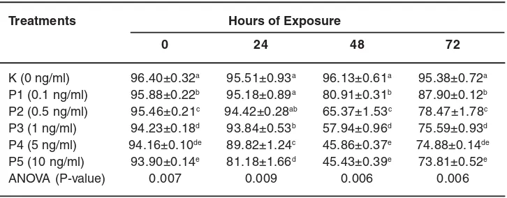
Quantification results of the flow cytometric (FCM) of the effects of mucoxin application on the proliferation of T47D cells of each exposure hour group are presented in Table 1. To determine whether the exposure hour has an effect on the proliferation, one way ANOVA also applied for comparing the average value of proliferation between the groups in which the results are shown in Table 2.
Based on the ANOVA and post hoc test shown in Table 1 and Table 2 it can be assumed that the mucoxin doses as well as the exposure hour have effects on T47D cell proliferation. In all exposure groups, the mucoxin treatment
significantly reduced the percentage of cell proliferation. The p-values of the comparative mean value of proliferation between treatment dose in exposure hour 0, 24, 48 and 72 respectively are 0.007, 0.009, 0.006, 0.006. The sharpest decline (>50%) occurred in the group of 48-hour exposure by the mucoxin dose of 5ng/ml (P4) and 10ng/ml (P5).
Effect of Mucoxin on Apoptosis
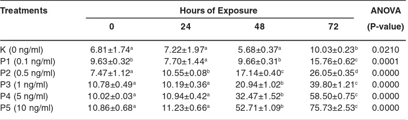
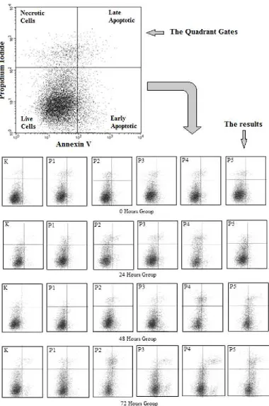
Gating strategy for quantification of the T47D cell apoptosis after mucoxin treatment in each hour group is ilustrated in Figure 1. The quantitative results from the FCM graph readings of each treatment group, the ANOVA results followed by LSD test against mean values of the effect of the mucoxin doses on the apoptosis of T47D cells of each exposure hour group are presented in Table 3. For comparing the average value of apoptotic cells between the exposure hour groups, the one way ANOVA has also applied and result in the data shown in Table 4.
Referring the data in Table 3 and Table 4, it was also clear that both mucoxin doses and exposure hour have effects on T47D cell apoptosis. In all exposure groups, the mucoxin treatment significantly increase the percentage of apoptotic cells. The p-values of the comparative mean value of apoptosis between treatment dose in exposure hour 0, 24, 48 and 72 respectively are 0.008, 0.012, 0.005, 0.005. However, the high increases (>50%) Table 1: Effect of mucoxin application on the proliferation
of T47D cells of each exposure hour group
Treatments Hours of Exposure
0 24 48 72
K (0 ng/ml) 96.40±0.32a 95.51±0.93a 96.13±0.61a 95.38±0.72a P1 (0.1 ng/ml) 95.88±0.22b 95.18±0.89a 80.91±0.31b 87.90±0.12b P2 (0.5 ng/ml) 95.46±0.21c 94.42±0.28ab 65.37±1.53c 78.47±1.78c P3 (1 ng/ml) 94.23±0.18d 93.84±0.53b 57.94±0.96d 75.59±0.93d P4 (5 ng/ml) 94.16±0.10de 89.82±1.24c 45.86±0.37e 74.88±0.14de P5 (10 ng/ml) 93.90±0.14e 81.18±1.66d 45.43±0.39e 73.81±0.52e
ANOVA (P-value) 0.007 0.009 0.006 0.006
Table 2: Effect of exposure hour on the proliferation of T47D cells given mucoxin of six different concentration
Treatments Hours of Exposure ANOVA
0 24 48 72 (P-value)
K (0 ng/ml) 96.40±0.32a 95.51±0.93a 96.13±0.61a 95.38±0.72a 0.272 P1 (0.1 ng/ml) 95.88±0.22a 95.18±0.89a 80.91±0.31c 87.90±0.12b 0.000 P2 (0.5 ng/ml) 95.46±0.21a 94.42±0.28a 65.37±1.53c 78.47±1.78b 0.000 P3 (1 ng/ml) 94.23±0.18a 93.84±0.53a 57.94±0.96c 75.59±0.93b 0.000 P4 (5 ng/ml) 94.16±0.10a 89.82±1.24b 45.86±0.37d 74.88±0.14c 0.000 P5 (10 ng/ml) 93.90±0.14a 81.18±1.66b 45.43±0.39d 73.81±0.52c 0.000
Values are the mean ± SD percentage of proliferated cells (n=3); numbers in the same line that shared the same superscript was not statistically different at α=0.05 based on LSD test
Table 3: Effect of mucoxin application on the apoptosis of T47D cells of each exposure hour group
Treatments Hours of Exposure
0 24 48 72
K (0 ng/ml) 6.81±1.74a 7.22±1.97a 5.68±0.37a 10.03±0.23a P1 (0.1 ng/ml) 9.63±0.32bc 7.70±1.44a 9.66±0.31b 15.76±0.62b P2 (0.5 ng/ml) 7.47±1.12a 10.55±0.08b 17.14±0.40c 26.05±0.35c P3 (1 ng/ml) 10.78±0.49c 10.19±0.36b 20.94±1.02d 39.80±1.21d P4 (5 ng/ml) 10.02±0.03c 10.94±0.42b 32.47±1.52e 58.50±0.75e P5 (10 ng/ml) 10.86±0.68c 11.23±0.66b 52.71±1.09f 75.73±2.53f
ANOVA (P-value) 0.008 0.012 0.005 0.005
Values are the mean ± SD percentage of apoptotic cells (n=3); numbers in the same column that shared the same superscript was not statistically different at α=0.05 based on LSD test
Table 4: Effect exposure hour on the apoptosis of T47D cells given mucoxin of six different concentration
Treatments Hours of Exposure ANOVA
0 24 48 72 (P-value)
occurred in the group of 48-hour exposure by mucoxin dose of 10ng/ml (P4) and 72-hour eposure by mucoxin level of 5ng/ml (P4) and 10ng/ ml (P5).
DISCUSSION
Based on the data presented above it was revealed that the application of mucoxin presumably inhibit proliferation and increase apoptosis of cell T47D. Due to a lack of data describing the biological properties of mucoxin in cancer cells, so the best approach to explain the effect is by reference to a similar substance derived from plants of the same family, Annonaceae, i.e. acetogenins.
The anti proliferative effect of annonaceous acetogenin has revealed by previous studies. Reference12, by using acetogenin bullatacin, suggested that the annonaceous acetogenin is cytotoxic against multidrug-resistant human mammary adenocarcinoma cells. Another study suggested that a mono tetra hydrofuran acetogenin, annonacin, arrest cancer cells at the G1 phase and causes cytotoxicity13. Such effects may be due to inhibition properties of acetogenin on the activity of deoxyribonucleic acid (DNA) and DNA topoisomerase14. Moreover, the substances also affect mitochondrial complex I, block the electron transport chain and stop the production of adenosinetriphosphate (ATP). In addition, this substance also activates adenosine monophosphate-activated protein kinase (AMPK) and inhibits the signaling pathway of the mammalian target of rapamycin complex 1 (mTORC1) in colon cancer cells15.
Current findings also confirm the apoptotic effects of Annona muricata leaves ethyl acetate extract (AMEAE) against lung cancer A549 cells. The bioactives substance induced apoptosis through mitochondrial-mediated pathway and involvement of NF-κB. AMEAE effectivley reduce the activation of NF-κB signaling pathway by suppressing the induced translocation of NF-κB from cytoplasm to nucleus16.
More recent study showed that in addition to suppress NF-êB activity, the acetogenin treatment
inhibits protein kinase B (Akt) and cyclin D1 protein in human hepatocellular carcinoma17. Cyclin D1 is a potein frequently linked to various types of human cancer18. If annonaceous acetogenin can actually suppress NF-κB activity, the activity of cyclin D1 should also be reduced since it is known that inhibition of NF-kB causes the reduction of serum-induced cyclin D1-associated kinase activity and resulted in delayed phosphorylation of the retinoblastoma protein19.
The other genetic factors frequently interconnected with NF-kB and/or cyclin-D1 is protein p53. The p53 gene that encode p53 protein is a tumor suppressor. As a tumor suppressor, p53 plays a very important role to prevent excessive cell proliferation and maintain genomic integrity20. This gene will be activated by the cells in response to the internal or external stress signals. Stress signals can be either DNA damage due to viral infection, radiation as well as chemotherapy drugs, hypoxia, excessive expression of oncogene, nutritional deficiencies or ribosomal dysfunction. The stress signals could induce various upstream mediators such as 14ARF and Mdm2 that make p53 stable and active21. In this study the stress signals, most likely and should be, originating from mucoxin.
Genes known to be activated by p53 for its transcription are WAFI/CIP1/p21, GADD45, 14-3-3, Bax, Bak, Puma, and Noxa. WAFI/CIP1/p21 is a gene that encodes a protein CDK inhibitor which will cause hypo-phosphorylated of Rb so that E2F inactive. The GADD45 gene that encode GADD45 protein function in arresting cell cycle by enhancing p21 performance as the CDK inhibitor. Protein p43, the product of 14-3-3 gene, acts as a negative regulator that arrest cell cycle at the G2/M phase. The protein Bax and Bak, on other hand, are the propoptotic protein that directly increase the permeability of mitochondria. Whereas Puma and Noxa are genes that encode proteins BH3 which also play a role in the intrinsic pathway for apoptosis22.
CONCLUSION
breast cancer cells and, thus, mucoxin deserved classified as a promising anticancer agent.
ACKNOWLEDGEMENTS
This project supported by the Faculty of Medicine, University of Lampung.
Conlict of Interest
The authors declare no conflicts of interest.
REFERENCES
1. Ng CH, Pathy NB, Taib NA, et al. Comparison of breast cancer in Indonesia and Malaysia aclinico pathological study between Dharmais Cancer Centre Jakarta and University Malaya Medical Centre, Kuala Lumpur. Asian Pacific J Cancer Prev 2011; 12: 2943 2946.
2. Ellis IO, Schmit SJ, Garau XS, et al. Invasive Breast Carcinoma. In (Fattaneh A, Tavassoli, Deville P). WHO clasification of tumours of the breast and Female genital Organ. 3th ed, Lyon: IARC press 2003; pp 14–17.
3. Kumar V, Abbas AK, Fausto N. Robbins and Cotran pathologic basis and disease, 7th ed. Philadelpia: Elsevier Sounders, 2005; pp 269–342, 1129–1149.
4. Hortobagyi GN. Toward individualized breast cancer therapy: translating biological concepts to the bedside. J Clin Oncol 2012; 17: 577 584.
5. Wang LQ, Nakamura N, Meselhy MR, Hattori M, Zhao WM, Cheng KF, et al. Four mono tetrahydrofuran ring acetogenins, montanacins B E, from Annona montana. Chem Pharm Bull 2000; 48(8):1109 1113. 6. Cerella C, Radogna F, Dicato M, Diederich
M, 2013. Natural compounds as regulators of the cancer cell metabolism. J Cell Biol 2013; 1–16.
7. Formagio ASN, Kassuya CAL, Neto FF, Volobuff CRF, Iriguchi EKK, Vieira MC, Foglio MA. The flavonoid content and anti proliferative, hypoglycaemic, anti inflammatory and free radical scavenging activities of Annonadioica St. Hill. J Altern Complement Med 2013; 13(14):1 8.\ 8. Rachmawati E, Karyono S, Suyuti H. The
effect of Annona muricata leaf on proliferation and appoptosis of HeLa cells
mediated by p53. JKB 2012; 27(1):28 32. 9. Narayan RS, Borhan B. Synthesis of the
proposed structure of mucoxin via regio and stereoselective tetrahydrofuran ring forming strategies. J Org Chem 2006; 71(4):1416 1429.
10. Shi G, Kozlowski JF, Schwedler JT, Wood KV, MacDougal JM, McLaughlin JL. Muconin and mucoxin: additional nonclassical bioactive acetogenins from Rollinia mucosa. J Org Chem. 1996; 61: 7988–7989
11. Garzan A. I. Synthesis of ether linked analogs and SAR studies of Annonaceous acetogenins II. Asymmetric electrophilic halocyclization reactions. A Ph.D. Dissertation Michigan State University. 2012. pp: 333.
12. Oberlies NH, Croy VL, Harrison ML, McLaughlin JL. The annonaceous acetogenin bullatacin is cytotoxic against multidrug-resistant human mammary adenocarcinoma cells.Cancer Lett 1997; 115: 73
13. Yuan SSF, Chang HL, Chen HW, Yeh YT, Kao YH, Lin KH, Wu YC, Su JH. Annonacin, a mono tetra hydrofuran acetogenin, arrests cancer cells at the G1 phase and causes cytotoxicity in a Bax and caspase 3 related pathway. Life Sci 2003; 72(25):2853 2861. 14. Matsui Y, Takefumi T, Yonezawa YK, Takemura
M, Fugawara F, Yoshida H, et al. The relationship between the molecular structure of natural acetogenins and their inhibitory activities which affect DNA polymerase, DNA topoisomerase and human cancer cell growth. ExpTher Med 2010; 1:19 26. 15. Liu YQ, Cheng X, Guo LX, Mao C, Chen YJ,
as an AMPK activator and autophagy inducer in colon cancer cells. Plos One 2012; 7(10):1 11.
16. Moghadamtousi S.Z., Kadir H.A., Paydar M., Rouhollahi E., and Karimian H. Annona muricata leaves induced apoptosis in A549 cells through mitochondrial-mediated pathway and involvement of NF-κB. BMC Complementary and Alternative Medicine 2014, 14: 299.
17. Qian JQ, Sun P, Pan ZY, Fang ZZ. Annonaceous acetogenins reverses drug resistance of human hepatocellular carcinoma BEL-7402/5-FU and HepG2/ ADM cell lines. Int J Clin Exp Pathol 2015; 8(9): 11934-44
18. Diehl, J.A. Cycling to cancer with cyclin D1. Cancer Biol. Ther. 2002; 1: 226-231
19. Hinz M., Krappmann D., Eichten A., Heder A., Scheidereit C. And Strauss M. NF-kappaB function in growth control: regulation of cyclin D1 expression and G0/G1-to-S-phase transition. Mol Cell Biol. 1999; 19(4):2690-8 20. Yang H.Y., Wen Y.Y., Chen H., Lozano G. and Lee H.M.,14-3-3σ Positively Regulates p53 and Suppresses Tumor Growth. Mol Cell Biol. 2003; 23(20): 7096–7107
21. Christopher L. Brooks CL, Gu W. p53ubiquitination: Mdm2 and beyond. Mol Cell 2006; 21(3): 307–315.