Legume Nodulating Bacterium,
Achromobacter xylosoxidans
Found in
Tropical Shrub Agroecosystem, Sumatera, Indonesia
Sri Wedhastri
1, Dinar Mindrati Fardhani
1, Siti Kabirun
1, Jaka Widada
1,
Donny Widianto
1*, Rusdi Evizal
2, and Irfan Dwidya Prijambada
11Department of Agricultural Microbiology, Faculty of Agriculture, Universitas Gadjah Mada, Yogyakarta, Indonesia
2Department of Agrotechnology, Faculty of Agriculture, Universitas Lampung, Bandar Lampung, Indonesia
Abstract
Legume nodulating bacteria (LNB), known also as rhizobia, are soil bacteria, which are able to form root nodules and fi x nitrogen in the leguminous plants. The LNB availability in the soil depends on the type of
agroecosystem, where plant grows. In this study, we isolated LNB from the shrub agroecosystem in Sumatera, Indonesia, and obtained four selected bacterial strains. Among them, the isolate UGM48a formed root nodule in Macroptilium atropurpureum and showed highest number of nitrogenase activity. UGM48a also contains nifH
and nodA genes. An analysis of the PCR-amplifi ed 16S rDNA and BLASTn analysis showed that UGM48a
displayed 96% similarity with Achromobacter xylosoxidans. In addition, UGM48a were successfully nodulated
Glycine max (L.) merr var. wilis. This is the fi rst report detecting A. xylosoxidans as nodule-forming species for Glycine max possesing the positive copy of nodA gene.
Keywords :Legume Nodulating Bacteria, shrub agroecosystem, Achromobacter xylosoxidans, nodA, Glycine max
*Corresponding author: Donny Widianto
Department of Agricultural Microbiology, Faculty of Agriculture, Universitas Gadjah Mada, Yogyakarta, Indonesia, Phone +62 274 523065
E-mail: [email protected]
Introduction
Legume nodulating bacteria (LNB), known also as rhizobia, are soil bacteria able to form root nodules and fi x nitrogen in the leguminous plants. Leguminous plants usually establish a symbiosis with rhizobia and benefi t from its ability to fi x atmospheric nitrogen, which allows them to grow more effi ciently on nutrient-limited soils.
LNB availability in the soil may change due to history of land use. Various agroecosystems in Sumatera, Indonesia have different land use, with 87% used for coffee plantation. Shrub agroecosystem means the soil was in fallow for about 5 years after coffee plantation (Evizal et al., 2013). This
condition may infl uence the availability of LNB in the soil, because there are shrubbery that dominated this area.
This study reported four LNB based on cell and colony’s morphology, as well as the nodulation assay in Macroptilium atropurpureum. Based on sequence analysis of 16S rDNA gene and its nitrogenase activity in M. atropurpureum, it was identified as strain of Achromobacterium xylosoxidans. This bacterium is one of the genera belonging to betaproteobacteria, which have been reported as legume nodulating bacteria for the fi rst time by Benata et al. (2008). A. xylosoxidans formed root nodule in Prosopis julifl ora (Benata et al., 2008); Mucuna bracteata (Salwani et al., 2012); and cowpea (Guimarães et al., 2012). In this study, we also confi rmed the existence of nifH and nodA copy genes. Our fi nding also observed the nodulation ability of A. xylosoxidans in soybean
Materials and Methods
Bacterial Isolates
Four isolates used in this study were screened from shrub agroecosystem in Sumatera, Indonesia. The pure cultures were maintained on yeast-extract mannitol agar (YEMA) slants at 4o C using standard
procedures (Somasegaran, 1994).
Nodulation and N2-fi xation Assay
Four isolates were examined through nodulation and N2-fixation assay using Macroptilium atropurpureum. Surface-sterilized seeds of M. atropurpureum were germinated in petridishes. The seedling were transferred to the test chamber called minirhizotron which consists zeolite based medium (Joachim et al., 2006). The seedlings were watered daily with N-free nutrient solution as described by Somasegaran (1994). At the 40th day, the plants were analyzed
for the number of fresh nodules, and their nitrogenase activity was determined using Acetylene Reduction Assay (ARA).
The best isolate showing highest number of nitrogenase activity were subsequently observed in various plants. In this report, soybean (Glicyne max [L.] merr var. wilis) was used as tested plant, to observe the nodulation
ability of the selected isolate. The purpose of using this plant was investigate nodulating ability in soybean, since soybean can only be infected by few bacteria. Furthermore, Glicyne max (L.) merr var. wilis is one of the superior types of soybean released in Indonesia since 1983. Surface-sterilized seeds of wilis were germinated in petridishes after soaking the seeds overnight in a sterile water. Observation was performed 40 days after cultivation.
Morphological Observation
Bacterial colonies and colours were observed by using standard microbiological method as described by Yang et al. (2008), Vincent (1972; 1982), and Somasegaran and Hoben (1985). The colonies of pure cultures were maintained on YEMA slants at 4o C and
also stored at 4oC.
Genomic DNA Isolation
Total DNAs were extracted from 5 ml bacterial cultures grown in Luria Bertani. The cultures were centrifuged at 11000 rpm for 8 min and washed in TE buffer (10mM Tris, 1 mM EDTA; pH 8). The pellets were suspended in 30 μl SDS 10%, incubated at 37oC for 30 min, then 100 μl NaCl 5 M and 200
μl CTAB were added, and incubated at 65oC
for 15 min. The mixtures were centrifuged at 11000 rpm for 8 min. The top layer was transferred to a new tube. This aqueous phase containing DNA was precipitated with 500 μl isopropanol and then centrifuged at 11000 rpm for 8 min. The precipitated DNA was washed with 100 μl ethanol, vacuum dried, and subsequently dissolved in TE buffer. This method was modifi cation as described method by Ausubel (1995).
to amplify nodA gene fragments. Forward primer (NodA-1) : 5’-TGC RGT GGA ARN RNN CTG GG<3’ and reverse primer (nodA-2) : 5’-GGN CCG TCR TCR AAW GTC ARG-3’ were used for PCR amplification of the nodA gene (Kaisa et al., 1998). These were also degenerate primer with notation W=A or T; B=C, G or T; R=A or G; N=A, C, G, or T; and I=inosine. To amplify both gene fragments, each reaction was performed on a 25 μl sample of the PCR reaction mixture contained 21,5 μl Vivantis Kit PCR Mix, the template genomic DNA (50 ng. μl-1), 0,5 μl
Taq DNA Polymerase, and 25 pmoles each DNA primers. PCR was performed using PCR thermal cycler (T100TM Thermal Cycler,
Bio-Rad). PCR condition for amplifi cation of nifH gene was set as follows : pre denaturation at 95o C for 5 min, denaturation at 94o for 30
second, annealing at 56o C for 30 second,
elongation at 72o C for 1min (35 cycles), and
fi nal elongation at 72o for 10 min. In addition
PCR cycling program for NodA amplifi cation was set as follows : 5 min pre denaturation at 94o C, followed by 30 cycles of 30 seconds
denaturation at 94o C, 1 min annealing at 55o
C, and 1 min polymerization at 68o C. Final
elongation was 10 min at 68o C. Amplifi ed bands were resolved by electrophoresis in a 1,5% (w/v) agarose gel in TBE buffer and visualized by ethidium bromide staining in UV-transiluminator. Size was compared by using 100 kb Vivantis DNA Ladder.
PCR amplification and Analysis of 16S rDNA Sequences
The 16S rDNA sequences were amplifi ed from the genomic DNA of the isolates using the
universal primer 27F (5’-AGA GTT TGA TCC TGG CTC AG-3’) and 1492R primer (5’-GGT TAC CTT GTT ACG ACTT-3’) as described by Lane (1991). PCR was performed using PCR thermal cycler (T100TM Thermal Cycler,
Bio-Rad) with total volume 25μl PuRe Taq Ready-To-Go™ PCR Beads consist of : 22μl dH2 O (nuclease free water), 1μl DNA template (50 ng. μl-1), 1 μl each DNA primers (25 pmoles).
The PCR condition was set as follows : pre denaturation at 94o C for 4 min, denaturation
at 94o for 30 second, annealing at 55o C for 1
minute 30 second, elongation at 72o C for 30
seconds (30 cycles), and fi nal elongation at 72o
for 10 min. Amplifi ed bands were resolved by electrophoresis in a 1,5% (w/v) agarose gel in TBE buffer and visualized by ethidium bromide staining in UV-transiluminator. Size was being compared by using 1 kb Vivantis DNA Ladder.
Results and Discussiom
Morphological observation
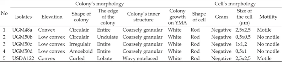
Based on morphological observations of both cells and colonies, the four isolates (Table 1) had similar colonies morphology on YEMA plates (picture not shown). They were transparent to creamy (white) colonies with 0,5-2 mm in diameter after 3 days incubation at room temperature. All bacterial isolates were Gram negative and rod-shaped with different shape of edge, elevation, and inner cell’s structure (Table 1).
Nodulation and N-fixation assay of four isolates on M. atropurpureum
The nodulation assay of four isolates on legume M. atropurpureum from shrub
Table 1. The observation of colony and cell morphology
No
Colony’s morphology Cell’s morphology
Isolates Elevation Shape of
colony
The edge of the colony
Colony’s inner structure
Colony growth on YMA
Shape
of cell Gram
Size of the cell (μm)
Motility
1 UGM48a Convex Circulair Entire Coarsely granular White Rod Negative 2,5x2,5 Motile
2 UGM50b Low convex Circulair Undulate Coarsely granular White Rod Negative 0,5x0,5 No motile
3 UGM50c Low convex Irregulair Entire Coarsely granular White Rod Negative 1x1,2 No motile
4 UGM50d Low convex Amoeboid Entire Coarsely granular White Rod Negative 0,5x1 No motile
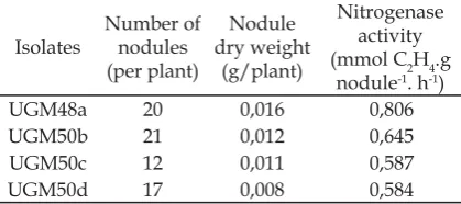
agroecosystem in Sumatera, Indonesia were obtained by using minirhizotron method. All bacterial isolates successfully formed nodule with different number of nodules. The fresh nodules were used for measuring the nitrogenase activity. Among them, UGM48a isolates showed the highest number of nitrogenase activity in 0,806 mmol C2H4.g nodule-1.h-1 with the highest nodule dry
weight (Table 2).
Minirhizotron is zeolite based medium in the chamber (Joachim et al., 2006). The seedlings in the chamber were watered daily with N-free nutrient solution. Based on fi ndings by Busch et al. (2006), Rhizotron method is the easiest reliable method to plant and to harvest the belowground biomass in distinct soil layers because of the simple access to the intact soil and root system at harvest. The tip of lateral root spreads out to all part of the chamber of rhizotron, include the surface. It is easier for isolates to initiate fl avanoid (compound that triggers the secretion of Nod factors, which in turn are recognized by the host plant and can lead to root hair deformation and several cellular responses, such as ion fl uxes and the formation of a root nodule). At harvest, which was the 40th day
after cultivation, the plants were analyzed for the number of fresh nodules and their nitrogenase activity was determined using Acetylene Reduction Assay (ARA). The result showed that UGM48a isolates have the highest number of nitrogenase activity in 0,806 mmol C2H4.g nodule-1.h-1. This isolate
was used as the selected isolate, which we observed thoroughly.
Analysis of the 16S rDNA sequences
Pure DNA genome of selected isolate, UGM48a, was used in 16s rDNA sequence analysis. This is one of the most effective tools for identifying bacteria. The 16S rDNA fragments of UGM48a was successfully amplifi ed. Based on BLASTn result at NCBI (http://blast.ncbi), UGM48a has 97% in similarity with Achromobacter xylosoxidans with identity 1272 bp query length. A. xylosoxidans is one of genera belong to betaproteobacteria that has been reported as legume nodulating bacteria for the fi rst time by Benata et al. (2008).
NifH and NodA amplifi cation
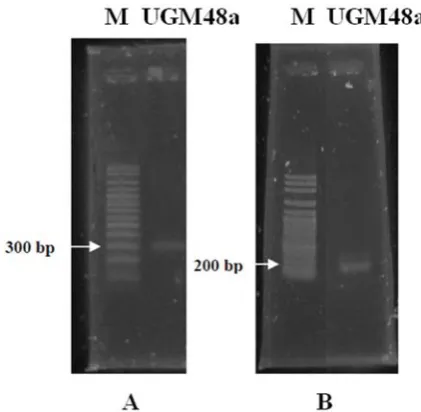
The existence of NifH and NodA gene copy were also amplified. The molecular weight of the amplified NifH and NodA gene were about 300 bp and 200 bp (Figure 1), whereas the molecular weight of the amplifi ed NifH genes was between 371-464 bp (Burgmann et al., 2004). The amplifi cation proved that the bacterium has the potential to fi x up free nitrogen. The other publications also mentioned that extensively NifH gene amplification in nitrogen-fixing bacteria, non-symbiotic, or associative. Achouak et al. (1999) successfully amplify NifH genes in bacteria Paenibacillus polimyxa, P. macerans, and P. azotofi xans with a molecular weight of about 370 bp. In the other hand, Mirza et al. (2006) successfully amplify NifH genes in Pseudomonas sp strain K1 with a molecular weight of 360 bp.
UGM48a isolates were amplifi ed using primers degenarated NodA : NodA-1 (5’ –TGC RGT GGA ARN RNN CTG GG -3’) and primer NodA-2 (5’- GGN CCG TCR TCR AAW GTC ARG - 3’) with a molecular weight of 200 bp. According to Haukka et al. (1998) the expected amplifi cation product is a band with a molecular weight of 666 bp. But the results showed other bands besides the expected band. Generally, the additional band does not preclude the measurement between the bright and the smear band. However, degenerate primer performed Table 2. Number, Dry Weight, and Nitrogenase
Activity of Nodules Formed by Bacterial Isolates in M. atropurpureum
in the attachment of the primary side is not suitable because of the low annealing temperature. In many experiments with degenerate primer, the expected molecular weight from amplifi ed products of PCR can not be predicted with certainty (McPherson and Møller, 2006).
Orthologs of the nodA gene, one of the key nod genes encoding an acyl transferase (Kamst et al., 1998), have been discovered in symbiotic Methylobacterium sp. (Sy et al., 2001) and Burkholderia sp (Moulin et al., 2001). Major nod factor-triggered responses include the formation and deformation of root hairs, intra- and extracellular alkalinization, membrane potential depolarization, changes in ion fluxes, induction of early nodulin gene expression, and formation of nodule primordia (Broughton et al., 2000; Perret et
al., 2000). So that, the NodA gene has been shown to be a good nodulation marker indeed (Boivin and Giraud, 1999).
Nodulation assay in Glycine max (L.) merr var. wilis
The last observation was the ability of UGM48a to form root nodule in soybean to ascertain the extent of bacterium ability to form root nodules on different host plants. The soybean is an important food commodity in Indonesia. 40 days after cultivation of soybean, observation showed that UGM48a formed nodule (Table 3). A. xylosoxidans has previously been reported having ability to nodulate Prosopis julifora (Benata et al., 2008); cowpea (Guimarães et al., 2012); and Mucuna bracteata (Salwani et al., 2012). This is the fi rst fi ndings that A. xylosoxidans were able to nodulate soybean. There are only seven species capable of symbiosis with soybean (Khan et al., 2010) : Bradyrizhobium japonicum (Jordan, 1982), B. elkanii (Kuykendall et al., 1992), B. liaoningenese (Xu et al., 1995), B. canariense, B. yuanmingense (Vinuesa et al., 2008), and E. fredii, Ensifer xinjiangensis (Young, 2003).
Acknowledgement
This work was funded in part by the Project of Below-Ground Biodiversity (BGBD) Indonesia and by the Directorate General of Higher Education, Republic of Indonesia. We are grateful to Dr. Denisa Kera for her critical reading on the manuscript.
References
Achouak, W., P. Normand and T. Heulin., 1999. Comparative phylogeny of rrs and NifH genes in the Bacillaceae., Int. J. Syst. Bacteriol., 49 : 961-967
Ardley, J., Parker, M., DE Meyer, S., Trengove, R., O’Hara, G.., Reeve, W., Yates, R., Dilworth, M., Willems, A. and Howieson, J., 2012. Microvirga lupini sp. nov., Microvirga lotononidis sp. nov. and Microvirga zambiensis sp. nov. are alphaproteobacterial root-nodule bacteria that specifi cally nodulate and fi x nitrogen Figure 1. The Agarose Gel Electrophoresis of UGM48a
Amplifi ed nifH (A) and nodA (B) genes.
Table 3. Nodulation assay in Glicyne max (L.) merr var. wilis
Isolates Nodulation assay
UGM48a +
USDA 122 +
Uninnoculated
with geographically and taxonomically separate legume hosts. Int. J. Syst. Evol. Microbiol., 62, 2579-2588.
Ausubel, F.M., R. Brent, R.E. Kingston, D.D. Moore, J.G. Seidman, J.A. Smith, and K. Struhl. (1995) Short protocols in molecular biology. 3rd edition. John Wiley & Sons Inc. California, USA
Benata, H., O. Mohammed, B. Noureddine, B. Abdelbasset, H. Abdelmoumena, R. Muresu, A. Squartini, and M. M., El Idrissi., 2008. Diversity of bacteria that nodulate Prosopis julifl ora in the eastern area of Morocco. Syst. and Appl. Microbiol.,
31 : 378–386
Benhizia, Y., et al. 2004. Gamma proteobacteria can nodulate legume of the genus Hedysarum. Syst. Appl. Microbiol., 27(4), 462-468
Boivin, C. and Giraud, E. (1999) Molecular symbiotic characterization of rhizobia: towards a polyphasic approach using Nod factors and nod genes. In: Highlights of Nitrogen Fixation Research (Martinez, E. and Hernandez, G., Eds.), pp. 295-299. Plenum Press, New York.
Burgmann et al., 2004. “New molecular screening tools for analysis of free-living diazotrophs in soil”, Appl. Environ. Microbiol, 70, 240–247
Busch, J., I.A. Mendelssohn, B. Lorenzen, H. Brix, S. Miao., 2006. A rhizotron to study root growth under flooded conditions tested with two wetland Cyperaceae. Flora,
201, 429–439
Euzeby’s, J. P., 2006. “Taxonomy of legume nodule bacteria (rhizobia) and agrobacteria : spesies with standing in nomenclature”. <http://edzna.ccg.unom.mx/rhizobial-taxonomy>. (Acessed 12.07.2010).
Evizal, R., I.D. Prijambada, J. Widada, D. Widianto and Tohari., 2013. Diversity of legume nodulating bacteria as key variable of coffee agro-ecosystem productivity. Int Res Journal of Agric Sci and Soil Sci., 3(4), pp. 141-146
Guimarães, A., P. Jaramillo, R. Nóbrega, L. Florentino, K. Silva and F. Maria de S.
Moreira., 2012. Genetic and symbiotic diversity of nitrogen-fixing bacteria isolated from soils under agriculture use in the Western Amazon using cowpea as the trap plant. Appl. Environ. Microbiol. doi:10.1128/AEM.01303-12
Haukka, K., K., Lindstro, and J. P.W., Young., 1998. Three phylogenetic groups of nodA and nifH genes in sinorhizobium and mesorhizobium isolates from leguminous trees growing in Africa and Latin America. Appl. Environ Microbiol.,
64 (2), 419-426.
Joachim et al., 2006. A rhizotron to study root growth under fl ooded conditions tested with two wetland Cyperaceae, Flora, 201, 429–439.
Jordan, D. C., 1982. Transfer of Rhizobium j a p o n i c u m B u c h a n a n 1 9 8 0 t o Bradyrhizobium gen. nov., a genus of slow-growing, root nodule bacteria from leguminous plants. Int Journal of Syst Bacteriol., 32(1), 136–139.
Kaisa, H. et al., 1998. Three phylogenetic groups of nodA and nifH genes in sinorhizobium and mesorhizobium isolates from leguminous trees growing in Africa and Latin America, Appl. Environ. Microbiol., 64 (2), 419-426
Kamst, E., Spaink, H.P., and Kafetzopolus, D., 1998. Biosynthesis and secretion of rhizobial lipochitin-oligosacharide signal molecules. In : Plant-microbe interactions – subcellular biochemistry (Biswas, B.B. and Das, H.K., eds.) pp. 29 – 71. Plennum Press, New York.
Khan, M.S., A. Zaidi, J. Musarrat. (2010) Microbes for Legume Improvement. Springer Wien NewYork.
Kuykendall, L.D., B. Saxena, T.E. Devine, S.E. Udell., 1992. Genetic diversity in Bradyrhizobium japonicum and a proposal for Bradyrhizobium elkanii sp. nov. Can J Microbial., 38, 501–505
Lin, D-W. et al., 2008. Shinella kummerowide sp. Nov., asymiotic bacterium isolated from root nodules of the herbal legume Kummerowia stipulacea. Int J Syt Evol Microbiol., 58, 1409-1413.
McPherson, M.J. and S.G. Møller. (2006) PCR Second Edition. Taylor and Francis Group, New York.
Mirza, M. S., S. Mehnaz, P. Normand, C. Prigent, C.Y.M., Loccoz, R. Bally, K.A., Malik., 2006. Molecular characterization and PCR detection of a nitrogen fi xing Pseudomonas strain promoting rice growth. Biol. Fertil. Soils., 43, 163-170 Moulin, L., A. Munive, B. Dreyfus, and C.
Bolvin-Masson., 2001. Nodulation of legumes by members of the β-subclass of proteobacteria. Nature Pub., 411, 948-950.
Perret, X., Staehelin, C., and Broughton, W.J., 2000. Molecular basis of symbiotic promiscuity. Microbiol. Mol. Biol. Rev., 64, 180 – 201
Salwani, S., Amir, H. G. and Najimudin, N., 2012. Evidence of Diazotrophic Symbionts in the Leguminous Cover Crop Mucuna bracteata. Pertanika J. Trop. Agric. Sci., 35
(3), 537 – 552
Sheu SY, Chou JH, Bontemps C, Elliott GN, Gross E, Dos Reis Junior FB, Melkonian R, Moulin L, James EK, Sprent JI, Young JP, Chen WM., 2012. Burkholderia diazotrophica sp. nov., isolated from root nodules of Mimosa spp., Int J Syst Evol Microbiol.
Shiraishi, A., Matsushita, N., and Hougetsu, T., 2010. Nodulation in black locust by the Gammaproteobacteria Pseudomonas sp. and the Betaproteobacteria Burkholderia sp. Sci Direct, 33, 269-274.
Somasegaran, P., Hoben, J.H. (1994) Handbook for Rhizobia: Methods in Legume-Rhizobium Technology. Springer-Verlag, New York Vinuesa, P., K. Rojas-Jime´nez, B.
Contreras-Moreira, S. K. Mahna, B. N. Prasad, H. Moe, S. B. Selvaraju, H. Thierfelder, and D. Werner., 2008. Multilocus sequence analysis for assessment of
the biogeography and evolutionary genetics of four bradyrhizobium species that nodulate soybeans on the Asiatic continent. Appl. Environ. Microbiol, 74
(22), 6987–6996
Xu, L.M., C. Ge, Z. Cui, J. Li, H. Fan., 1995. Bradyrhizobium liaoningense sp. nov., isolated from the root nodules of soybeans. Int J Syst Bacteriol., 45, 706–711