Research Report
Caspase 3 and Survivin Expressions as a Predictor of Response
to Radiation Therapy in Advanced Stage Cervical Cancer
Ekspresi Caspase 3 dan Survivin sebagai Prediktor Respons Radiasi pada Kanker Serviks Stadium Lanjut
Fitriyadi Kusuma1, Laila Nuranna1, Primaria D. Rustamadji2, Bambang Sutrisna3
1Division of Oncology Ginecology, Department of Obstetrics and Gynecology 2Department of Pathology Anatomi, Medical Faculty of Indonesia University
Dr. Cipto Mangunkusumo General Hospital, Jakarta 3Faculty of Public Healths University of Indonesia, Jakarta
INTRODUCTION
Cervical cancer is the most common cancer in women in developing countries.1,2 International Agency for
Research of on Cancer (GLOBOCAN 2008) estimated that 529,000 new cases and 275,000 deaths from cer-vical cancer occur in the world every year and only 5% of women had been screened for cervical can-cer.1,2 The 5-year survival rate of cervical cancer was
50% for stage 1, 40% for stage II, 20% for stage III, and 0% for stage IV.3 Management of cervical cancer
includes surgery, radiation, chemoradiation and che-motherapy, depending on the clinical stage, and this greatly affects the survival and the possibility of local control and loco-regional control.4,5 Radiation therapy
and chemo-radiation are treatment options for advanced-stage cancer.5,6
Abstract
Objective: To identify prognostic factors of advanced stage cer-vical cancer which was treated by radiation therapy, so that they can be implemented to support treatment in order to increase the success of therapeutic response in advanced stage cancer.
Methods: Observational analytic historical cohort method study was conducted in Dr. Cipto Mangunkusumo General Hospital and 38 cases of stage IIB and IIIB cervical cancers receiving a complete radiation therapy were involved. Data were retrieved from medical records and cervical biopsies examination results. Paraffin blocks of cervical biopsies were examined histopathologically using HE stain-ing followed by IHC. The expression of caspase 3 and survivin were then assessed by IRS scoring. Case group included 5 samples with negative therapeutic response, which means the therapeutic response other than complete response. Control group included 33 samples with positive therapeutic response which have complete response to therapy. The collected data were analyzed by univariate, bivariate, and multivariate analysis.
Results: Demographic and clinicohistopathologic data were not significantly related to the occurrence of negative response to radia-tion therapy. Negative cytoplasmic caspase 3 expression has a pre-dictive value of 90.75% for the occurrence of a negative response to radiation therapy (RR=9.81, CI 95%=1.231-78.286, p=0.019). Posi-tive cytoplasmic survivin has a predicPosi-tive value of 86.75% for the occurrence of a negative response to radiation therapy (RR=6.55, CI 95%=2.659-16.119, p=0.000). Nuclear caspase 3 and survivin ex-pressions have no significant relation to the occurrence of a negative response to radiation therapy. Clinical response to radiation is influ-enced by clinicohistopathologic factors, caspase 3, and survivin ex-pression altogether.
Conclusion: Negative cytoplasmic caspase 3 expression and positive cytoplasmic survivin have predictive value for the risk of a negative response to radiation therapy to occur.
[Indones J Obstet Gynecol 2011; 35-3: 139-45]
Keywords: advanced stage cervical cancer, clinicohistopathol-ogy, caspase 3, survivin, response to radiation therapy.
Abstrak
Tujuan: Untuk mengidentifikasi faktor-faktor prognosis pada kanker serviks stadium lanjut yang dilakukan terapi radiasi se-hingga dapat menunjang penatalaksanaan untuk meningkatkan ke-berhasilan respons terapi pada kanker stadium lanjut.
Metode: Penelitian dengan metode historikal kohort analitik ob-servasional di RSCM terhadap 38 kasus kanker serviks stadium IIB dan IIIB yang telah dilakukan terapi radiasi lengkap. Data diambil dari rekam medis dan blok parafin biopsi serviks diperiksa histopa-tologinya dan dilakukan pewarnaan HE dan dilanjutkan dengan IHC untuk melihat ekspresi kaspase 3 dan survivin dengan penilaian skor IRS. Kelompok kasus sebanyak 5 sampel adalah kelompok dengan respons terapi negatif yaitu respons stabil dan progresif. Kelompok kontrol sebanyak 33 sampel adalah kelompok dengan respons terapi positif yaitu kelompok respons komplit dan respons parsial. Data yang terkumpul dianalisis dengan analisis univariat, bivariat dan multivariat.
Hasil: Dari data demografi dan klinikohistopatologi tidak memiliki hubungan bermakna untuk terjadinya respons terapi radi-asi negatif. Ekspresi kaspase 3 sitoplasma negatif mempunyai nilai prediksi sebesar 90.75% untuk terjadinya respons terapi radiasi ne-gatif (RR=9.81, IK 95%=1.231-78.286, p=0.019) dan survivin sito-plasma positif mempunyai nilai prediksi sebesar 86.75% untuk ter-jadinya respons terapi radiasi negatif (RR=6.55, IK 95%=2.659-16.119, p=0.000). Ekspresi kaspase 3 inti dan survivin inti tidak memiliki hubungan bermakna untuk terjadinya respons terapi radi-asi negatif. Respons klinik radiradi-asi dipengaruhi oleh faktor-faktor klinikohistopatologi, ekspresi kaspase 3 dan survivin secara ber-sama-sama.
Kesimpulan: Ekspresi kaspase 3 sitoplasma negatif dan survivin sitoplasma positif mempunyai nilai prediksi untuk risiko terjadinya respons terapi radiasi negatif.
[Maj Obstet Ginekol Indones 2011; 35-3: 139-45]
Kata kunci: kanker serviks stadium lanjut, klinikopatologi, ek-spresi kaspase 3, ekek-spresi survivin, respons terapi radiasi
Up to now, it is known that the prognostic factors of cervical cancer are influenced by clinico-his-topathologic factors.7 Tumor markers have been
de-veloped to determine prognostic factors of cervical cancer such as serum-associated antigene, angiogene-sis factors, and apoptotic factors.8,9
Dysregulation of apoptosis in cell plays an impor-tant role in the development and progression of ma-lignancies in humans. Defects of apoptosis will lead to neoplastic processes through various mechanisms. Caspase is a family of protease which has an impor-tant role in pro-apoptotic process.10,11 Some studies
which focused on apoptotic regulation-expression analysis showed the reduced levels of caspase in cer-vical cancer compare to the one in precancerous le-sions.12-5
Beside the pro-apoptotic factors, there are also anti-apoptotic genes that are developed and known as inhibitors of apoptosis (IAPs).16,17 Survivin is an IAP
which has 142 amino acids and molecular weight of 16.5 kDa. It contains a single baculovirus IAP repeat (BIR) which has capability to regulate cell prolifera-tion and cell death. in the cell cycle BIR is expressed at the G2/M phases.18-20 Survivin has an important
role in tumorigenesis of colorectal cancer and virus-induced carsinogenesis.19 Suzuki et al, in their
re-search found a correlation between survivin expres-sion and locoregional control of cervical cancer which had under gone radiation therapy.21
Survivin in gynecologic cancer has not been much studied. It is expected that the new findings about the ratio of pro-apoptotic caspase 3 and anti-apoptotic survivin expressions can be used to predict the suc-cess of chemoradiation therapy for early and ad-vanced stage cervical cancer. It is also expected that in the future the results of this study can be used as a reference for the study on gene targeted therapy for cancer. To identify prognostic factors of advance stage cervical cancer which was treated by radiation therapy, so that they can be implemented to support treatment in order to increase the success of therapeu-tic response in advanced stage cancer is the aim of this study.
METHODS
The study was conducted at the Division of Gynecol-ogy OncolGynecol-ogy, Department of Obstetrics and Gyne-cology, Faculty of Medicine, University of Indonesia, Dr. Cipto Mangunkusumo General Hospital Jakarta, and Department of Pathology Faculty of Medicine, University of Indonesia.
Population of this study was all advanced-stage cervical cancer patients (stage IIB, IIIA, and IIIB) who visited the Gynecology Oncology Clinic in Dr. Cipto Mangunkusumo Hospital Jakarta and had un-dergone radiation therapy from 2005 to 2010. Sam-ples were taken from the case group and control group consecutively. Any subjects who met the inclusion criteria were included.
Secondary data was obtained from existing medical records of advanced-stage cervical cancer patients (stage IIB, IIIA, and IIIB) and was selected using the inclusion criterias stated. Course of the disease and radiation therapy accepted were observed.
Tissue that was taken by biopsy was preserved in 10% buffered formalin solution, and then sent to De-partment of Anatomic Pathology, Faculty of Medi-cine, University of Indonesia, where it, and embedded in paraffin. The paraffin block was processed by con-ventional histopathologic examination using Hema-toxilin and Eosin staining (HE) and proceeded with the immunohistochemistry (IHC).
The primary caspase 3 antibody used in this study was rabbit monoclonal caspase 3 antibody (3CSP03): sc-56046 Santa Cruz Biotechnology, In. The primary survivin antibody used was mouse monoclonal sur-vivin antibody Clone 12C4 reagent provided Code M3624 (Dako, Denmark). Detection instrument (de-tection kit) used was the Star Trek Universal Detec-tion System HRP (Biocare Medical, LLC, Concord CA, USA) containing a Trekkie Universal Link; Trek avidin-HRP; Betazoid DAB chromogen; Betazoid DAB Substrate Buffer; Background Sniper.
Rabbit monoclonal caspase 3 antibody (3CSP03): sc-56 046 with a 1:600 dilution and Mouse mono-clonal survivin antibody clone 12C4 with a 1:50 di-lution were added into the paraffin block.
Both immunohistochemical staining were per-formed by incubation in a closed place at room tem-perature for 60 minutes and then rinsed with normal serum (PBS) 2 times, 5 minutes each. To bind the biotinylated secondary antibody using the imunoper-oxidase method, a primunoper-oxidase (HRP) labeled septravid-ine (Trekavidin-HRP) was added. Diaminobenzidseptravid-ine tetrahydrochloride /DAB (Betazoid DAB Chromogen) was added and given time to react for 2-5 minutes until it formed a brown chromogenic bond, followed with dehydration, cleaning, and fixation process into the object glass. Positive controls were taken from cervical carcinoma tissues with positive expressions of caspase 3 and survivin, while the negative controls were taken from the same cervical tissues which its primary antibody had been replaced with PBS.
Observations were made on 500 tumor cells from five different large fields of view (magnification of 400 times) ramdomly. Each field represented 100 tu-mor cells. Evaluation was carried out and analyzed by researchers and Anatomical Pathology specialists. The degree of positivity was determined by staining scoring using Immunoreactive Scoring System (IRS) modification, which was used by Remmele et al to analyze the expression of survivin.22 This scoring
sys-tem can also be used to analyse caspase 3 expression. This scoring system was modified and determined by multiplying the intensity of staining with the percent-ages of positive cells. The percentage of positive cells was rated as follows: 0, 1-5% positive cells; 1, 5-25%; 2, 25-50%; and 3, > 50% positive cells. Staining in-tensity was scored as 1, weak; 2, moderate, and 3, intensive. Scores for percentage of positive cells and scores for expression intensities were multiplied to calculate an immunoreactive score (IRS), 0 = no staining; 1-2 = weak staining; 3-6 = moderate stain-ing; > 7 = strong staining. It was classified as positive expression if the IRS > 3 for cytoplasmic survivin, cytoplasmic and nuclear caspase 3, and if the IRS > 1 for nuclear survivin.
re-sponse to radiation therapy which was classified into complete, partial, stable, or progressive response.
RESULTS
Through the processes of selection according to in-clusion and exin-clusion criterias, 38 cases of advanced stage cervical cancer that had been treated with ra-diation therapy and examined for caspase 3 and sur-vivin expressions by the Department of Anatomical Pathology Faculty of Medicine, University of Indone-sia, Jakarta, were taken. In this historical cohort study, the case group included 5 samples of patients with positive response to radiation, whereas the control group were 33 samples of patients with negative re-sponse to radiation therapy.
From the demographic and clinic-histopathologic data analysis, it was found that they have no signifi-cant relation to the occurrence of a negative response to radiation therapy.
From the analysis of cytoplasmic caspase 3 and response to therapy, it was obtained that the propor-tion of negative cytoplasmic caspase 3 was greater than positive caspase 3 in negative response to ther-apy (other than complete response), which was 36.36% compared to 3.70% with RR 9.81, CI 95% 1.231-78.286, and p=0.019 by Fisher’s Exact test. Be-cause negative survivin expression was not found in the unexposed group (complete response), a correc-tion of number from 0 to 0.5 was done in the statis-tical calculation in order to do the bivariate test. The proportion of positive cytoplasmic survivin was
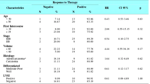
Table 1. Relations Between Patient’s Characteristics and Response to Radiation Therapy
Characteristics
*Endometrioid Adenocarcinoma + Adenocarcinoma + Adenosquamous Carcinoma
Table 2. Relations Between Caspase 3 and Survivin Expressions and Response to Radiation Therapy
greater than negative cytoplasmic Survivin in negative response to therapy, which was 20.45% compared to 3.12% with RR 6.55, CI 95% 2.659-16.119, and p = 0.000 in Fisher’s Exact test.
Nuclear caspase 3 and nuclear survivin did not have significant relations with negative response to therapy.
DISCUSSION
Modality of treatment for advanced cervical cancer (stage IIB and IIIB) is external radiation of whole pelvic followed by internal radiation, and in addition is concurrent chemotherapy with platinum base.5
Ra-diotherapy plays an important role in determining therapy for all stages of cancer.23 It was believed that
the combination of chemotherapy and radiation can control the disease locally, whereas chemotherapy alone controls the sub-clinical metastases that occur outside the radiation field.6
Tumor resistancy is a serious problem in the treat-ment of patients with neoplastic agents. Although it is still in controversy, the accumulation of experimen-tal evidences showed that the initial damage caused by chemotherapeutic agents assembles into common apoptotic pathway. Upregulation of the protein inhibi-tor of apoptosis would be advantageous for tumor cells.24 Several studies have found that in the tumor
resistancy, there is enhancement of the expression of apoptosis inhibitor protein, especially survivin.25
There was no difference in the risk of negative therapeutic response occurence among various demo-graphic data.
According to FIGO, one of the most important prognostic factors in cervical cancer is the clinical stage. Kleinberg et al, found that stage IB, IIA, and IIB have a significantly better survival rate compared to stage III or IVA treated by chemoradiation.26 This
study observed only stage IIB and IIIB. There were no significant differences for the risk of negative re-sponse occurence with p = 0.592 between the two
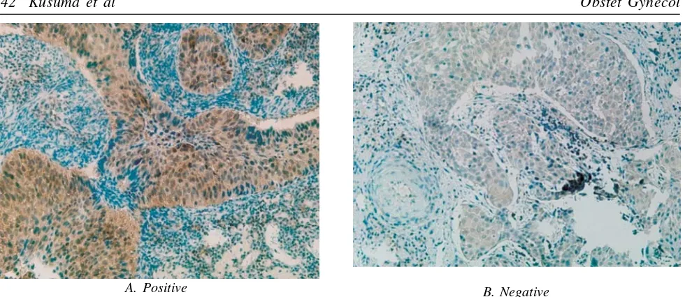
B. Negative
Figure 2. Cytoplasmic Survivin Expression in slide with immunohistochemical staining with 400x enlargement. A. Positive Expression. B. Negative Expression
A. Positive
Figure 1. Cytoplasmic Caspase 3 Expression in slide with immunohistochemical staining with 400x enlargement. A. Positive Expression. B. Negative Expression.
were, but the proportion of negative prognosis of therapeutic response was greater in the stage IIIB group than in stage IIB, with a relative risk of 0.54 with CI 95% 0.104-2.754.
Based on ROC analysis, the cut-off point of tumor size was 60 cm3. The proportion of negative response
to radiation therapy was higher in tumor size that greater than or equal to 60 cm3 (22.22%) than less of
60 cm3 (5.00%), with a relative risk of 4.44 CI 95%
0.546-36.176, although it was not statistically signifi-cant (p = 0,170). Assessment of therapeutic response through physical examination has limitations because of intratumor necrosis and fibrosis that occur in the cervix. CT scans and physical examination also has the limitations because they can not differentiate be-tween the tumor residue from post therapy fibrosis and the tumor residue from post inflammatory fibro-sis. This can only be differentiated by MRI.
Nakanishi et al stated that tumor size is a signifi-cant prognostic factor in stage IB cervical cancer with cut-off point 4 cm.27 Huang et al also reported
sig-nificant differences of response in 80 cases treated by radiation therapy, where the complete response (local control) occured in the mean tumor volume of 66 cm3
(3.0 - 342.0) while negative response (local failure) occured in the mean tumor volume of 129.5 cm3 (41.0
- 700.0) with the mean regression ratio for 0.6% and 19.4%.28
The most common histology type of cervical can-cer is squamous cell carcinoma. Based on FIGO, his-tology type can affect the prognosis. In several stud-ies, adenocarcinoma and adenosquamose affect 5-year survival rate.29 In this study, the most frequent type
was squamous cell carcinoma (71.05%). The fre-quency of endometrioid adenocarcinoma, adenocarci-noma and adeno-squamous carciadenocarci-noma was respec-tively 2.63%, 18.42% and 7.89%. There were no sig-nificant differences in the occurrence of a negative response to radiation therapy between the groups of squamous carcinoma and the groups of adenocarci-noma (adenocarciadenocarci-noma, adeno-squamous, and en-dometrioid adenocarcinoma), with RR 1.64, CI 95% 0.315-8.487, p = 0,615.
Similar results were obtained by Long et al, squamous cell carcinoma 75%, adenocarcinoma 20%, and adenosquamous 2 - 3%. According to clinical his-topathology analysis by Kleinberg et al, there were no significant differences between the histology of squamous cell carcinoma, adenocarcinoma and ade-nosquamous.26 Peters et al in the Gynecologic
Oncol-ogy Group (GOG) 109/Southwest OncolOncol-ogy Group (SWOG) 8797 found that patients with adenocarci-noma or adenosquamous treated with radiation alone had a poor progression-free survival rates. They also found that there was no difference in prognosis be-tween radiation alone and chemoradiation therapy.30
Other histopathologic factors showed no significant predictive value for the occurrence of a negative re-sponse to radiation therapy.
Caspase 3 and Survivin Expressions
Immunohistochemical examination showed that in ad-vanced cancer, positive of caspase 3 were found more
frequently in the cytoplasm (71%) than in the nucleus (55.2%). Similarly, survivin were found more fre-quently in the cytoplasm (56.8%) than in the nucleus (5.26%). Similar result had been reported by several studies.31-3 Caspase 3 is one of the effector caspase
activated by initiator caspase (caspase 8 and 9). Cy-tochrome C that is released from the intermembran chamber will bind the Apaf-1 and caspase 9. This process will activate the proteolytic caspase 3. SMAC/diablo will also be released from the mito-chondria at the same time. It can be inverted that the process of apoptosis occurs in the cytoplasm.34
While working as an inhibitor of Bax and Fas (signal of apoptosis), survivin also inhibits the process of caspase 3 and 7 (pro-apoptosis). All these processes also occur in the cytoplasm. It was relevant with this study, immunohistochemical staining of caspase 3 and survivin positive occurs more frequently in the cyto-plasm.35 Positive result in the nucleus occurs because
the apoptotic process involves the cell cycle.
Cytoplasmic caspase 3 has strong relation with the response to radiation therapy. The response is influ-enced by the expression of caspase which increase the apoptosis process. Characteristics of apoptosis in stereotyped morphologic picture are chromatin con-densation, nucleus fragmentation, and membrane clo-sure of apoptotic body. The changes of this morphol-ogy are executed by the caspase family. Caspase 3 as an executioner caspase activates this apoptotic func-tion. Radiation exposure will induce damage of DNA via extrinsic pathway by forming a caspase arcade and will start the process of apoptosis. FADD stimu-lation will alter a pro-caspase 8 to caspase 8 which will stimulate apoptosis. Caspase 8 and 9 alone will stimulate the pro-caspase 3 to become caspase 3 and then caspase 3 will further regulate apoptosis.36-7 Cell
death occurs during the transition from metaphase to anaphase is characterised by the activation of caspase 2 as the initial activation of DNA damage response and by the release of cell death effectors such as caspase 9 and 3 as well as an activator of cytochrome C. High expression of caspase 3 in the cytoplasma activates apoptosis. Cells that fail to execute the apop-totic program with the failure of mitosis will have asymmetric division in the next cell division and will become aneuploid cells. This will non-activates apop-totic program and causes chromosomal disorders.38
In this study, the cut-off point of ROC analysis was > 3. We found that the specificity of positive cytoplasmic caspase 3 was 80% while the sensitivity was 62.50% (ROC area = 0.7542). Of the 38 samples, we found that there was a significant negative relation between cytoplasmic caspase 3 and response to ther-apy with a value of p = 0.031. Negative expression of cytoplasm caspase 3 increases the risk of a negative response to radiation therapy with RR 4.09 and CI 95% 1.174-14.249. There is no significant relation be-tween expression of nuclear caspase 3 and radiation response with p = 0.426, although the proportion of negative nuclear caspase 3 (29.41%) was greater than positive nuclear caspase 3 (14.29%) for the occur-rence of negative responses with a relative risk of 95% CI 2058 0572 -7414.
squamous cell carcinoma) and mice breast cancer treated by radiation therapy shows that positive caspase 3 has better survival rate compared with the negative caspase 3 (p = 0.0114 and p = 0.0006).39 In
the study by Cheung et al, the expression of caspase 3 was reported. In the study by Shi et al to be related to cervical cancer in which reduced expression of caspase 3 inhibits apoptosis process.31
Survivin protein serves as an inhibitor of caspase activation that plays an important role in mitosis and negative regulation of apoptosis. The survivin expres-sion is also increased by cell cycle regulation and is only expressed in the G2-M phase. The location of survivin is at the mitotic spindle that interacts with tubulin during mitosis.40 Tamm et al in their study
also found that survivin blocked apoptosis directly in 293 cells which had been transfected by over-expres-sion of caspase 3 and 7 and plasmids encoding sur-vivin. It was showed that survivin inhibits second process of caspase in the active form only. Survivin might only prevent the breakage of amplification ac-tivation cascade which decreased apoptosis process.35
Survivin was thought to have a relation with radiation resistancy and hypoxia, so that over-expression of survivin causes resistance to therapy and poor prog-nosis.17 Bache in his study found that over-expression
of survivin significantly decreased the 5-year overall survival with odds ratio 3.3 (p = 0.02) in bivariate analysis and 3.2 (p = 0.03) in univariate and Cox re-gression multivariate analysis.41 In the study by Shi
et al, survivin is an independent prognostic indicator that affected survival rates for cervical cancer. There was shorter survival rates in groups with positive sur-vivin expression (p < 0.05).31
Of the 38 samples, we found that cytoplasmic vivin expression is more positive than the nuclear sur-vivin, which were 57.89% (22/38) and 18.42% (7/38). Suzuki et al found similar results which were positive cytoplasmic survivin 47% and nuclear survivin only 14%.21 We found that there was a significant positive
relation between cytoplasmic survivin and response to therapy with a value of p = 0.031. Positive expres-sion of cytoplasm survivin increases the risk of a negative response to radiation therapy with RR 6.55, CI 95% 2.659-16.119, and p = 0.000 in Fisher’s Exact test.
It can be concluded that cytoplasmic caspase 3 and cytoplasmic survivin has a predictive value for the response to radiation therapy by using score > 3, whereas nuclear caspase 3 and nuclear survivin can not be used as a predictor because caspase 3 is not activated in the nucleus but for the mitochondrial membrane. Survivin is expressed in the phase G2 and M of cell cycle and is located at the mitotic spindles so it should be expressed in the nucleus, but in our study it was not significantly related.
REFERENCES
1. Ferlay J, Shin HR, Bray F, Forman D, Mathers C, Parkin DM. Estimates of worldwide burden of cancer in: GLO-BOCAN 2008. Int J Cancer. 2010;127(12):2893-917 2. IARC. World cancer report. IARC. 2008.
3. Aziz MF. Gynecology cancer in Indonesia. J Gynecol On-col. 2009; 20(1):8-10.
4. Monk Bj. Chemotherapy for advanced and recurrent cervi-cal carcinoma in the new era of upfront chemotherapy and radiation: Have we arrived. The Women’s Oncology Re-view. 2005; 5(2):77-9.
5. LongIII HJ, Laack NNI, Gostout BS. Prevention, Diagno-sis, and Treatment of Cervical Cancer. Mayo Clin Proc. 2007; 82(12):1566-74.
6. Rose PG. Chemoradiation. Drug. 2000; 60(6):1239-44. 7. Andrijono. Kanker Serviks. 3rd ed. Andrijono, editor.
Ja-karta: Pustaka Spirit; 2009.
8. Gadducci A, Tana R, Fanucchi A, Genazzani AR. Bio-chemical prognostic factors and risk of relaps in patients with cervical cancer. Gynecol Oncol. 2007; 107(1(supp 1)):S23-S6.
9. Zusterzeel PLM, Span PN, Dijsterhuis MGK, Thomas CMG, Sweep FCGJ, Massuger LFAG. Serum vascular en-dothelial growth factor: a prognostic factor in cervical can-cer. J Cancer Res Clin Oncol. 2009; 135:283-90.
10. Porter AG, Janicke RU. Emerging roles of caspase-3 in apoptosis (review) Cell Death & Diff. 1999(6):99-104. 11. Wolf BB, Green DR. Suicidal tendecies : apoptotic cell
death by caspase family proteinases. J Bil Chem. 1999; 274:2049-52.
12. Cheung T-H, Chung TK-H, Lo KW-K, YU M-Y, Kra-jewski S, Reed JC, et al. Apoptosis-Related Proteins in Cer-vical Intraepithelial Neoplasia and Squamous Cell Carci-noma of the Cervix. Gynecol Oncol. 2002; 86:14-8. 13. Chung TKH, Cheung TH, Lo WK, Yim SF, Yu MY,
Kra-jewski S, et al. Expression of apoptotic regulators and their significance in cervical cancer. Cancer Letter. 2002; 180:63-8.
14. Ledwaba T, Diamini Z, Naicker S, Bhoola K. Molecular genetics of human cervical cancer : role of papilomavirus and the apoptotic cascade. Biol Chem. 2004; 385:671-82. 15. Zanotti S, Eckhoff AF, Mannherz HG. Changes in
topo-logical expression of markers of differentiation and apop-tosis in defined stages of human cervical dysplasia and can-cer. Gynecol Oncol. 2003(89):376-84.
16. O’Driscoll L, Linehan R, Clynes M. Survivin: Role in Nor-mal Cells and in Pathological Conditions. Current Cancer Drug. 2003; 3:131-52.
17. Altieri DC. Survivin, versatile modulation of cell division and apoptosis in cancer. Oncogene. 2003; 22:8581-9. 18. Wang Z, Fukuda S, Pelus LM. Survivin regulates the p53
tumor suppressor gene family. Oncogene. 2004; 23:8146-53.
19. Li F, Ling X. An Update of "What is the Next Wave". J Cellul Physiol. 2006; 208:476-86.
20. Xu Y, Fang F, Ludewig G, Jones G, Jones D. A Mutation Found in the Promoter Region of the Human Survivin Gene is Correlated to Overexpression of Survivin in Cancer Cells. DNA & Cell Biol. 2004; 23(7):419-29.
21. Suzuki Y, Oka K, Yoshida D, Shirai K, Ohno T, Kato S, et al. Correlation between survivin expression and locore-gional control in cervical squamous cell carcinomas treated with radiation therapy. Gynecol Oncol. 2007; 104:643-6. 22. Remmele W, Stegner HE: Recommendation for uniform
definition of an immunoreactive score (IRS) for immuno-histochemical estrogen receptor detection (ER-ICA) in breast cancer tissue. Pathologe 1987; 8:138-40
23. Gonzalez-Cortijo L, Carballo N, Gonzalez-Martin A, Cor-raliza V, Chiva LM, Sastre FL, et al. Novel chemotherapy approaches in chemoradiation protocols. Gynecol Oncol. 2008; 110(supplement):45-8.
24. Espinosa M, Cantu D, NormaHerrera, Lopez CM, Garza JGDl, Melendez-Zajgla J, et al. Inhibitors of apoptosis pro-teins in human cervical cancer. BMC Cancer. 2006; 6(45):1-10.
25. Schimmer AD. Inhibitor of apoptosis proteins: translating basic knowledge into clinical practice. Cancer Res. 2004; 64:7183-90
26. Kleinberg MJ, JR JMS, Alvarez RD. Concurrent chemo-therapy and radiation for advanced cervical cancer. CME J Gynecol oncol. 2007:92-8.
stage IB cervical cancer. Can a cut-off figure be deter-mined? Int J Gynecol Cancer. 2000; 10(5):397-401 28. Huang Z, Mayr NA, Yuh WTC, Lo SS, Montebello JF,
Grecula JC, et al. Outcome prediction of cervical cancer: kinetic model of tumor regression during radiation therapy. Cancer Res. 2010; 70:463-70.
29. Hacker NF. Cervical cancer. 4th ed. Berek JS, Hacker NF,
editors: Lippincott Williams & Wilkins; 2005.
30. Peters WA, Liu PY, Barret RJ, JR WG, Stock R, Berek JS, et al. Concurrent chemotherapy in pelvic radiation ther-apy composed to pelvic radiation alone as adjuvant therther-apy following radical surgery in high risk stage cancer of the cervix. J Clin Oncol. 2000; 18:1606-13.
31. Shi L, Baohua Z, Zehua W. Expression of survivin, cy-clinD1, p21 wan , caspase-3 in cervical cancer and its re-lation with prognosis. Journal of Huazhong University of Science and Technology 2005; 25(1):78-81.
32. Jiang H, Gong M, Cui Y, Ma K, Chang D, Wang TY. Upregulation of caspase-3 expression in esophageal cancer correlates with favorable prognosis: an immunohistochemi-cal study from a high incidence area in northern China. Diseases of the Esophagus. 2010; 23:487-92.
33. Ding W, Hu W, Wang X, Dong X. Expressions of survivin and caspase-3 in human hepatocellular carcinoma and the relationship with prognosis. Chinese-German J Clin Oncol. 2010; 9(11):628-32.
34. Pirnia F, Schneider E, Betticher DC, Borner MM. Mitomy-cin C induces apoptosis and caspase 8 and 9 processing through a caspase 3 and Fas-independent pathway. Cell Death Differ. 2002; 9(9):905-14.
35. Tamm I, Wang Y, Sausville E, Scudiero DA, Vigna N, Oltersdorf T, et al. IAP-Family Protein Survivin Inhibits Caspase Activity and Apoptosis Induces by Fas (CD95), Bax, Caspase, and Anticancer Drugs. Cancer Res. 1998; 58:5315-20
36. Devarajan E, Sahim AA, Chen JS, Krishnamurthy RR, Ag-garwal N, Brun A-M, et al. Down-regulation of caspase 3 in breast cancer: a possible mechanism for chemoresis-tance. Oncogene. 2002; 21:8843-51.
37. Kang HJ, Lee Y-M, Jeong YJ, Park K, Jang M, Park SG, et al. Large-scale preparation of active caspase-3 in E. coli by desinging its thrombin-activatable precursors. BMC Biotech. 2008; 8(92)
38. Castedo M, Prefettini J-L, Roumier T, Andreau K, Medema R, Kroemer G. Cell death by mitotic catastrophe : a moleculer definiton. Oncogene. 2004; 23:2825-37. 39. Huang Q, Li F, Liu X, Li W, Shi W, Liu F-F, et al.
Caspase-3 mediated stimulation of tumor cell repopulation during cancer radiotherapy. Nat Med. 2011; 17:860-6. 40. Sah NK, Khan Z, Bisen PS. Structural, functional and
therapeutic biology of survivin. Cancer Lett. 2006; 244(2):164-71