The anthropological aspects of dentofacial deformities: A comparison between
Indonesian and Dutch cohorts
(Aspek antropologi kelainan bentuk dentofasial:
Sebuah perbandingan antara kelompok Indonesia dan Belanda)
1,2
M. Ruslin,2T. Forouzanfar,3I.A. Astuti,4E.S. Soemantri,2D.B. Tuinzing
1
Department of Oral and Maxillofacial Surgery, Faculty of Dentistry Hasanuddin University, Makassar, Indonesia 2
Department of Oral and Maxillofacial Surgery/Oral Pathology, VU University Medical Center/Academic Center for Dentistry Amsterdam (ACTA), Amsterdam, The Netherlands
3
Department of Oral and Maxillofacial Surgery, Faculty of Dentistry Padjadjaran University, Bandung, Indonesia 4
Department of Orthodontic, Faculty of Dentistry Padjadjaran University, Bandung, Indonesia
ABSTRAK
Tujuan dari penelitian ini adalah untuk mengetahui prevalensi kelainan dentofasial dalam kelompok Indonesia dibandingkan dengan kelompok Belanda dan untuk mempelajari aspek antropologis kelainan dentofasial. Kelompok Indonesia meliputi 36 laki-laki dan 50 perempuan dari Bandung. Kelompok Belanda meliputi 1.003 laki-laki dan 1.623 perempuan dari Amsterdam. Sebagian dari kelompok Indonesia berusia kurang dari 30 tahun (93%) dibandingkan dengan 61,5% dari kelompok Belanda. Distribusi usia 31-40 tahun terdiri pada kelompok Belanda dibandingkan pasien Indonesia (p<0,01). Prognatisma mandibula dengan open bite adalah kelainan yang paling umum (46,5%) di antara kelompok Indonesia dengan usia rata-rata 22,78 (SD;6,34). Defisiensi mandibula adalah deformitas umum sedikit (2,3%) dengan usia rata-rata 31 (SD;12,73). Sebaliknya, defisiensi mandibula dengan mandibula sudut bidang normal atau rendah adalah kelainan yang paling umum (55,9%) di antara kelompok Belanda dengan usia rata-rata 30,48 (SD; 1075) dan prognatisma mandibula dengan open bite adalah deformitas paling lazim (3,3%) dengan usia rata-rata 22,49 (SD;6,20). Dibandingkan dengan penduduk Belanda, penduduk Indonesia lebih banyak prognatisma mandibula
(p<0,01)dan lebih sedikit defisiensi mandibula(p<0,01).Pada kelompok Indonesia,pasien muda mencari bedah ortognatik paling sering untuk memperbaiki masalah fungsional yang berhubungan dengan prognatisma mandibula dengan open bite. Disimpulkan bahwa tingkat keparahan terbesar kelainan dentofasial diamati pada pasien Asia Tenggara.
Kata kunci: antropologi, bedah ortognatik, Asia Tenggara, Eropa Barat
ABSTRACT
The aim of the present study was to investigate the prevalence of dentofacial deformities in an Indonesian cohort compared with a Dutch cohort and to study the anthropological aspects of dentofacial deformities. The Indonesian cohort included 36 male and 50 female patients from Bandung. The Dutch cohort included 1,003 male and 1,623 female patients from Amsterdam. The majority of the Indonesian cohort was less than 30 years old (93%) compared with 61.5% of the Dutch cohort. The age distribution of 31-40 years consisted of more Dutch than Indonesian patients (p<0.01). Mandibular prognathism with an open bite was the most prevalent deformity (46.5%) among the Indonesian cohort with a mean age of 22.78 (SD;6.34). Mandibular deficiency was the least prevalent deformity (2.3%) with a mean age of 31 (SD;12.73). In contrast, mandibular deficiency with a normal or low mandibular plane angle was the most prevalent deformity (55.9%) among the Dutch cohort with a mean age of 30.48 (SD;1075) and mandibular prognathism with an open bite was the least prevalent deformity (3.3%) with a mean age of 22.49 (SD;6.20). Compared to the Dutch population, the Indonesian population consisted of more mandibular prognathism (p<0.01) and less mandibular deficiency (p<0.01). In the Indonesian cohort, young patients seek orthognathic surgery most frequently to correct a functional problem associated with mandibular prognathism with an open bite. It was concluded that the greatest severity of dentofacial deformities that are observed in Southeast Asian patients.
Key words:anthropological, orthognathic surgery, Southeast Asian, Western European
Koresponden:Muh. Ruslin. E-mail: [email protected]
INTRODUCTION
Dentofacial deformities, which include the malfunctions of the jaws and teeth, result from an uneven growth pattern between the mandible and maxilla.Such dental deformities appear as mandibular
protrusion, maxillary retrusion, or both.1-3
Orthognathic surgery is performed on a daily basis across the world to correct dentofacial deformities. Many treatment strategies have been identified in
the literature.4,5
The prevalence of the number and type of malocclusions vary among the racial groups and
nationalities.2 Class III malocclusions are more
prevalent in Southeast Asian populations. Previous studies have estimated that 15-23% of individuals from Asian Mongoloid populations exhibit a class
III malocclusion.1,6 The increased prevalence of
Class III malocclusions in these populations is
likely to be genetically determined.7,8 In contrast,
1-5%) of Class III malocclusions in American,
European and African Caucasian populations.1,2,7,9
The prevalence of the different types of dentofacial deformities has not been studied in an Indonesian population. Therefore, the aim of the present study was to investigate the prevalence of dentofacial deformities in the Indonesian population. The prevalence of dentofacial deformities in an Indonesian cohort was compared with the prevalence of the same deformities within a Dutch cohort to study the anthropological aspects of dentofacial deformities.
MATERIAL AND METHODS Indonesian and Dutch cohorts
The Indonesian (Bandung) cohort was generated from the hospital and outpatient records from 88 patients who were treated for dentofacial deformities with orthognathic surgery at the Department of Oral and Maxillofacial Surgery, Faculty of Dentistry, Padjadjaran University, Hasan Sadikin General Hospital in Bandung, Indonesia. While the Dutch (Amsterdam) cohort was generated from the hospital and outpatient records from 3,473 patients who were treated for dentofacial deformities with orthognathic surgery at the Department of Oral and Maxillofacial Surgery/Oral Pathology, VU University Medical Center and Academic Centre for Dentistry Amsterdam, The Netherlands; from January 1990 to December 2011.
The inclusion criteria for the study cohorts were the presence of a nonsyndromic dentofacial deformity and a signed informed consent. The exclusion criteria were facial asymmetry and cleft lip and palate. Data from each patient, including gender, age, and deformity classification, were collected from medical records and reviewed and analyzed retrospectively. From the original cohort of patients, 2 Bandung patients and 847 Amsterdam patients were excluded from further study because they did not meet the inclusion/exclusion criteria.
Treatment strategy
In 1984,the Department of Oral and Maxillofacial
Surgery at Hasan Sadikin General Hospital in Bandung, Indonesia and the Department of Oral and Maxillofacial Surgery/Oral Pathology in Amsterdam, The Dutch established a collaboration to apply the same treatment strategies for dentofacial deformities. Approximately 95% of the patients who seek orthognathic surgery for the treatment of the dentofacial deformities exhibit one of five deformities, as described in detail below. Each deformity has a unique treatment strategy with
predictable results about the stability and behavior of the temporomandibular joints (TMJ) and tongue following treatment as well as the potential
psychological implications.10
Dentofacial deformity definitions
The5dentofacialdeformities thatareexhibited by 95% of patients treated with orthognathic surgery are mandibular prognathism, mandibular prognathism with an open bite, mandibular deficiency, relative mandibular deficiency, and absolute mandibular deficiency. The naming convention used for these deformities suggest that all deformities concern the mandible. However, each type includes deformities in other dentofacial structures in addition to the mandible. The specific dentofacial deformities determine the orthognathic surgical strategy as well
as the outcomes and expectations.10,11
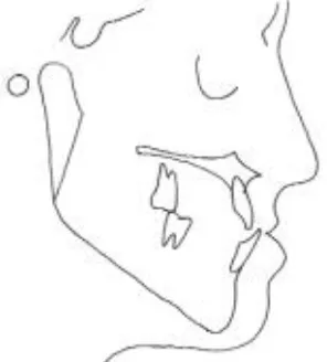
Mandibular prognathism is characterized by a reversed overjet that may arise from a maxillary
deficiency,a mandibular prognathism, or both (Fig.1).
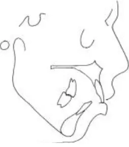
Mandibular prognathism with an open bite; this deformity is characterized by mandibular
prognathism,as described above,with both a reversed
overjet and an open bite that may be due to a maxillary deficiency, a mandibular prognathism, or both. The intraoral magnitude of the reversed overjet and open bite is important.In addition, the transverse relation of the arches is relevant in connection with the function of the tongue (Fig.2).
Mandibular deficiency; this kind of deformity is characterized by a mandibular retrognathism that presents with a convergent facial appearance, an increased overjet, or deep overbite (Fig.3).
Relative mandibular deficiency; this kind of deformity is characterized by a mandible of normal
size that is retruded secondary to a vertical maxillary hyperplasia (Fig. 4).
Absolute mandibular deficiency; this kind of deformity is characterized by absolute mandibular
deficiency,include a divergent facial appearance, an
increased sagittal overbite, and the tendency of an open bite,differsignificantly fromrelative mandibular deficiency. Therefore, the differentiation between
these two conditions must be made early in the classification and treatment plan. To distinguish this condition from relative mandibular deficiency, the lateral cephalostat film must be taken in the correct position with the condyles within the articular fossae and with the lips in repose (Fig. 5).
Statistical analysis
Statistical analyses were conducted with the Statistical Package for Social Sciences (SPSS) version 15.0. The distribution of ages according to gender and the distribution of deformities by gender and age in the Indonesian cohort were analyzed with Mann-Whitney tests. Chi-square tests were used analyzed the distribution of deformities by gender and age in the Dutch cohort.
RESULTS
The study population consisted of 86 Indonesian patients and 2,626 Dutch patients. The Indonesian cohort included 36 (41.9%) males and 50 (58.1%) females and the Dutch cohort included 1,003 (38.2%) males and 1,623 (61.8%) females.
Table 1 presents the age distribution according to gender among the Indonesian cohorts and Dutch cohorts. No significant differences could be found concerning the gender distribution. However, in the Dutch population, there were significantly more patients in the age group 31-40 years compared to the Indonesian population (p<0.001). The majority of the Indonesian cohort (93%) was under 30 years old, whereas only 61.5% of the Dutch cohort was under 30 years old.
The distribution of the prevalence of each deformity within the Indonesian and Dutch cohorts are reported in Table 2 and stratified by both gender and age aspect no significant (p>0.05). Mandibular Figure 2Characteristic deformities of
mandibular prognathism with an open bite
Figure 3Characteristic deformities of mandibular deficiency
Figure 4Characteristic deformities of relative mandibular deficiency
prognathism with an open bite deformity was the most prevalent deformity among the Indonesian cohort (46.5%) with a mean age of 22.78 (SD;6.34). Mandibular deficiency deformity was the least prevalent deformity among the Indonesian cohort (2.3%) with a mean age of 31.00 (SD;12.73). In contrast, mandibular deficiency deformity was the most prevalent deformity among the Dutch cohort (55.9%) with a mean age of 30.48 (SD;10.75) and mandibular prognathism with an open bite deformity was the least prevalent deformity (3.3%) with a mean age of 22.49 (SD;6.20) (Table 3). Compared to the Dutch population, the Indonesian population cohort consisted of significantly more mandibular prognathismwith an open bite deformity (p<0.01) and significantly less mandibular deficiency deformity (p<0.01).
DISCUSSION
The study of human dentofacial deformities is a fascinating area of a research not only from the
perspectives of surgical treatments, complications, and psychological aspects but also because of the anthropological implications. There are studies of anthropological aspects of disease, including the effects of gender,age, ethnicgroup, and morphology, provides important information for improving patient
care, treatment, and health care access.12-16
Modig et al17reported that functional problems
were the principal reason that patients seek surgical treatment for dentofacial deformities. In contrast,
Finlay et al18reported that facial appearance was a
more significant motivation for seeking surgical treatment (52%) compared with functional problems (31%). A similar result was reported in an Indian population, in which patients cited aesthetics as the
primary motivation for orthognathic surgery.19
Furthermore, a study by van Steenbergen et al20
reported that the majority of patients who seek orthognathic surgery are young and affluent.
Our comparison between Indonesia (developing nation) and the Netherlands as a developed nation
Table 2The distribution of deformities in the Indonesian cohort by gender and age
Deformities
A mandibular prognathism 32 37.2 12 (33.3) 20 (40) 21.03 (3.89)
0.430 B mandibular prognathism with an open bite 40 46.5 23 (63.9) 17 (34) 22.78 (6.34)
C mandibular deficiency 2 2.3 - 2 (4) 31.00 (12.73) D relative mandibular deficiency 6 7 1 (2.8) 5 (10) 24.83 (3.82) E absolute mandibular deficiency 6 7 - 6 (12) 21.67 (4.89) Mann Whitney test. P < 0.05 : significant
Table 1The distribution of ages according to gender among the Indonesian and Dutch cohorts
Range
Male Female Total Male Female Total
n % N % n % n % n % N %
Total 36 100 50 100 86 100 1003 100 1623 100 2626 100 Mann-Whitney test; p<0.05: significant
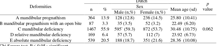
Table 3The distribution of deformities in the Dutch cohort by gender and age
Deformities
A mandibular prognathism 364 13.9 128 (12.8) 236 (14.5) 25.80 (10.41)
0.062 B mandibular prognathism with an open bite 87 3.3 35 (3.5) 52 (3.2) 22.49 (6.20)
shows differences in age and morphology in patients undergoing orthognathic surgery. Because the kind of surgery is categorized as an aesthetic treatment that is not covered by insurance, the majority of Indonesian patients who receive surgical treatment
are young,affluent,and/or report functional problems.
They have probably high socioeconomic status compared with the patients not seeking surgical treatment. However the present data we are not able to prove this issue.
The gender distribution between the Indonesian cohort and Dutch cohort were similar with a higher prevalence of female patients. These results are
similar to the results reported by Modig et al17 in
which 55% of the patients were female. Watanabe
et al8 found that mandibular prognathism in the
proband was reported more frequently in females (60 patients) compared with males (45 patients). A
similar study by Finlay et al18reported results from
a population with 61% female patients, of which 81% were single (19% married). A study by van
Steenbergen et al20also found that women comprised
the majority of respondents (75.9%). The gender disparity in thesestudies does not necessarily indicate that dentofacial deformities are more prevalent in females compared with males. These results may suggest a selection bias because women may be more concerned with aesthetics and health and may disproportionately seek solutions for their dentofacial
deformites compared with males.2,8
Although the gender distribution between the Indonesian and Dutch cohorts was similar, the age distribution was significantlyskewed toward younger individuals (under 30 years old) in the Indonesian cohort. The ages of the Dutch cohort were more evenly distributed between younger (under 30 years old) and older (over 30 years old) patients. The age ranges of the patients in these cohorts were similar
to the age ranges reported by Finlay et al18 who
reported an age range between 18 and 60 years old.
The age distribution in the Finlay et al18study was
also skewed such that 45% of the patients were less than 20 years old and 35% of the patients were between 21 and 30 years old. Furthermore, in that study, most young males reported seeking treatment due to functional problems, whereas most young females reported seeking treatment to improve their self-confidence. In another study, the older female patients reported an improvement in self-esteem following treatment, whereas older male patients reported no changes in either self-esteemor depression
following a surgical intervention.21
Previous studies1,6,7,9 have suggested that the
severity of dentofacial deformities could be predicted
by racial group or nationality. A critical finding of the present study was the differences in theprevalence of five defined dentofacial deformities between the Indonesian and Dutch cohorts. This study reported that mandibular prognathism with an open bite (deformity B) was the most prevalent deformity and mandibular deficiency with a normal or low mandibular plane angle (deformity C) was the least prevalent deformity in the Indonesian cohort. In contrast, mandibular deficiency with a normal or low mandibular plane angle was the most prevalent deformity and mandibular prognathism with an open bite was the least prevalent deformity in the Dutch cohort.
These results show a correlation with respect to the severity and type of dentofacial deformities between Southeast Asian and Caucasian populations.
A study byWatanabe et al8showed a high aggregation
of Class III mandibular prognathism in Japanese
families,whichsuggestsasignificantgeneticinfluence.
The normal lateral view of the facial structure among Southeast Asian individuals appears flat compared with the normal lateral view among Caucasian
individuals, which appears concave.1,22Chang et al1
compared the cranial base configuration on
cephalographs between European-American and
Taiwanese,Japanese,Korean and Chinese populations
and reported a relatively low prevalence of Class III malocclusions among Caucasians compared with a
higher prevalence among Asians. Khadka et al3
reported a square face with a wide mandible and/or a wide and prominent zygoma among East Asian
populations. Ishii et al23 reported that a Class II
Division 1 maxillary protrusion in Japanese girls mayrepresent a vertical problem due to the backward rotation of the mandible. In contrast, a Class II Division 1 maxillary protrusion in Caucasians may indicate a horizontal problem due to the anterior
position of the maxilla. Furthermore, Ayoub et al24
reported that there are significant differences within each subgroup of the same race. For example, the craniodentofacial size of the Chinese in Taiwan is significantly larger in males compared with females but their craniodentofacial morphology is similar.
According to, Rauso et al25surgical treatment results
in the widening of the alar base, which is found to occur almost universally following maxillary osteotomies. Widening of the alar baseis an important consideration for Asian patients because their noses are wider and flatter than the noses of Europeans
and Americans.26,27
the results could not be stratified over the same age ranges between the two cohorts. In Indonesia, there are four university hospitals with a Department of Oral and Maxillofacial Surgery. Each of these university hospitals treats patients with dentofacial deformities; however, the number of treated patients are not equally distributed among the hospitals. Most patients are treated at the Bandung university
hospital,which has been offeringorthognathic surgery
since 1984. Therefore, our results may not be generalizable to the whole population of Indonesia. Because this study was retrospective, the analysis
may include an information bias.However,the results
presented in this study are similar to the reports from other studies. Furthermore, the analysis in this report provides important data for improving the treatment plans for orthognathic surgery.
It is concluded that in the Indonesian cohort, young patients seek orthognathic surgery most frequently to correct a functional problem associated with mandibular prognathism with an open bite. These results may reflect the greatest severity of dentofacial deformities that are observed in Southeast Asian patients.
REFERENCES
1. Ayoub F, Rizk A, Yehya M, Cassia A, Chartouni S, Atiyeh F, et al: Sexual dimorphism of mandibular angle in a Lebanese sample. J Forensic Leg Med 2009; 16:121-4.
2. Bell WH. Modern practice in orthognathic and reconstructive surgery. Vol. 3. Philadelphia: Saunders, 1992:1979-2110
3. Boeck EM, Lunardi N, Pinto AS, Pizzol KE, Boeck NRJ. Occurrence os skeletal malocclusions in Brazilian patients with dentofacial deformities. Braz Dent J 2011; 22:340-45.
4. Chang HP, Tseng YC, Chang HF. Treatment of mandibular prognathism. J Formos Med Assoc 2006;105:781-790 5. Chung C, Lee Y, Park KH, et al. Nasal changes after surgical correction of Skeletal Class III malocclusion in
Koreans. Angle orthod 2008;78:427-432
6. Epker BN, Wolford LM. Dentofacial deformities; surgical-orthodontic correction. St Louis: Mosby, 1980:41-93 7. Farkas LG, Katic MJ, Forrest CR. Comparison of craniofacial measurements of young adult African-American and
North American white males and females. Ann Plast Surg 2007;59:692-698
8. Finlay PM, Atkinson JM, Moos KF. Orthognathic surgery: patient expectations; psychological profile and satisfaction with outcome. Br J Oral Maxillofac Surg 1995;33:9-14
9. Flint M. Anthropological perspectives of the menopause and middle age. Maturitas 1982;4:173-180
10. Ishii N, Deguchi T, Hunt NP. Morphological differences in the craniofacial structure between Japanese and Caucasian girls with Class II devision 1 malocclusion. Eur J Orthod 2002;24:61-67.
11. Khadka A, Liu Y, Li J, Zhu S, Luo E, Feng G, Hu J. Changes in quality of life after orthognathic surgery: a comparison based on the involvement of the occlusion. Oral Surg Oral Med Oral Pathol Oral Radiol Endod 2011; 112:719-25.
12. Kitagawa Y, Manabe Y, Oyamada J, Rokutanda A. Morphological deciduous lower first molar teeth.Arch Oral Biol1996; 41:387-91.
13. Lee LH, Jun JH, Danganan M, Pogrel MA, Kushner H, Lee JS. Orthognathic surgery for the Asian patient and the influence of the surgeon’s background on treatment. Int J Oral Maxillofac Surg 2011; 40:458-63.
14. Manabe Y, Oyamada J, Kitagawa Y, Rokutanda A, Kato K, Matsushita T. Dental morphology of the Dawenku Neolithic population in North Cina; implication for the origin and distribution of sinodonty. J Hum Evol 2003; 45:369-80.
15. Milella M, Belcastro MG, Zollikofer CPE, Mariotti V. The effect of age, sex, and physical activity on entheseal morphology in a contemporary Italian skeletal collection. Am J Phys Anthropol 2012; 148:379-88.
16. Modig M, Andersson L, Wardh I. Patients’ perception of improvement after orthognathic surgery: pilot study. Br J Oral Maxillofac Surg 2006;44:24-27
17. Naffah, J. Dermatoglyphic analysis: anthropological and medical aspects. Bull N Y Acad Med 1977;53:681-692 18. Narayanan V, Guhan S, Sreekumar K, et al. Self-assessment of facial form oral function and psychosocial function
before and after orthognathic surgery: a retrospective study. Indian J Dent Res 2008;19:12-16
19. Nicodemo D, Pereira MD, Ferreira LM. Effect of orthognathic surgery for class III correction on quality of life as measured by SF-36. Int J Oral Maxillofac Surg 2008;37:131-134
20. Rauso R, Gherardini G, Santillo V, et al. Comparison of two techniques of cinch suturing to avoid widening of the base of the nose after Le Fort I osteotomy. Br J Oral Maxillofac Surg 2010;48:356-359
21. Singh GD, McNamara JA Jr, Lozanoff S. Finite-element morphometry of soft tissues in prepubertal Korean and European-Americans with Class III malocclusions. Arch Oral Biol 1999;44:429-436
22. Tang EL. The prevalence of malocclusion amongst Hong Kong male dental students. Br J Orthod 1994;21:57-63 23. Tuinzing DB, Greebe RB, Dorenbos J, Becking AG. Surgical orthodonthics: classification, diagnosis and treatment.
Maarssen: Elsevier; 2005. p.23-33.
Amsterdam: VU Uitgeverij; 1990. p.37-67.
25. Van Steenbergen E, litt MD, Nanda R. Presurgical satisfaction with facial appearance in orthognathic surgery patients. Am J Orthod Dentofacial Orthop 1996;109:653-659
26. Van Vuuren C. A review of the literature on the prevalence of class III malocclusion and mandibular prognathic growth hypotheses. Aust Orthod J 1991;12:23-28