Abstract. Objective: To present surgical treatment options, meniscal suturing techniques and surgery results. Materials and methods:Over a period from September 2008 till August 2014, we performed 203 meniscal suturing procedures in our hospital. We used outside-in, inside-out and all-inside repair techniques (Rapid Loc and FasT-Fix). In the research conducted in our hos-pital, we prospectively monitored 175 patients who were 27 (14-53) years old, 120 male (69 %) and 55 female (31 %). Of these 175 patients, 88 (50.3 %) participated in recreational sports and 42 (24 %) were actively engaged in sports. Almost 50 percent (87 patients) also had anterior cruciate ligament tear. We monitored the patients for 36 (7-66) months through regular medi-cal examinations, and with Lysholm and Tegner functional tests before and after surgeries.
Re-sults: Before surgery, the Lysholm score showed the average result of 59, which increased to 92 after surgery. The Tegner questionnaire value was 6.3 before surgery, and 5.4 after it on aver-age. Out of 135 patients who were active in sports before their surgeries, 68 (50 %) returned to the same activity level. Subsequent arthroscopies were performed on 27 patients (15.4 %), and 15 of those patients (56 %) had new knee injuries after meniscal repair. Meniscectomy had to be done on 24 patients and meniscus was to be fixed by one Omni span system on two pa-tients. There were 84.6 % of patients with no complications. Conclusions: Meniscal repair is a procedure that allows healing of some torn menisci and thus prevents the occurrence of early osteoarthritis. Meniscal repair shows very good results, especially if the suturing is performed along with the anterior cruciate ligament reconstruction.
Key words: anterior cruciate ligament reconstruction; knee; meniscus
Sažetak. Cilj:Prikazati mogućnosti kirurškog liječenja, operacijske tehnike šivanja meniska i re-zultate operacijskog liječenja. Materijali i metode: U našoj bolnici u razdoblju od rujna 2008. do kolovoza 2014. godine u 203 pacijenta učinjeno je šivanje meniska. Koristili smo se tehnikama šivanja out-in, in-out te all-in (RapidLoc i FasT-Fix). U istraživanju koje je provedeno u našoj bol-nici prospektivno smo pratili 175 pacijenata prosječne životne dobi 27 godina (u rasponu od 14 do 52 godine), od toga 120 muškaraca (69 %) i 55 žena (31 %). Sportom se rekreativno bavilo 88 pacijenata (50,3 %), a aktivno 42 pacijenta (24 %). Ruptura prednje ukrižene sveze bila je prisut-na kod 87 pacijeprisut-nata (50 %). Pacijente smo pratili 36 mjeseci (7 – 66), praćeni su prospektivno redovitim ambulantnim kontrolama, a funkcionalno testiranje pomoću Lysholm i Tegner upitni-ka izvršili smo prije i nakon operacijskog zahvata. Rezultati: Lysholm upitnik je prije operacije iznosio u prosjeku 59, a nakon operacije 92. Tegner upitnik je prije operacije u prosjeku iznosio 6,3, a nakon operacije u prosjeku 5,4. Od 135 pacijenata koji su se prijeoperacijski bavili spor-tom, njih 68 (50 %) vratilo se potpuno istoj razini aktivnosti u sportu. Kod 27 pacijenata (15,4 %) učinjena je naknadna artroskopija. Od tih 27 pacijenata, njih 15 (56 %) imalo je novu povredu koljena nakon šivanja meniska. U 24 pacijenta učinjena je meniscektomija, a u dva pacijenta učinjena je ponovna fiksacija meniska sa jednom Omni span kopčom. Postotak pacijenata bez komplikacija iznosio je 84,6 %. Zaključci:Šivanje meniska je postupak koji omogućuje izlječenje kod određenih ruptura meniska koljenskog zgloba i na taj način sprječava nastanak ranog osteo-artritisa. Šivanje meniska pokazuje izrazito dobre rezultate, osobito ukoliko se učini zajedno s rekonstrukcijom prednje ukrižene sveze.
Ključne riječi: koljeno; menisk; rekonstrukcija prednjeg ukriženog ligamenta
*Corresponding author:
Denis Tršek, MD
AKROMION, Hospital for Orthopeadic Surgery
Ljudevita Gaja 2, 49 217 Krapinske Toplice, Croatia
e-mail: [email protected] AKROMION, Hospital for Orthopeadic Surgery, Krapinske Toplice, Croatia
Received: 31.07.2014 Accepted: 3.11.2014
INTRODUCTION
Its semilunar shape and specific fibrocartilagenous structure enables the meniscus to be a multifunc-tional part of the knee joint. An intact meniscus is of crucial importance for cartilage preservation and knee function, namely, prevention or slowing down of osteoarthritis. The aim of this study is to present the basics of anatomy and histology of the meniscus of the knee joint, its biomechanical prin-ciples, surgical treatment options, meniscal sutur-ing techniques and surgery results.
Meniscus – anatomical and functional characteristics
The knee menisci are two elastic, semilunar-shaped, fibrocartilagenous structures which pro-vide conformity of the articular surfaces of the femur and the tibia. The menisci are concave on the top, which allows their smooth articulation with convex femoral condyles, and flat on the bottom, which corresponds to the flat surface of the tibia. The anterior and posterior horns of the meniscus are attached to the tibial plateau. The external rim of the meniscus is thick and linked to the articular capsule, whilst it grows thinner to its midpart and forms a thin free edge, which is why the menisci are wedge-shaped in their cross section1-6. The peripheral part of each meniscus is attached by coronary ligaments to the superior surface of the tibia. The medial meniscus has a firm attachment to the deep portion of the medi-al collatermedi-al ligament, which limits its mobility compared to the lateral meniscus. This limited mobility of the medial meniscus results in its more frequent injuries. Morphologically, the lat-eral meniscus varies more in its size, thickness, shape and mobility. The posterior horn of the lat-eral meniscus is attached to the femur by front (Humphrey) and back (Wrisberg) meniscofemoral ligaments3. The anterior horns of the medial and lateral menisci are intertwined by the transverse ligament. All these meniscal links prevent its ex-trusion under load.
The menisci are composed of 70 % water and 30 % organic matter. This organic matter mostly consists of fiber (75 %) which is mainly type I col-lagen, as distinct from the cartilage, which is
composed of type II collagen2,3,7. The collagen bundles of the surface layer are radial; in the in-ner part they are transverse or circular, while in the peripheral part they form a network. Such orientation of the collagen fibers together with their waviness and cross-striation makes the me-nisci strong and elastic1,2.
The meniscus is vascularized by the upper and lower branches of the genicular artery which form a perimeniscal capillary plexus. It is entirely
Meniscus tear is the most frequent injury and accounts
for up to 75 % of knee joint intraarticular injuries.
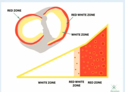
Figure 1. Meniscus vascularization zones
vascularized at birth, till the age of ten, vasculari-zation decreases to 10-25 % of in the lateral me-niscus periphery, and 10-30 % of in the medial meniscus periphery. Thus, Arnoczky8 divides the meniscus into three zones: the red (or Henschen pericapsular) peripheral vascularized meniscus zone, the red-white transitional zone, and the white inner avascular meniscus portion, nutrition of which is performed by synovial nutrients diffu-sion only1,2,3,9 (Figure 1).
Alongside with the blood vessels in the knee joint, and consequently in the meniscal periph-ery, there are branches of posterior tibial, femo-ral and obturator nerves, which together with the three types of mechanoreceptors (Ruffini,
Pa-cini and Golgi) perform the proprioceptive role of the meniscus3.
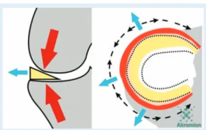
The semilunar shape and the specific fibrocartila-genous structure enable the meniscus to be a multifunctional part of the knee joint. The me-nisci can stretch radially, which allows equal dis-tribution of load and absorption of pressure (Fig-ure 2).
They disperse pressure onto the whole femorotibi-al joint surface, absorb shocks, facilitate joint movement, improve lubrication of joint parts and consequently also the cartilage nutrition, prevent
knee joint by preventing tibial plateau frontal dis-location11 and are crucial for decreasing the pres-sure on the condyle cartilage10.
Therefore, the menisci slow down the occurrence of osteoarthritis, which develops faster in an un-stable knee joint after meniscectomy3,9,11,12 . Medial meniscus removal results in reduction of the contact surface by 50 to 70 %, and a load in-crease at the place of contact by 100 %. Total lat-eral meniscectomy cuts the contact surface by 40 to 50 % and leads to twice or three times greater pressure at the contact place (Figure 3).
Partial meniscectomy at the rate of only 10 % of the meniscal surface raises pressure at the con-tact place by 65 %. The pressure rise also de-pends on the type of meniscectomy. After menis-cectomy along the whole length, the load on the cartilage is bigger than in cases when only the anterior or posterior horn was removed in equal percentage13.
Therefore, it is obvious that an intact meniscus is of crucial importance for cartilage preservation and knee function, namely, prevention or slowing down of osteoarthritis2,3,13,14.
Meniscal tear is one of the most frequent knee injuries. It accounts for almost 75 % of intraartic-ular knee pathology15,16. There are 60 to 70 me-niscal injuries per 100,000 knees annually, men suffering meniscal injuries four times as often as women. Vertical tears are the most frequent, es-pecially in patients in their forties, while horizon-tal tears are more frequent in their fifties17. It is proved that excessive weight and knee varus in-crease the risk of medial meniscus tear18. A meniscus tear is a result of degeneration, inju-ry, or a combination of both. The medial menis-cus, and in particular its posterior horn, suffers four times more often than the lateral5. A menis-cus tear may be a sole injury or appear together with other knee traumas (e.g., unhappy triad of the knee: injuries of the medial collateral
liga-Figure 2. Radial meniscal expansibility
Figure 3. Contact surface reduction and load increase on the place of contact between two condyles after meniscectomy. The knee joint before (a) and after (b) meniscectomy.
ment, the medial meniscus and the anterior cru-ciate ligament). A posterior horn tear often oc-curs with an anterior cruciate ligament rupture, while flap tears are usually linked to condyle de-generative changes17.
Meniscus tear treatment
Although total meniscectomy up to the vascular-ized area had been the only treatment of torn meniscus for decades, it was realized that the menisci do not restore but only heal and may grow up to one third of their normal volume, which is not enough for normal meniscus func-tion1,19,20 (Figure 4).
All this leads to early degenerative changes in the knee joint. For that reason, meniscectomy should be avoided whenever possible, and if not, then only the minimum necessary part of the menis-cus should be removed.
Meniscus suture
Torn meniscus treatment calls for special individ-ual approach. There are several factors that must be taken into consideration before deciding on meniscus suturing: subjective complaints, the pa-tient’s activity level, their age, expectations and possible accident injuries.
The first open meniscus repair was performed by Thomas Annandale in 188321,22.
However, meniscus suture was not popular till late 1970s. DeHaven23 popularized open menis-cus repair as an alternative to meniscectomy. Development of arthroscopic techniques and other technical prerequisites has enabled a mini-mum invasive treatment of a torn meniscus. The first arthroscopic suture was performed by Hiro-sho Ikeuchi in 1969. Over time, this method has become a widely accepted way of treatment11,24. Unfortunately, not every meniscal tear is suitable for suturing. It is crucial to understand meniscus vascularization for its repair. As the blood supply is a key factor for meniscus healing, it is evident that the red zone is the best for suture (Figure 5), while the red-white zone is less suitable. Despite this fact, Kurzweil et al.25 found reports of 98 hor-izontal tears, 78 % of which healed even though they included the white avascular meniscus zone as well. This result is almost identical to that of
Figure 4. After total meniscectomy, the meniscus can grow up to one third of its volume only
Figure 5. Recent meniscal tear in the red zone
Figure 6. Longitudinal tear in the vascularized zone at the meniscocapsular junction is the most suitable for suturing.
meniscus sutures in the red and red-white zones26.
The type of tear is also important for a decision about suturing. Longitudinal 1-2 cm long tears in the vascularized zone and tears on the menisco-capsular junction are the most suitable for repair (Figure 6). Longer longitudinal tears of a bucket-handle shape also may be sutured and very suc-cessfully fixed. Indications are questionable in case of horizontal tears, flap tears and various
degenerative tears. The quality of the meniscus itself is also important, as it must not be torn apart or degeneratively changed (Figure 7). Younger patients (i.e., younger than 40 years of age, but even younger than 50 according to some authors) with recent tear (less than two months) are more eligible for meniscus suturing. Anterior cruciate ligament reconstruction during the same surgery is highly recommended.
In spite of the fact that knee stability is a pre-con-dition for meniscus repair, some authors leave the possibility of meniscus suture in patients with an anterior cruciate ligament rupture without its reconstruction15,27.
Kimura et al.28 showed that in case of a lateral meniscus tear in the popliteal tendon zone, it is better to perform subtotal meniscectomy or me-niscus suture rather than partial meniscectomy. The reason is that partial meniscectomy increas-es lateral meniscus mobility, thus increasing knee instability and internal knee structures friction. Surgical meniscus suture techniques
There are three basic meniscus suture techniques – outside-in, inside-out and all-inside. Each of them has its advantages and disadvantages. It is useful to be able to perform several surgical tech-niques and be equipped for them, as they
fre-quently combine. The same technique cannot be used for all parts of the meniscus, hence all three techniques are usually used during longitudinal tear repair which involves the posterior horn, the body and the anterior meniscus horn (bucket han-dle). It is recommended to use the outside-in tech-nique for the anterior horn suture, the inside-out for the meniscus body, and the all-inside one for the posterior horn.
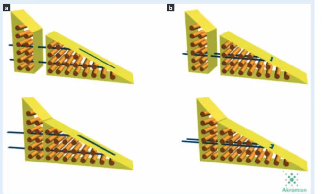
Because of the horizontal collagen fibers inside the meniscus, vertical stitches are more durable than horizontal ones and they are today’s gold standard in the meniscus repair practice29 (Fig-ure 8). It is important to refresh the tear edge right before suturing to open blood vessels and enable blood supply of the sutured part, as the blood contains growth factors, fibrin and throm-bocytes, which is necessary for tear healing. This is performed by using a rasp or powered in-strument, and the other possibility is meniscal percutaneous trephination with a needle. In case of isolated meniscus tears, it is recom-mended to place a fibrin clot inside the tear it-self. A fibrin clot is prepared from the patient’s peripheral blood3,4,11,24,30.
Formerly, resorbable suture materials (PDS) were used in order not to damage the joint parts, but they proved to resolve too fast to ensure tear
Figure 7. Meniscus tears not suitable for suture: a) transverse tear; b) flap tear; c) cross tear; d) horizontal tear; e) degenerative tear; f) combined tear
healing. Organism reaction to resorbable sutures may result in subacute seroma. That is why non-resorbable suturing materials have been used more frequently recently, as they remain in the sutured meniscus and additionally strengthen it. The rule is to put a stitch at every 5 mm of the tear24,30. Tests have shown that stitches are more durable than meniscus suture implants31.
Outside-in technique
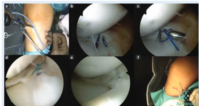
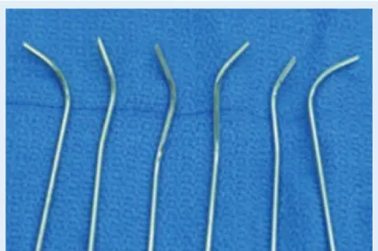
This surgical technique was introduced by War-ren, Morgan and Casscalles4. It is suitable for pos-terior horn suturing of both menisci and is slight-ly less suitable for the meniscus body repair. Furthermore, it is the cheapest and best available technique due to the fact that it may be per-formed with two regular injection needles 1.2 mm in diameter (Figure 9). The meniscus is fixed in two points, which allows us to place both verti-cal and horizontal stitches at our option.
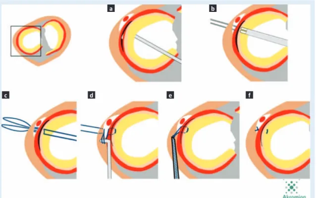
Under arthroscopy control, the first needle (Fig-ures 10a and 11a) together with suturing materi-al is inserted percutaneously throughout the me-niscus tear, and next to it another needle with suturing material is placed (Figures 10b and 11b). Using the instruments, the first suture is
broached through the loop of the other suture (Figures 10c and 11c). The needle with the loop and the end of the other suture inside the loop is pulled outside the knee (Figures 10d and 11d). This is the way to form a stitch which may be placed horizontally, vertically or on the cross (Fig-ures 10e and 11e). After slight skin incision, the subcutaneous tissue is prosected, the suture thrums are taken out by a probe and tied, and
Figure 8. The vertical stitch, due to its position, embraces collagen fibers bundles, whilst the horizontal stitch is placed abreast with collagen fibers layers. Therefore, the vertical stitch is more durable.
Figure 9. Two ordinary injection needles 1.2 mm in diameter used for the outside-in suture technique.
Figure 10. The outside-in suture technique with two needles.
Figure 11. A schematic drawing of the outside-in suture technique with two needles.
the knot is placed subcutaneously through the joint capsule (Figures 10f and 11f) 24,30,32.
The disadvantage of this technique is that dur-ing suturdur-ing of the meniscal anterior parts, the neurovascular bundle in the popliteal space may be damaged, especially the peroneal nerve
dur-ing lateral meniscus suturdur-ing, and the saphen-ous nerve during medial meniscus repair. This is why it is important to observe the rule of not placing of subcutaneous stitches behind the back edge of the medial and lateral collateral ligaments3,4,11,24,30.
d e f
a b
Inside-out technique
This technique was introduced by Henning in 198024. The inside-out technique needs special surgical instruments. The authors use instru-ments by Smith & Nephew, which contain single and double cannulas for needle introduction, usually paired (Figure 12).
A cannula may be modeled at different angles as needed, which is the advantage over other sutur-ing techniques. Cannula modelsutur-ing allows us to adjust to the anatomy of every knee.
After the cannula is placed under arthroscopy control onto the place where the stitch is to be put (Figures 13a and 14a), both needles are alter-nately stretched through the meniscus (Figures 13b and 14b), the subdermis and the skin outside the joint (Figures 13c and 14c). After that, the su-ture material which is inserted into both needles’ eyes is stretched through the meniscus, the sub-dermis and the skin (Figures 13d and 14d). By do-ing so, the suture thrums stay outside the knee and the knot is tied on the outer side of the cap-sule subcutaneously after a slight skin incision (Figures 13e and 14e).
This technique is suitable for meniscus body re-pair. The meniscus is fixed in two points and the stitches may also be placed vertically, horizontal-ly or on the cross (Figures 13f and 14f).
Just as with the outside-in technique, the appli-cation of this technique for the posterior horn with subcutaneous suturing may result in saphe-nous and peroneal nerves injuries.
In case of a lateral meniscus tear in the popliteal tendon zone and its suturing with the inside-out technique, we cannot place subcutaneous stitches and thus fix lateral meniscus onto the joint capsule or the popliteal tendon due to the specific anato-my and mobility of the lateral meniscus. There-fore, we are to modify the inside-out technique
Figure 12. Adjustable single and double cannulas which direct and lead needles.
Figure 13. The inside-out suture technique
a b c
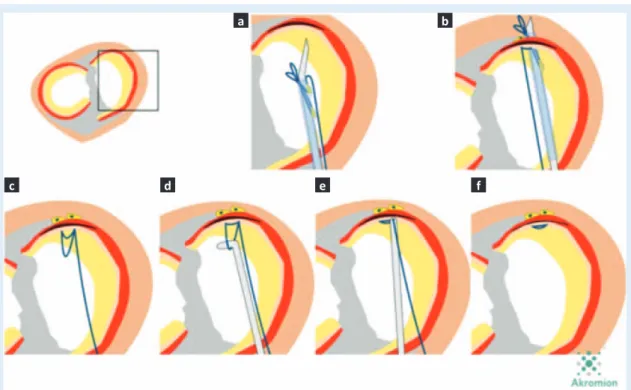
and place the stitch in front of the popliteal ten-don, between the meniscus and the capsule. In the group of pictures numbered with 15, a lon-gitudinal tear of the lateral meniscus in front of the popliteal tendon can be seen (Figures 15a and 16a). Using a modular cannula, we define the place where we want to place the stitches (Figures 15b and 16b). The thrums of both
su-tures are broached through the meniscus, the subdermis and the skin and pulled out of the knee (Figures 15c and 16c). Hereupon we find both thrums between the meniscus and the cap-sule, and bring them back to the joint. After-wards, both sutures are taken out through the lateral portal (Figures 15d and 16d). Finally, by placing a slide knot behind the meniscus (but in
Figure 14. A schematic drawing of the inside-out suture technique
Figure 15. A modified inside-out technique for fixation of a lateral meniscus tear in front of the popliteal tendon
c
d e f
a b c
front of the popliteal tendon) (Figures 15e and 16e), we fix the meniscus tear and leave the lat-eral meniscus mobile (Figures 15f and 16f).
All-inside technique
In order to simplify meniscus repair and reduce the risk of avascular damage, in the first genera-tion of suturing using the all-inside technique Morgan12,24 introduced intraarticular meniscus repair in 1991. This technique consists of placing the stitches intraarticularly through the meniscus tear by using protective curved hooks inserted into the popliteal space. This technique is a com-plex one and needs additional posteromedial and posterolateral portals along with a vast experi-ence of the surgeon.
Later on, special instruments were developed for placing implants through the tear and fixing them with arthroscopic knots. The advantage was that the stitches could be placed by using standard anterior arthroscopic portals only, with minimal risk of damaging the neurovascular structures. The disadvantage of such implants lay in the ne-cessity of placing arthroscopic knots, which in-creased the possibility of hondral damage and
rendered the knots tightening after their placing impossible.
The third generation consists of various resorba-ble implants, arrows, screws and braces (Figure 17). These were durable rigid implants which were placed through the tear itself and stabilized the damaged meniscus. Because of resorbable implants and suture materials, frequent compli-cations after this meniscus fixation technique oc-curred. Frequent sinovitis, inflammatory process-es and cysts onset were registered12. The majority of complications, like migration of implants and
Figure 16. A schematic drawing of a modified inside-out suture technique for fixation of a lateral meniscus tear in front of the popliteal tendon.
Figure 17. The third generation of resorbable material for fixation of a torn meniscus (all-inside repairs) (Courtesy of De Puy Mitek,
Johnson&Johnson)
a b
cartilage damage, occurred when applying re-sorbable arrows33,34.
Such complications and the fact that implants of the third generation could not provide the nec-essary durability at the very place of tear fixa-tion caused the development of implants which are widely used nowadays. These are adjustable implants which are based on stitches, have a
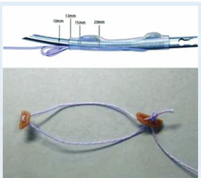
hospital are RapidLoc and Omni span (DePuy Mitek) and FasT Fix (Smith & Nephew). Omni span consists of two anchors linked be a non- resorbable suture material with a slide knot on it (Figure 18).
By using the inserter, the first anchor is placed under the tear (Figures 19a and 20a). The second anchor is placed 5 mm from the first one (Figures 19b and 20b). By pulling the suture, we drag and tighten the slide knot onto the meniscus (Figures 19c and 20c) along with preventing the suture from tangling by a probe (Figures 19d and 20d). Finally, the knot is tightened (Figures 19e and 20e) and the suture is cut (Figures 19f and 20f). The advantage of this implant is the double-point fixation, which allows placing stitches both hori-zontally and vertically.
RapidLoc (Figure 21) enables easier placing as it consists of one anchor with the suture linked with a top hat. After the anchor is placed, the pusher is used to place the top hat onto the inner part of the meniscus and the previously tied slide knot is
Figure 18. The Omni span meniscal repair system (Courtesy of De Puy Mitek, Johnson&Johnson)
Figure 19. The all-inside meniscus suture technique with the Omni span system. Placing of two 5 mm anchors. Tightening of a sliding stitch between two anchors.
a b c
tightened. The disadvantage of RapidLoc instru-ments is the single-point fixation (Figure 22). The all-inside technique is used for suturing pos-terior horns tears of both menisci as it reduces the possibility of popliteal neurovascular struc-ture damage. These implants are not suitable for repairs on the meniscocapsular junction place (ramp tear) as they demand intact rare edge of the torn meniscus for its anchorage3,4,12,24,30. In the group of pictures numbered with 23, one can see an example of a successfully repaired bucket handle tear (Figure 23a). For successful meniscal suture all three techniques were used
Figure 20. A schematic drawing of the all-inside suture technique. Placing of two anchors at a 5 mm distance. Slide stitch tightening between two anchors.
Figure 21. The RapidLoc meniscal repair system (Courtesy of De Puy Mitek, Johnson&Johnson)
Young patients’ longitudinal meniscal tears and recent
tears in the meniscus peripheral vascularized red zone
are the most suitable for suturing, especially if
per-formed along with the anterior cruciate ligament
re-construction.
Figure 22. Meniscus fixation with the RapidLoc system
a b
Figure 23. Successful usage of all three suture techniques and medial meniscus bucket handle tear healing ten months from the surgery (f).
Figure 24. a) An example of poor RapidLoc system positioning; b) Loose body after the backstop breaks; c) The anchor falling out of an Omni Span inserter before pulling through the meniscus.
Figure 25. a) Loosening of a slide knot on the RapidLoc system; b) Pulling out of the anchor in the Omni span system through the meniscus after placing both anchors; c) Loosening of a subcutaneous knot at an in-out stitch during tightening and suture cutting.
d e f
a b c
Complications of meniscal repair
Speaking about complications, injuries of saphe-nous and peroneal nerves, arthrofibrosis, septic arthritis, reaction to foreign matter and cartilage damage are to be emphasized. Some of the pos-sible complications include a too-low placed im-plant (Figure 24a), break of a plastic RapidLoc backstop (Figure 24b), the anchor falling out of an Omni span inserter (Figure 24c), slide knot loosening on a RapidLoc system (Figure 25a), de-tachment of an anchor of the Omni span system after both anchors were placed (Figure 25b), and loosening of a subcutaneous knot of an inside-out stitch during tightening and cutting of a su-ture (Figure 25c).
Many studies have shown that reruptures of a sutured meniscus frequently appear two years after the suturing.
Rehabilitation
There are significant differences between reha-bilitation after meniscectomy and rehareha-bilitation after meniscal repair. Rehabilitation after menis-cectomy is fast, usually almost painless and, if there are no additional joint injuries, the patients go back to their work and sports activity level 3 to 4 weeks after the surgery.
Rehabilitation after meniscus suture lasts longer, complaints are bitter, it continues for up to three months or even up to six months for sports which involve fast knee rotations. Unfortunately, this is the reason why professional sportsmen do not opt for this treatment method.
According to our postsurgical protocol, rehabilita-tion consists of flexion limitarehabilita-tion and knee rota-tion prevenrota-tion. Mobility is limited to 0/30˚ by an orthesis during the first three weeks, and to 0/90˚ after that. Four weeks later, full gradual flexion is allowed with a prohibition of squats and kneeling. Full weight bearing is possible right after the sur-gery. Running with no abrupt turns can be prac-ticed after three months, and contact sports with rotation are allowed six months after the surgery. Another rehabilitation possibility is using crutch-es during six weeks without an orthcrutch-esis. The sub-sequent procedure is the same.
In case the anterior cruciate ligament is recon-structed along with meniscus suture, the fast
re-habilitation procedure after the cruciate ligament reconstruction is practiced, and orthesis is not needed3,4.
Meniscal transplantation
Due to the fact that the importance of the menis-cus for the knee joint is significant, new surgical techniques are designed to replace the damaged tissue. In case of total meniscectomy, meniscal transplantation is imposed24. This is performed with a deep-frozen or a fresh allograft.
There are three basic meniscus suturing techniques.
The same technique cannot be used on every part of
the meniscus. It is recommended to perform the
out-side-in suture for the anterior horn and the inside-out
technique for the meniscus body, while the posterior
horn is mostly repaired by the all-inside technique.
A synthetic meniscus may also be used instead of an allograft, the indications for its use being simi-lar35.
A meniscal transplantation candidate is a patient with a first pain after total meniscectomy, young-er than 50 years old, without any obvious arthrit-ic changes evident on knee X-rays, with a mini-mum of 2 mm space between the tibia and the femur on the X-ray in 45-degree flexion, with a normal knee axis, and minimal defects of the car-tilage of the tibia and the femur.
Counterindications include advanced knee ar-throsis with femoral condyle attrition, or en-hanced tibial condyle concavity with edge osteo-phytes, all of which may be prevented if the meniscus is normally positioned in its place, and also injury of the anterior cruciate ligament, var-us or valgvar-us of knee joint, excessive weight, or body mass index greater than 30. That is why it is crucial to take care of all other diseases and inju-ries (cruciate ligament reconstruction, physiologi-cal axis securing).
Stone et al.36 performed meniscal transplantation together with cartilage reparation and achieved an average time of durability of 9.9 years with significant pain reduction and knee function im-provement, especially during the first two years after the transplantation. The meniscus can be
During the same period of time, 203 meniscal re-pairs were performed, which accounts for 9.9 % of all knee arthroscopies or 15.4 % of all meniscus tears. We used the outside-in technique for ante-rior horn suturing, the inside-out technique for the meniscus body and part of the posterior horn, and the all-inside technique for posterior horn su-turing, all with RapidLoc and FasT-Fix. We placed on average 2.8 stitches on every tear. During this study we questioned 175 available patients (Table 1). Out of them 120 were male and 55 female. The average age of the patients was 27 years (range, 14 to 53 years). There were 135 patients that prac-ticed sports, 88 (50.3 %) of them recreatively and 42 (31 %) were active sportsmen. Five patients were professional sportsmen and opted for menis-cus suture despite the prolonged rehabilitation. Out of these sports-active patients, 65 played soc-cer, 19 went in for basketball, 18 preferred skiing, 9 wrestling and 7 practiced handball. The
remain-performed suturing of both menisci. There were 44 bucket handle tears on the medial meniscus, and 10 bucket handle tears on the lateral menis-cus. An anterior cruciate ligament rupture was found in 87 (50 %) patients and all of them under-went its reconstruction. We monitored the pa-tients for 36 (7-66) months on average. Approxi-mately seven months (5 days to 36 months) had passed from the moment of injury till surgery.
RESULTS
The Lysholm score showed 59 before the surgery and 92 after it on average (Figure 26). According to it, before the surgery 3 patients (2 %) had a perfect result, 14 (8 %) had a good result, 58 (33 %) had a satisfying result and 100 (57 %) had a poor result. According to the Lysohlm scale, af-ter the surgery 111 (63 %) patients had a perfect result, 42 (24 %) good, 16 (9 %) satisfactory, and 6 (4 %) patients had a poor result.
Table 1. Overview of our results after meniscal tear suturing
№ of repairs 175
Age / years (range) 26 (14-53)
Gender (№ M/№ F) 120/55
Follow up / months (range) 36 (7-66)
Laterality (№ medial/№ lateral / № both) 155/22/2
Bucket handle (№ medial / № lateral) (44/10)
ACL reconstruction (№) 87
Sport before injury (№ active / № recreational / № professional) (88/42/5)
№ Type of sport football 65 basketball 19 skiing 18 karate/judo 9 handball 7 other 17
Time between injury and repair / days (range) 2013 (5-1098)
Time between repair and rerupture (months) 21
Reruptures (№ meniscectomy / № refixation) (24/2)
Stitches removal 1
The Tegner questionnaire showed 6.3 before sur-gery and 5.4 after it on average (Figure 27). By adhering to all the above mentioned indica-tions, our study showed that in practice only 203 out of 1321 meniscal tears (15.4 %) were suitable for suturing.
Out of the 135 sports-active patients, 68 (50 %) returned to the same activity level. Thirty three of these patients (48 %) also underwent the an-terior cruciate ligament reconstruction together with meniscus repair, and in 35 (52 %) only the meniscal repair was performed.
Additional arthroscopy was performed on 27 (15.4 %) patients, 15 (55 %) of them had a new knee injury after the meniscus repair. Meniscec-tomy was made on 24 (89 %) patients and two patients underwent recurrent meniscus fixation with one Omni span system because there was only a part of the meniscus which had not healed completely. We also performed lateral meniscus stitches releasing in front of the popliteal tendon of one patient, thus enabling full mobility of the lateral meniscus.
Two patients suffered subcutaneous seroma on the very place of medial meniscus stitches. Along with seroma drainage, in one patient we re-moved subcutaneous stitches and fixed the me-niscus with one Omni span system as the bigger part of the meniscus had already healed. We have had no new subcutaneous seroma cases from the moment we started to use non-resorba-ble suture materials.
Due to the inflammatory response, in one patient we removed all stitches two weeks after the sur-gery and performed meniscectomy and flowing drainage (swab results were sterile).
If we take meniscus healing into consideration only, we may state that the success rate of me-niscus repair is 85.1 % (26 reruptures, 24 menis-cectomies and 2 refixations), or, if we regard all patients with no complications, than the success rate is 84.6 % (26 reruptures and one lateral me-niscus stitches release).
DISCUSSION
The meniscus has a very important role in the knee joint, so it is to be preserved, if possible, or healed, which is always the goal of any disease or
trauma curing. Fairbank was the first to emphasize the importance of meniscus preservation, which he described in his study of 1948. He noticed radi-ological changes inside the knee joint, lipping processes, joint space narrowing and medial fem-oral condyle attrition after performing total medial meniscectomy38.
Orlić et al.20 have shown fast development of de-generative changes inside the knee joint after me-niscectomy.
Compared to meniscectomy, meniscal repair is technically a more complicated procedure which demands longer surgery, use of several suturing techniques and availability of special surgical in-struments. Also, rehabilitation after suturing is significantly prolonged.
Figure 26. The Lysholm score before and after surgery
out its volume1-3,9,39.
In addition to vascularization, there are other fac-tors which contribute to meniscus healing: con-joined anterior cruciate ligament reconstruction and fibrin clot formation, tear edge refreshing just before suturing together with synovial mem-brane proliferation. By a histological analysis of the meniscus of a hare, Okuda et al.40 showed that tear edge refreshing with a rasp caused revascularization and synovial proliferation at the place of the tear.
Based on that study, Ochio et al.41 conducted their own study in which they performed tear edge rasping on 48 patients without meniscus suturing.
healed completely without suturing. Such studies show that tears in poorly vascularized meniscus zones may also be repaired.
Treatment results may be divided into complete healing, incomplete healing or no healing at all. It is hard to evaluate the level of success, as it may be practically proved by a recurrent arthroscopy only, given that the MR does not give reliable re-sults. Complete or incomplete healing after me-niscal repair occurs in 20 to 100 % cases. Treat-ment results based on a review of various studies are shown in Table 226.
Surgery success depends on the size and type of tear, the suturing technique and coexisting
dis-Table 2. Overview of the results after meniscal tear suturing according to literature32
Authors
Follow-up (mean) (year)
Operative Technique Meniscus Repairs Healed
Procedure Marrow stimulation № %
Vanderhave 2.2 I-O 31/33 94
Ra 2.5 I-O Rasping, Fibrin clot 11/12 92
O´Shea 4.3 I-O Trephination 10/11 91
Rubman 3.5 I-O Rasping 159/198 80
Bombelli 2.2-4 I-O Rasping 12/15 80
Asahina 1.3* I-O Rasping 35/44 80
Papachristou 3 I-O 7/10 70
Noyes 16.8 I-O Rasping 18/29 62
Zhang 3.9 I-O Trephination 34/36 94
Biedert 2.1 I-O
None 21/28 75
Access channel 9/10 90
Fibrin clot 3/7 43
Choi 3 A-I(5) or I-O(9)** Rasping 14/14 100
Quinby 2.8 A-I: RapidLoc Rasping 43/47 91
Barber 2.5 A-I: RapidLoc Rasping, Trephination 19/21 90
Billante 2.5 A-I: RapidLoc 25/28 89
Barber 2.5 A-I: FasT-Fix 22/26 85
Tachibana 1.1* A-I: FasT-Fix Rasping 23/28 82
Miao 2.1 A-I 27/31 87
Kalliakmanis 2 A-I: RapidLoc, Fast-Fix 90/106 85
A-I, all inside; I-O, inside out * Second-look arthroscopy.
** A-I(5) or I-O(9) – meniscal repair is performed with the use of the all-inside technique on 5 patients, and with
eases and injuries. Younger patients with smaller recent tears in the peripheral part of the menis-cus in the red zone are the most suitable for me-niscus suturing especially when it is performed along with anterior cruciate ligament reconstruc-tion3,4,11,12,42,43.
Adhering to all the above-stated indications, in our practice we came to the conclusion that ap-proximately only 15.4 % of traumatically torn menisci are suitable for repair. This figure might be greater if the scope of indications were ex-panded. Although everyone agrees that vascular-ization is crucial for healing, some authors prove that healing may occur in avascular white zone as well39. Though there is a higher risk of reruptures after meniscus repair in an avascular zone, the benefit from a completely healed meniscus ex-ceeds the possible reoperation risks33.
In their meta-analysis which embraced 60 studies and 767 meniscus sutures, Barber-Westin and Noyas26 showed that meniscus red-zone tear re-pair was successful in 83 % of cases. The average monitoring time was 4 years.
Ahn et al.15 carried out clinical and second-look evaluation of 140 patients after ACL reconstruc-tion and medial meniscus repair. Due to the tear place, only 5 out of 58 patients with tears in the red-white or in combined red and red-white zones did not have their menisci healed, whilst all pa-tients (82) with meniscus tears in the red zone had their menisci healed completely.
Many studies show perfect results and a high percentage of meniscus healing after its repair was performed along with anterior cruciate liga-ment reconstruction5,44-46. Analyzing the cases when a rerupture of sutured meniscus occurred, Bach et al.42 proved that those patients who had underwent anterior cruciate ligaments recon-struction together with meniscal repair had a re-rupture on average 37 months after the surgery. Reruptures of a meniscus which was sutured with no additional interventions occurred ap-proximately 16 months from the surgery.
Thus, hemarthrosis which appears in the knee af-ter anaf-terior cruciate ligament reconstruction con-tributes to the meniscus healing. Yagishite and colleagues47 also proved this in their study, as their results show that stable meniscus tears up
to 15 mm long may be left in situ during the ACL reconstruction. Their second-look evaluation showed that 79 % of lateral meniscus and 63 % of medial meniscus tears had healed.
From our own experience and available scientific sources, we see that sportsmen return to their activity level. The problem is that professional sportsmen rarely or never decide for meniscus suturing due to its longer rehabilitation period. The majority of authors consider knee stability to be a prerequisite for meniscus suturing.
Howev-By adhering to all the above-mentioned indications, our
study showed that in practice only 203 out of 1321
me-niscal tears (15.4 %) were suitable for suturing, whilst
the treatment was successful in 84.6 % of the 175
mon-itored patients.
er, Levy et al.48 showed in their in vitro study that in a knee joint with a damaged anterior cruciate ligament, the medial meniscus might limit dislo-cation of the tibial bone to the front, and that its excision could compromise the knee stability by enabling additional tibial frontal dislocation49. Hanks et al.27 performed 23 meniscal repairs in the joint with an anterior cruciate ligament rup-ture and only 3 (13 %) patients suffered rerup-ture and consequent meniscectomy. Steen-brugge et al.15 came to a conclusion that despite the higher incidence of meniscal reruptures in unstable knees (18 %, vs. 5 % in stable knees), meniscal suturing was not counterindicated in case of an anterior cruciate ligament rupture. Due to multiple functions of the meniscus in the knee, they encourage meniscal repair even with-out ACL reconstruction15,50.
During healing, the binding tissue goes through the phase of inflammation, the proliferant phase and the remodeling phase. For tissue to heal suc-cessfully, it is crucial that is contains own cells. Meisha et al.51 showed that patients after 40 years of age had fewer cells than younger pa-tients and were thus under a higher risk of de-generative processes and reruptures. This point is supported by Mintzer et al.52, who states that better healing is to be expected in children and adolescents than in adults. He had 100 % success
who do not have particular expectations about high knee loading. Meisha et al.51 also stated that the cell number is lower in cases of inveterate rup-tures, which means that the time between injury and surgery negatively affects the meniscus heal-ing process. Steenbrugge et al.15 had been moni-toring patients for 9 years and also concluded that the time between injury occurrence and surgery influenced the patients’ satisfaction and results. All patients who underwent meniscal repair within two weeks of injury had good and perfect results. Popescu et al.53 showed that healing was possible at inveterate ruptures as well. He performed su-turing of a chronically torn meniscus on 25 pa-tients who had waited for their surgeries for 27 (6-80) months on average, and 21 of these menisci (84 %) healed.
In their study, Kotsovolos et al.54 states that there is no significant difference in the results between patients who undergo meniscal repair within or after three weeks of injury. They came to that conclusion after treating 61 patients with FasT Fix for meniscus fixation and monitoring them for 18 months. The healing rate was 96 % for those pa-tients that had meniscus sutured within 3 weeks of injury, and 84 % who came after 3 weeks. All tears were in the red or the red-white zones. Based on these results we may conclude that al-though meniscus suture is recommended as soon after the injury as possible, a chronically torn me-niscus is also to be sutured.
Comparing more than 20 studies and meta-analy-ses, Neple et al.55 realized that there was no big difference in the possibility of rerupture regardless of whether a patient had orthesis after the surgery or not, and whether he had limited weight bearing during the first month or not. In all groups rerup-tures happened in 21.7 %-28.6 % of cases five and more years after the surgery.
Along with complications that appear in case of arthroscopic or open knee surgeries, such as
in-According to Lombardo and Eberly and Yoo et al.59, after introduction of the arthroscopic me-niscus suture technique, neurovascular complica-tions occurred in almost 45 % of cases. However, development of surgical techniques and technol-ogy has reduced the possibility of such complica-tions to 0.01 %. Anatomic structures that may suffer during meniscal repair are saphenous, per-oneal, femoral and posterior tibial nerves, pop-liteal blood vessels and genicular branches of the popliteal artery. Vascular complications usually occur because of intraoperational damage, but may also happen due to compartment syndrome emersion after ample liquid extravasation. Meniscal cysts or cartilage damage are the com-plications specific to meniscal repair and are con-nected with various implants57. Some authors state that meniscal cysts appear in 10 % of me-niscal suturing cases58,59.
Aside from complications which are linked to the surgery itself and to the materials used, authors describe neurovascular complications connected to the patient’s position, Esmarch’s bandage us-age, and leg holder usage56. In our hospital, the meniscus repair success rate is 84.6 %, which cor-responds to other authors’ results26 (Table 1).
CONCLUSION
Meniscal repair enables healing of defined groups of meniscus tears thus preventing or sig-nificantly slowing down early secondary osteoar-thritis onset. Although it requires the surgeon’s experience, special instruments and prolonged rehabilitation in comparison with meniscectomy, meniscus repair shows positive results. Meniscus suture is especially recommended in case of an anterior cruciate ligament injury as long as the meniscal repair and reconstruction of the liga-ment are performed during the same surgery.
Conflicts of interest statement: The authors report no conflicts of interest.
REFERENCES
1. Keros P, Pećina M. Funkcijska anatomija lokomotornog sustava. Medicinska biblioteka. Zagreb: Naklada Ljevak, 2006;262-73.
2. Weinstein SL, Buckwalter JA. Turek’s Orthopaedics, Princeples and their application. 6th Edition.
Philadel-phia: Lippincott Williams & Wilkins, 2005;589-95. 3. Brindle T, Nyland J, Johnson DL. The meniscus: Review
of basic principles with application to surgery and reha-bilitation. Journal of athletic training 2001;36:160-9. 4. Barber FA, McGarry JE. Meniscal repair techniques.
Sports Med Arthrosc 2007;15:199-207.
5. Ahn JH, Lee YS, Yoo JC, Chang MJ, Koh KH, Kim MH. Clinical and second-look arthroscopic evaluation of re-paired medial meniscus in anterior cruciate ligament-reconstructed knees. Am J Sports Med 2010; 38:472-7. 6. Pećina M, Hašpl M. Koljeno i potkoljenica. In: Pećina M
et al. Ortopedija. Zagreb: Naklada Ljevak, 2004;341-50. 7. Maffulli N, Longo UG, Campi S, Denaro V. Meniscal
tears. Open Access Journal of Sports Medicine 2010;1: 45-54.
8. Arnoczky SP, Warren RF. Microvasculature of the hu-man meniscus. Am J Sports Med 1982;10:90-5. 9. Gray JC. Neural and vascular anatomy of the menisci of
the human knee. J Orthop Sports Phys Ther 1999;29: 23-30.
10. Harner CD, Mauro CS, Lesniak BP, Romanowski JR. Bio-mechanical consequences of a tear of the posterior root of the medial meniscus. Surgical technique. J Bone Joint Surg Am 2009;91 Suppl 2:257-70.
11. Seil R, VanGiffen N, Pape D. Thirty years of arthroscopic meniscal suture: What’s left to be done?. Orthop Trau-matol Surg Res 2009;95(8 Suppl 1):S85-96.
12. Turman KA, Diduch DR, Miller MD. All-inside meniscal repair. Sports health 2009;1:438-44.
13. Atmaca H, Kesemenli CC, Memişoğlu K, Özkan A, Celik Y. Changes in the loading of tibial articular cartilage fol-lowing medial meniscectomy: a finite element analysis study. Knee Surg Sports Traumatol Arthrosc 2013;21: 2667-73.
14. Pećina M. Koljeno – Primjenjena biomehanika. Zagreb: JUMENA, 1982;199-216.
15. Steenbrugge F, Van Nieuwenhuyse W, Verdonk R, Ver-straete K. Arthroscopic meniscus repair in the ACL-defi-cient knee. Int Orthop 2005;29:109-12.
16. Pećina M, Orlić D. Kliničko-statistički prilog poznavanju ozljeda meniska koljena. Acta orthop Jugosl 1974;5/1-2:61-74.
17. Dandy DJ. The arthroscopic anatomy of symptomatic meniscal lesions. J Bone Joint Surg Br 1990;72-B:628-33. 18. Hwang BY, Kim SJ, Lee SW, Lee HE, Lee CK, Hunter D et
al. Risk factors for medial meniscus posterior root tear. Am J Sports Med 2012;40:1606-10.
19. Pećina M, Orlić D. Gonartroze nakon meniscektomije. Acta orthop Jugosl 1975;6/2-3:377-84.
20. Orlić D, Pećina M, Antičević D. Uzroci i učestalost nas-tanka gonartroza nakon meniscektomija. Športnomedi-cinske objave 1984;21/9-12:302-4.
21. Annandale T. An operation for displaced semilunar car-tilage. Br Med J 1985;1:779.
22. Di Matteo B, Tarabella V, Filardo G, Viganò A, Tomba P, Marcacci M et al. Thomas Annandale: the first menis-cus repair. Knee Surg Sports Traumatol Arthrosc 2013; 21:1963-6.
23. DeHaven KE. Rationale for meniscus repair or excision. Clin Sports Med 1985;4:267-73.
24. Chow JCY. Advanced artroscopy. New York: Springer-Verlag, 2001;329-66.
25. Kurzweil PR, Lynch NM, Coleman S, Kearney B. Repair of horizontal meniscus tears: a systematic review. Ar-throscopy 2014;30:1513-9.
26. Barber-Westin SD, Noyes FR. Clinical healing rates of meniscus repairs of tears in the central-third (red-white) zone. Arthroscopy 2014;30:134-46.
27. Hanks GA, Gause TM, Handal JA, Kalenak A. Meniscus repair in the anterior cruciate deficient knee. Am J Sports Med 1990;18:606-13.
28. Kimura M, Shirakura K, Hasegawa A, Kobayashi Y, Uda-gawa E. Anatomy and pathophysiology of the popliteal tendon area in the lateral meniscus: 2. clinical investiga-tion. Arthroscopy 1992;8:424-7.
29. Chang HC, Nyland J, Caborn DN, Burden R. Biomechani-cal evaluation of menisBiomechani-cal repair systems: a comparison of the Meniscal Viper Repair System, the vertical mat-tress FasT-Fix Device, and vertical matmat-tress ethibond sutures. Am J Sports Med 2005;33:1846-52.
30. Strobel MJ. Manual of arthroscopic surgery. New York: Springer-Verlag, 2001;99-200.
31. Rankin CC, Lintner DM, Noble PC, Paravic V, Greer E. A biomechanical analysis of meniscal repair techinques. Am J Sports Med 2002;30:492-7.
32. Gulan G, Salamon R, Rubinić D, Sušanj R, Jurdana H, Legović D et al. Artroskopsko šivanje meniska tehnikom “izvana-prema-unutra”. Medicina 2007;43:246-9. 33. Barber FA. Articular cartilage damage, peripheral
migra-tion, and device failure as meniscus arrow complica-tions: case report. Am J Knee Surg 2000;13:234-6. 34. Ross G, Grabill J, McDevitt E. Chondral injury after
me-niscal repair with bioabsorbable arrows. Arthroscopy 2000;16:754-6.
35. Noyes FR, Barber-Westin SD. Meniscus transplantation: indications, techniques, clinical outcomes. Instructional course lectures 2005;54:341-53.
36. Stone KR, Adelson WS, Pelsis JR, Walgenbach AW, Turek TJ. Long-term survival of concurrent meniscus allograft transplantation and repair of the articular cartilage: a prospective two- to 12-year follow-up report. J Bone Joint Surg Br 2010;92:941-8.
37. Verdonk PC, Verstraete KL, Almqvist KF, De Cuyper K, Veys EM, Verbruggen et al. Meniscal allograft trans-plantation: long-term clinical results with radiological and magnetic resonance imaging correlations. Knee Surg Sports Traumatol Arthrosc 2006;14:694-706. 38. Fairbank TJ. Knee joint changes after meniscectomy. J
Bone Joint Surg Br 1948;30B:664-70.
39. Rubman MH, Noyes FR, Barber-Westin SD. Arthroscopic repair of meniscal tears that extend into the avascular zone. A review of 198 single and complex tears. Am J Sports Med 1998;26:87-95.
40. Okuda K, Ochi M, Shu N, Uchio Y. Meniscal rasping for repair of meniscal tear in the avascular zone. Arthros-copy 1999;15:281-6.
44. Paša L, Višna P. Suture of meniscus. Scripta medica (Brno) 2005;78:135-50.
45. Toman CV, Dunn WR, Spindler KP, Amendola A, Andrish JT, Bergfeld JA et al. Success of meniscal repair at ante-rior cruciate ligament reconstruction. Am J Sports Med 2009;37:1111-5.
46. Logan M, Watts M, Owen J, Myers P. Meniscal repair in the elite athlete: results of 45 repairs with a minimum 5-year follow-up. Am J Sports Med 2009;37:1131-4. 47. Yagishita K, Muneta T, Ogiuchi T, Sekiya I, Shinomiya K.
Healing potential of meniscal tears without repair in knees with anterior cruciate ligament reconstruction. Am J Sports Med 2004;32:1953-61.
48. Levy IM, Torzilli PA, Warren RF. The effect of medial me-niscectomy on anterior-posterior motion of the knee. J Bone Joint Surg Am 1982;64:883-8.
49. Pećina M, Bilić R. Nestabilnosti koljena nakon meniscek-tomija. Športnomedicinske objave 1984;21/9-12:283-5. 50. O’Shea JJ, Shelbourne KD. Repair of locked
bucket-han-dle meniscal tears in knees with chronic anterior cruciate ligament deficiency. Am J Sports Med 2003;31:216-20.
54. Kotsovolos ES, Hantes ME, Mastrokalos DS, Lorbach O, Paessler HH. Results of all-inside meniscal repair with the FasT-Fix meniscal repair system. Arthroscopy 2006;22:3-9.
55. Nepple JJ, Dunn WR, Wright RW. Meniscal repair out-comes at greater than five years: a systematic literature review and meta-analysis. J Bone Joint Surg Am 2012; 94:2222-7.
56. Kim TK, Savino RM, McFarland EG, Cosarega AJ. Neu-rovascular complications of knee arthroscopy. Am J Sports Med 2002;30:619-29.
57. Tingstad EM, Teitz CC, Simonian PT. Complications as-sociated with the use of meniscal arrows. Am J Sports Med 2001;29:96-8.
58. Lombardo S, Eberly V. Meniscal cyst formation after all-Inside meniscal repair. Am J Sports Med 1999;27: 666-7.
59. Yoo JH, Yoon JR, Lee SJ. Parameniscal cyst formation af-ter arthroscopic meniscal repair with biodegradable meniscal arrow: case report. Knee Surg Sports Trauma-tol Arthrosc 2008;16:815-7.