MUTATION ON WD DIPEPTIDE MOTIFS OF THE p48 SUBUNIT OF CHROMATIN
ASSEMBLY FACTOR-1 CAUSING VIABILITY AND GROWTH OF DT40 CHICKEN B CELL
LINE
Ahyar Ahmad
1.*and Harningsih Karim
2 1Biochemistry and Biotechnology Lab., Department of Chemistry, Faculty of Natural Sciences, Hasanuddin University, Makassar 90245, Indonesia
2
Department of Pharmacy, School of Pharmacy YAMASI, Jl. Mapala 2 Blok D5 No.10 Makassar 90222, Indonesia
Received January 12, 2009; Accepted October 5, 2009
ABSTRACT
Chromatin assembly factor-1 (CAF-1), a protein complex consisting of three subunits, p150, p60, and p48, is highly conserved from yeast to humans and facilitated nucleosome assembly of newly replicated DNA. The p48 subunit, CAF-1p48 (p48), with seven WD (Trp-Asp) repeat motifs, is a member of the WD protein family. The immunoprecipitation experiment revealed that ß-propeller structure of p48 was less stringent for it’s binding to HDAC-1, but more stringent for its binding to both histones H4 and CAF-1p60 but not to ASF-1, indicating that the proper ß-propeller structure of p48 is essential for the binding to these two proteins histone H4 and CAF-1p60. Complementation experiments, involving missense and truncated mutants of FLAG-tagged p48, revealed that mutations of every of seven WD dipeptide motifs, like both the N-terminal and C-terminal truncated mutations, could not rescue for the tet-induced lethality. These results indicate not only that p48 is essential for the viability of vertebrate cells, although the yeast p48 homolog is nonessential, but also that all the seven WD dipeptide motifs are necessary for the maintenance of the proper structure of p48 that is fundamentally important for cell viability.
Keywords:Chromatin assembly factor-1, complementation experiments, viability
INTRODUCTION
In the nucleus, DNA is packaged into a nucleoprotein structure known as chromatin. The basic repeating unit of chromatin, the nucleosome, consists of approximately two turns of DNA wrapped around an octamer of core histone proteins. Classical activators contain distinct activation domains and functions through the interaction either directly or indirectly of nuclear proteins with components of the basal transcription machinery. On the other hand, alterations in chromatin structure through the acetylation of core histones have been thought to be of fundamental importance in numerous DNA-utilizing processes [1-2]. The acetylation and deacetylation of core histones are catalyzed by HATs and HDACs, respectively. A number of HATs, HDACs, and CAF-1 have been identified in the past several years [3]. These findings led us to expect that the members of the HAT and HDAC families play individual, particular roles, and then participate in combination with one another in the processes of acetylation and/or deacetylation of core histones for the regulation of numerous cell functions.
Chromatin assembly factor-1 (CAF-1) was originally purified from human cells as a factor to promote de novo chromatin assembly on replicating SV40 DNA in the presence of a cytosol replication
system [4-5]. CAF-1 is a complex of three polypeptides of 48 kDa (p48), 60 kDa (p60) and 150 kDa (p150) [6]. During DNA replication, CAF-1 assembles new nucleosomes through a two-step reaction [7-8]. As the first step, coupled to DNA replication, acetylated histones H3 and H4 are deposited through a reaction that is dependent upon CAF-1, but histones H2A and H2B are added later to this immature nucleosome precursor, even in the absence of CAF-1 [6,9]. These results indicate that CAF-1 interacts preferentially with newly synthesized, acetylated forms of H3 and H4, whereas NAP-1 binds to H2A and H2B [8,10].
immunoprecipitation and complementation experiments, involving truncated and missense p48 mutant proteins as to the seven WD dipeptide motifs, revealed that all of them, in addition to both the 1-32 N-terminal and 406-420 C-terminal sequences, are essential for the viability of DT40 chicken B cell line.
EXPERIMENTAL SECTION
Materials
The pCite-4a and pApuro plasmids and Single Tube ProteinTM System 3 were purchased from Novagen. [35S]-Met and fluorographic reagent was purchased from Amersham Bioscience. Anti-HA beads and anti-FLAG antibody were purchased from Santa Cruz, Biotech. Inc. DMEM medium and fetal bovine serum was purchased from GIBCO, Invitrogen Life Technologies.
Procedure
Gene Constructs
Using the chicken p48 cDNA as a probe [16], the genomic DNA clone specific for p48 was isolated on the screening of a DT40 genomic DNA library in a Lambda FIX II as described [17]. The organization of the p48 genomic gene obtained was determined by the PCR and partial sequencing. For the in vitro translation, the FLAG-tagged full length and double missense mutations of p48 cDNA was generated by the PCR amplification, using Pfu turbo (Stratagene), and then inserted into pCite-4a (Novagen). Various 5' and 3'-truncated fragments of p48 cDNA were generated by PCR, and cloned into pCite-4a plasmid. For the complementation assay, DNA fragments containing FLAG-tagged p48 sequence and its derivatives were inserted into pApuro plasmid, carrying chicken ß-actin promoter at the upstream of the cloning site and a marker gene, puro-resistant gene driven by SV40 promoter at the downstream. All gene
pipet tip and incubated at 30 °C for 30 min. Next, 5L (15 mCi/mL) [35S]-Met (Amersham Bioscience) and
30 L in vitro STP3 translation kit were added to the transcription reaction products as mentioned above. Reactions were typically performed in 50 L volumes by adding 5 L nuclease-free water and translation reactions were incubate at 30 °C about 1 h. After the reaction was completed, the final products were saved as [35S]-Met-labeled protein and stored at –20 °C until used.
Immunoprecipitation experiment
The in vitro immunoprecipitation experiment was performed with 5L of [35S]-labeled FLAG-tagged p48, its derivatives and FLAG-tagged ASF-1, HDAC1, histone H4, CAF-1p60, and CAF-1p150 proteins in 200 L of bead-binding buffer (50 mM potassium phosphate buffer, pH 7.5, 450 mM KCl, 10 mM MgCl2, 10% glycerol, 1% Triton X-100, and 1% BSA). After standing for 60 min, 20 L of the reaction mixture was removed as an input sample, and the remaining mixture was added to 20 L of anti-HA beads (Santa Cruz, Biotech. Inc.) or anti-FLAG M2 beads (Eastman Kodak Co.), followed by gentle rotation for 60 min at 4 °C. The affinity beads were collected by centrifugation at 9,500 X g for 10 sec, and then washed with 1 mL of the bead-binding buffer containing 0.1% 4-(2-aminoethyl)-benzosulfonyl fluoride (AEBSF) three times. The beads were suspended in 30L of 2X SDS sample buffer and then boiled for 5 min. Aliquots (15L) of the resultant eluates were analyzed by 10 or 12% SDS-PAGE, and then the gels were washed with a fluorographic reagent (Amersham Bioscience), dried, and subjected to fluorography.
Western blotting
Fig 1.In vitrointeraction of Flag-chCAF-1p48 and its truncated mutant proteins with histone deacetylase 1 (HDAC-1), histone H4, and CAF-1p60
described [16,18].
Cell Growth and Complementation Assay
To determine the ability of p48 and its derivatives to rescue for the tet-induced lethality of the p48-deficient cells, a complementation assay was carried out as described [19]. To examine the growth of the p48-deficient cells expressing FLAG-tagged p48 and its derivatives, puro-resistant colonies were expanded, and then clones, which expressed almost equally FLAG-tagged proteins, were selected by Western blotting involving anti-FLAG antibody (Santa Cruz, Biotech. Inc.). These established cells were examined for their growth ability in the presence or absence of tetracycline at indicated time.
RESULT AND DISCUSSION
To determine the region(s) and/or motif(s) of p48 for its binding ability to chromatin-related proteins, HDAC-1, histone H4 and CAF-1p60 (p60), we employed the in vitro coimmunoprecipitation assays, involving the constructed mutant derivatives of p48. [35S]-labeled HDAC-1, histone H4 and p60, which had been translated in vitro, respectively, were incubated with the in vitro translated [35S]-labeled FLAG-tagged p48 and its mutant derivatives, followed by immunoprecipitation with anti-FLAG antibody-coupled beads. As shown in Fig 1, the parental p48 bound to all of HDAC-1, histone H4 and p60, and these results were consistent with those obtained by using different assay systems [6,17], whereas chp46 could bind to H4 but not to p60 (data not shown). Results obtained for the binding of p48 to HDAC-1 revealed that both 1-32 N-terminal and 376-405 C-terminal regions of p48 were necessary (Fig 1A), and these findings were somewhat distinct from our previous result, in which these regions were not required for its in
vitro binding activity [16]. This difference should be due to influence(s) of GST-tag and/or an abnormal folding of purified GST-HDAC fusion protein, since we used previously the in vitro GST pulldown affinity assay, wherein the fusion protein was purified from bacteria. Moreover, Ala substitutions as to first, second and seventh WD dipeptide motifs did not affect on the interaction of p48 with HDAC-1 but the remaining substitution mutations (as to third to sixth motifs) disrupted the interaction completely. In the case of the binding of p48 to histone H4, deletions of 1-32 N-terminal amino acids and 406-425 C-terminal amino acids, respectively, abolished its association with H4, indicating the necessity of the N-terminal and C-terminal regions (Fig 1B). On the other hand, CAF-1p60 was also known to carry five WD motifs, and its association with p48 was thought to be mediated through the interaction between the structures of WD repeat motifs of p48 and those of p60 each other [9]. Consistent with this idea, our mutation analyses showed that all of the mutations as to seven WD dipeptide motifs of p48 abolished its binding activity to p60 (Fig 1C).
These results indicated that p48 interacts mainly with these three proteins through central core ß-propeller structure and mediated differentially by portions of N-terminal and/or C-terminal tails. The requirement of ß-propeller structure was less stringent for it’s binding to HDAC1, but more stringent for its binding to both histones H4 and CAF-1p60, indicating that the proper ß-propeller structure of p48 is essential for the binding to these two proteins.
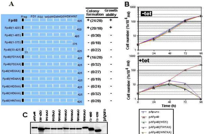
The complementation ability of these p48 mutant proteins was determined based on the number of surviving colonies in the presence of both tet and puro. The N-terminal truncated mutant, Fp48(33-425), together with other mutants (data not shown), exhibited no complementation ability, indicating that the region comprising amino acids 1-32 was necessary for cell viability. On the other hand, one C-terminal truncated mutant, Fp48(1-420), exhibited almost similar complementation ability toward the parental Fp48 construct. However, two other C-terminal truncated mutants, Fp48(1-405) and Fp48(1-375), exhibited no complementation ability. These findings indicated that the region comprising amino acids 406-420 was also necessary for the viability of DT40 chicken B cell line.
All of WD double missense mutants, Fp48(FD2AA), Fp48(WD3AA), Fp48(WD4AA), Fp48(WD5AA),
Fig 2. In vitro interaction of chCAF-1p48 and another histones chaperone with chromatin related proteins Anti-silencing factor 1 (ASF-1)
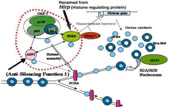
Fig 4. Model a role of p48 subunit of chromatin assembly factor 1 (CAF-1p48) as histone chaperons on DNA replication process for the maintenance of the cell viability
In addition, dead cells occurred at 72 h, and finally, almost all of the cells were dead by 96 h (Fig 3B), although all of transformants obtained expressed appropriate FLAG-tagged mutant proteins at almost same levels (Fig 3C). These results indicated that all of seven WD dipeptide motifs, including the first one, were essential for the cell viability and growth in DT40 chicken B cell line.
As illustrated in Fig 4, during DNA replication, CAF-1 containing p48 subunit assembles new nucleosomes through a two-step reaction. Coupled to DNA replication, as the first step, histones H3 and H4 are deposited through a reaction that is preferentially dependent upon CAF-1, but histones H2A and H2B are added later to this immature nucleosome precursor, even in the absence of CAF-1. These results indicate that CAF-1 interacts preferentially with H3 and H4, whereas NAP-1 (Nucleosome Assembly Protein 1) binds to H2A and H2B. On the other hand, ASF-1 was found to be a histone chaperones via interacts with CAF-1p60 and HIRA proteins that modulate the formation of nucleosomes. Thus, these CAF-1-participating interactions with HIRA and HDAC-1 (and probably other CAF-1-carrying complexes including ASF-1) should be involved directly or indirectly in chromatin assembly or maintenance and alterations of chromatin structure, involving gene replication and transcription to the maintenance and support of the cell viability. On the other hand, the ASF-1 activity suggested regulates the semi-conservative replication of nucleosomes in mammalian cells [24]. Our preliminary analysis of the HIRA-deficient conditional DT40 mutant and the essential role of HIRA protein in transcriptional regulation [25-26], together with the AFS-1 conditional mutant cells [27-28], will be of powerful tools for understanding the role of CAF-1 and the other histones
chaperones in relation to DNA replication and transcription regulations to the maintenance and support of the cell viability and growth in DT40 chicken B cell line.
CONCLUSION
Through the work presented in this paper, we studied functional analyses of CAF-1, and demonstrated not only that chCAF-1p48 binds to HDAC-1, histone H4, and chCAF-1p60, but also WD dipeptide motifs in the chCAF-1p48 are necessary for interaction with these proteins. Based on the results obtained for chCAF-1p48 in this study, we propose a molecular basis for the role of p48 subunit for the viability of DT40 cells. The fundamental function of the core regions should be to interact with other core regions to provide a scaffold on which to display the surface specializations, although that of the variable regions may be preferentially involved in protein-protein interactions relative to the maintenance of the proper structure to support the viability and growth of the chicken DT40 B-cell lymphoma cells line (and probably vertebrate cells).
ACKNOWLEDGEMENT
8. Krude, T., 1999,Eur. J. Biochem.,263, 1-5.
9. Verreault, A., Kaufman, P.D., Kobayashi, R., and Stillman, B., 1996,Cell,87, 95-104.
10. Ito, T., Bulger, M., Kobayashi, R., and Kadonaga, J.T. 1996,Mol. Cell. Biol.,16, 3112-3124.
11. Qian, Y.W., Wang, Y.C., Hollingsworth, R.E., Jones, D., Ling, N., and Lee, E.Y., 1993,Nature, 364, 648-652.
12. Marheineke, K. and Krude, T., 1998,J. Biol. Chem., 273, 15279-15286.
13. Taunton, J., Hassig, C.A., and Schreiber, S.L., 1996, Science,272, 408-411.
14. Tyler, J.K., Bulger, M., Kamakaka, R.T., Kobayashi, R., and Kadonaga, J.T. 1996, Mol. Cell. Biol., 16, 6149-6159.
15. Hassig, C.A., Fleischer, T.C., Billin, A.N., Schreiber, S.L., and Ayer, D.E., 1997,Cell, 89, 341-347.
Kaufman, P.D. 2001,Curr Biol.,11, 463-473. 22. Mello, J.A., Sillje, H.H., Roche, D.M., Kirschner,
D.B., Nigg, E.A., and Almouzni, G., 2002, EMBO Rep.,3, 329-334.
23. Munakata, T., Adachi, N., Yokoyama, N., Kuzuhara, T., and Horikoshi, M., 2000, Genes Cells,5, 221-233.
24. Eitoku, M., Sato, L., Senda, T., and Horikoshi, M. 2008,Cell Mol. Life Sci.,65, 3, 414-444.
25. Ahmad, A., Kikuchi, H., Takami, Y., and Nakayama, T., 2005, J. Biol. Chem., 280, 32090-32100.
26. Ahmad, A., 2008,Indo. J. Chem.,8, 3, 454-458. 27. Sanematsu, F., Takami, Y., Barman, H.K., Ono, T.,
Fukagawa, T., Shibaraha, K.I., and Nakayama, T. 2006,J. Biol. Chem.,281, 13817-13827.