VOLUME 49 January• 2009 NUMBER 1
Case Report
Role of multidetector spiral CT scanning for
pulmonary embolism confirmation in a child with
pulmonary hypertension: a case report
Heda Melinda Nataprawira
1, Sri Endah Rahayuningsih
1, Nono Sumarna Afandi
1,
Armijn Firman
1, Tan Siauw Koan
2ulmonary embolism (PE) is associated with considerable morbidity and mortality. Early diagnosis and prompt treatment is essential,1,2 however PE is rarely clinically diagnosed or treated in children. Most clinically significant PE is not recognized antemortem.3 While its diagnosis remains a challenge as the signs and symptoms can often be non-specific, an accurate diagnosis is essential for the management of this disease. It is known that a number of non-invasive diagnostic tools are available for its detection nowadays.1,2,4
Even though multi-detector spiral, also called helical, CT scanning is promising and has been proven to be useful in diagnosing this condition with high sensitivity and specificity,5
it is unavailable even in referral hospitals in Indonesia. The gold standard, pulmonary angiography, is considered as the procedure of choice to diagnose PE, but unfortunately it is invasive. Failure to diagnose PE accurately and promptly can result in excess morbidity and death due to pulmonary hypertension (PH) and recurrent venous thromboembolic events. Conversely, unnecessary anticoagulation therapy poses a risk without any benefit.2
Pulmonary embolism should be considered in the evaluation of unexplained PH, respiratory insufficiency, and disseminated intravascular coagulation in children.3A diagnosis of PH can be made when the
mean pulmonary artery pressure is greater than 25mmHg in a resting individual at sea level.6
Pulmonary hypertension refers to a group of conditions with multiple causes rather than a single condition. PH is caused by increased pulmonary blood flow as seen in congenital heart defects with large left-to-right shunts (hyperkinetic pulmonary hypertension), alveolar hypoxia, increased pulmonary venous pressure, primary pulmonary vascular disease and chronic thromboembolic disease.6,7
An extensive evaluation should be performed in children with severe PH, as the most successful strategy for managing PH involves treatment of any underlying disorders. A specific phosphodiesterase inhibitor, sildenafil, is known as a potent and selective pulmonary vasodilator that has been proven to be superior in decreasing the mean
From The Department of Child Health, Medical School, Padjajaran University, Hasan Sadikin Hospital, Bandung, Indonesia (HMN, SER, NSA, AF).1 From the Department of Radiology, Santo Borromeus Hospital,
Bandung, Indonesia (TSK).2
Reprint request to: Heda Melinda, MD, Department of Child Health,
Padjajaran University, Hasan Sadikin Hospital, Jl. Pasteur No. 38, Bandung Indonesia. Telp. 62-22-2034426. Fax. 62-22-203-5957.
pulmonary artery pressure. It is also equally effective and selective in reducing pulmonary vascular resistance compared to inhaled nitric oxide (iNO) treatment.6,8
The Case
An 11-year-old boy was referred to Hasan Sadikin Hospital with fatigue as the chief complaint. Eight months before admission he felt fatigue that was sometimes accompanied by shortness of breath. During the last two months before admission, these complaints had become worse, accompanied by sudden onset left chest pain. He also suffered from two episodes of syncope and had infrequent coughing symptoms. There were no symptoms of chronic pulmonary problems or disease. He was previously described as healthy and similar histories were not found in family members. Echocardiography examination revealed PH (pulmonary artery pressure
of 140 mmHg) with severe tricuspid insufficiency butno
cardiac defect was found (
Figure 1
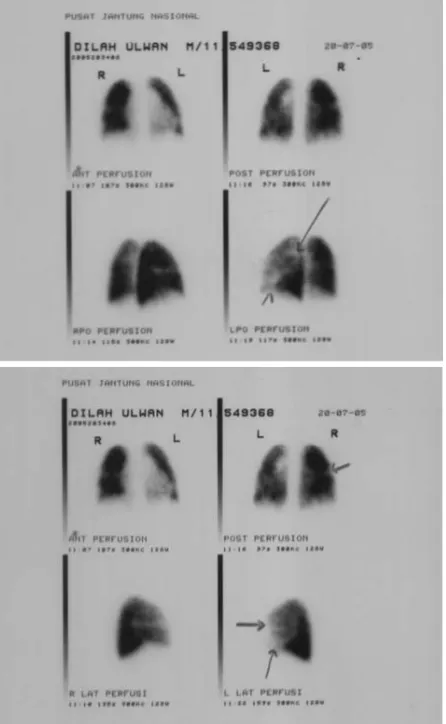
).He was referred to Cardiac Center at Harapan Kita Hospital where a lung perfusion scintigraphy examination was performed. This procedure revealed primary pulmonary hypertension (PPH) with suspected PE (
Figure 2
). He was then given oral sildenafil and the anticoagulant warfarin, to maintain the internationalnormalized ratio (INR) within the range of 1.5-2.0.
Afterwards, he received follow-up care at Hasan Sadikin Hospital and at a private clinic.
Figure 1. Echocardiogram revealed pulmonary hy-pertension with severe tricuspid insuf~ciency.
Figure 2. Lung perfusion scintigraphy showed primary pulmonary hypertension with observed pulmonary embolism.
On physical examination, he was fully alert;the pulse rate was 96 beats/minute and was regular. The respiratory rate was 24 times/minute, and blood pressure
was 120/80 mmHg. No cyanosis or clubbing of the
fingers was noted. On auscultation of the heart, the first sound was normal with a loud second heart
sound. A 3/6 systolic murmur in the third and fourth
intercostal spaces was noted. No liver enlargement was found. Laboratories findings were within normal
limit including an INR of 1.5-2.0. Chest X-raysshowed
Figure 3. Spiral CT-scan showing lack of filling defect in the subsegmental branches of the pulmonary arteries on both sides.
tricuspid regurgitation, and contrast regurgitation from the right atrium to the inferior cava vein and the hepatic vein. There was no sign of PE i.e. no filling defect in the subsegmental branches of the pulmonary arteries on both sides (
Figure 3
). Right and left lung examination revealed no disorder or abnormality explaining the cause of PH. Therefore, the warfarin was discontinued.One month after these examinations he was brought to the hospital again with a complaint of
shortness of breath that was accompanied by low grade fever and cough for one day. On examination he was alert but tachypneic (respiratory rate of 43/minute), with blood pressure of 110/70 mmHg, pulse rate of 107/minute with an irregular pulse (10 extrasystoles/ minute), and a temperature of 37.90
C. The sclera of the eyes was slightly icteric and crackles in the lung base were detected. On auscultation, the first heart sound was normal, a loud second heart sound was found in the pulmonary area, and a systolic murmur of tricuspid regurgitation was heard. The liver was enlarged and a slight peripheral edema was noted. He was then diagnosed as having right heart failure (RHF) with PH, tricuspid insufficiency, cardiac arrhythmia, bronchopneumonia and cardiac liver cirrhosis. He was given oral sildenafil, captopril, clarithromycin, and furosemide. Repeated echocardiogram revealed PH with decreasing pulmonary artery pressure (pulmonaryartery pressure 40 mmHg). A liver ultrasonogram showed cardiac liver cirrhosis.
Sildenafil and captopril were taken regularly. One month later, the patient still had jaundice and
the echocardiography showed decreasing
pulmonaryartery pressure (pulmonary artery pressure 40 mmHg). So forth, he never turned-up in the clinic or hospital.
Discussion
Pulmonary embolism (PE) remains a common cause of mortality and its diagnosis is often
missed. Although prompt diagnosis and
appropriate therapy have been shown to reduce the mortality from 30% to less than 10%,2 the diagnosis of PE is often not suspected in children. Even though most of the signs and symptoms, including chest pain (70%), tachypnea (70%), cough (40%), tachycardia (33%), shortness of breath (25%), signs of deep venous thrombosis (10%) and syncope (5%),6 are likely to be seen in PE patients, diagnosis based on the clinical manifestations is not reliable.3 It has been noted that in adults clinicaldiagnosis has a sensitivity of 85% but a specificity of 38%, reflecting the vast differential diagnosis foundin both adults and children.7 These clinical symptoms (chest pain, dyspnea, shortness of breath, syncope) also occur in PH.6
investigation performed in a cardiac or respiratory emergency. In acute pulmonary thromboembolism it may be normal or non-specific.9 Chest radiographs and electrocardiograms are often normal in young patients with PE.7 The use of several imaging
modalities such as ventilation-perfusion (V/Q) lung
scanning, spiral volumetric computed tomography (CT) scanning, magnetic resonance imaging (MRI), CT angiography and digital subtraction angiography suggests that one single technique is not reliable to confirm or exclude the diagnosis in a patient with clinically suspected PE.2,4
V/Q scanning is the most
frequently ordered diagnostic test in patients with clinically suspected PE.7 The obvious advantages of
V/Q scanning are its low cost, ease of performance andits
non-invasive nature. However, this type of scan can be problematic because it rarely provides a definitive
"high probability' or "normal' diagnosis.4In addition, this
diagnostic test is not available in this hospital. Even though it was established in the PIOPED study (Prospective Investigation of Pulmonary Embolism Diagnosis) that a normal or low probability scan has a high negative predictive value and a high probability scan has a high positive predictive value when the results are interpreted along with the clinical assessment of the likelihood of PE, one study has shown that in approximately 70% of patients, the results
of V/Q scanning were non-diagnostic.2Inpatients with a
non-diagnostic V/Q scan, pulmonaryangiography is the
next recommended imaging modality to establish the diagnosis of PE. However, pulmonary angiography is not often requested by clinicians because of its invasive nature.2,10 Pulmonary angiography is the
historical gold standard for diagnosing PE, with which all other imaging tests have been compared.4,7,11,12In our case study, lung perfusion scintigraphy performed on this patient revealed idiopathic or PPH with an observation of PE which was then treated with sildenafil and warfarin. As a confirmation of PE was needed to justify prescription of warfarin, a
multidetector helical/spiral CT scanwas performed. This
technique has been proven to be useful to diagnose PE, and is available in another hospital (Borromeus Hospital) in the Bandung area. The choice between
V/Q scanning and helical CT scanning tends to
dominate most current discussions about non-invasive PE diagnosis.10 Recent studies have reported the usefulness of spiral CT scanning for
the detection of central and segmental PE. It is non-invasive, easy to perform, quick and has been reported to have good specificity and sensitivity.2
Compared to V/Q scintigraphy or pulmonary angiography, the sensitivity of spiral CT has ranged from
53% to 100% and its specificity from 78% to 100%.7
Based on those findings, even though anti-coagulation may be required in cases of PH that are associated with low cardiac output, leading to sluggish blood flow through the pulmonary artery which may predispose the patient to the development of pulmonary thrombi, we decided to stop warfarin treatment as PE was not diagnosed by multidetector spiral CT scanning. In the treatment of PH, oral sildenafil is given to promote an increase of cGMP levels and thus cause pulmonary vasodilatation.6
Although we had some limitations in performing all tests for the diagnosis of PH as proposed by the WHO6
, this patient likely had an idiopathic PH/PPH,as
we could not determine other possible causes.6 Although PPH is a rare disease in childhood, and is associated with poor outcome and long term survival, the natural history of disease is heterogeneous with some patients dying within months of diagnosis and others living for decades.13
In present days, the treatment of PH is changing.3Most cases of PH are difficult to treat and are irreversible unless the cause can be eliminated.5 Previously, the main treatment was aimed only to control congestive heart failure (CHF).3The prognosis of PH is variable and represents a significant improvement compared to several years ago. Children who respond to short-term drug testing (responders)
have a five year survival rate of 90%, while those who
are nonresponders have a five year survival rate of
only33%. Many of the children in the later group may be
candidates for lung transplants.14,15
blood flow into terminal hepatic venules. Sinusoidal stasis results in accumulation of deoxygenated blood, parenchymal atrophy, necrosis, perisinusoidal collagen deposition, and ultimately, fibrosis.16
In conclusion, although the precise role of various non-invasive imaging techniques in the diagnosis of acute PE remains to be clarified, the result from using multidetector spiral CT as a diagnostic tool in the detection of PE is promising. It has high sensitivity and specificity, gives quick results and can help to provide alternative diagnosis, despite the fact that PE in this patient was unlikely to be confirmed based on multidetector spiral CT-scanning results.
References
1. Grandmaison, Lorin G, Durigon, Michel. Pulmonary embolism: a rare cause of sudden infant death. Am J Forensic Med Pathol. 2002;23(3):257-9.
2. Lingamanaicker J, Mukherjee JJ, Fock KM, Khoo TK. The
role of spiral computed tomogram in the diagnosis of acute
pulmonary embolism. Singapore Med J. 2001;42:455-9.
3. Donnerstein RL, Berg RA. Pulmonary embolism. In: Taussig LM, Landau LI, editors. Pediatric respiratory medicine. St.Louis: Mosby, 1999;p. 910-5.
4. Goldhaber SZ. Multislice computed tomography for
pulmonary embolism - A technological marvel. N Engl J Med. 2005;352:1812-4.
5. Park MK, Troxler RG. Pediatric cardiology for practitioners. 4th ed. St. Louis: Mosby. 2002.
6. Rashid A, Ivy D. Severe paediatric pulmonary hypertension: new management strategies. Arch Dis Child. 2005;90:9-8.
7. Carman TL, Deitcher SR. Advances in diagnosing and excluding pulmonary embolism: spiral CT and D-dimer
measurement. Cleveland Clin J Med. 2002;69:721-9.
8. Michelakis E, Tymchak, Lien D, Webster L, Hashimoto
K, Archer S. Oral sildenafil is an effective and specific pulmonary vasodilator in patients with pulmonary arterialhypertention. Circulation. 2002;105:2398-400.
9. Vaughan DJ. Pulmonary infarction. [cited on 26 July 20051.Available from:http://www.emedicine.com
10. Kohli A, Rajput D, Gomes M, Desai S. Imaging of pulmonary thromboembolism. Ind J Radiol Image. 2002;12:207-12.
11. Hartmann IJC, Hagen PJ, Melissant CF, Postmus PE, Prins MH. Diagnosing acute pulmonary embolism. Am J RespirCrit Care Med. 2000;162:2232-7.
12. Morris T. New options for pulmonary embolism diagnosis. [cited on 30 July 20051. Available from:http://www.chrowrd/ news/pulm emb dx.doc
13. McLaughlin VV, Presberg KW, Doyle RL, Abman SH, McCrory DC, Fortin T,et al. Prognosis of pulmonary arterial hypertension. ACCP evidence-based clinical practiceguidelines. Chest. 2004;126:78S-92S.
14. Primary pulmonary hypertension. Cincinnati children’s [cited on 30 July 20051. Available from:
http://www. cincinnatichildrens.org
15. Berger S. Pulmonary hypertension, primary. [cited on 27 May 20041. Available from:http://www.emedicine.com
16. Andrews AH, Holtzmuller KC. Cardiac cirrhosis. [cited on 2 Sept 20041. Available from:http://www.emedicine.com/med/