COMMON ALLERGIES AND ALLERGENS
IN ORAL AND PERIORAL DISEASES
Liborija Lugović-Mihić1,2, Ivana Ilić3, Jozo Budimir1, Nives Pondeljak1 and Marinka Mravak Stipetić4
1Department of Dermatovenereology, Sestre milosrdnice University Hospital Centre, Zagreb, Croatia; 2School of Dental Medicine, University of Zagreb, Zagreb, Croatia;
3Faculty of Dental Medicine and Health, Josip Juraj Strossmayer University of Osijek, Osijek, Croatia; 4Department of Oral Medicine, School of Dental Medicine, University of Zagreb, Zagreb, Croatia
SUMMARY – Allergic reactions sometimes participate in the development of perioral and oral diseases, indicating the need for appropriate allergen assessment. This review discusses current knowl-edge on the potential allergic reactions to different dental materials in patients with oral and perioral diseases. Aside from allergies to various dental materials, similar non-allergic, non-immune contact reactions (irritant or toxic) can occur. Among dental materials, the most frequent allergens are alloys, followed by rubber materials, polymers and acrylates. Allergic reactions to dental alloys that contain nickel, cobalt and amalgam are especially frequent since dentists use them for prosthetic and other restorations. There is a broad spectrum of clinical presentations of oral and perioral diseases possibly related to allergies, such as lichenoid reactions, cheilitis, perioral dermatitis, burning sensations, etc. Despite some limitations, patch test is crucial in the diagnosis and recognition of causative allergens because it reveals contact allergies, and is still superior in differentiating allergic and irritant contact reactions. It is important to examine patient medical histories (e.g., occurrence of symptoms after dental therapy or food consumption), and in consultation with their dentist, carry out allergy tests to specific dental allergens which are used or planned to be used in subsequent treatment.
Key words: Allergy; Burning mouth syndrome; Cheilitis; Gingivostomatitis; Oral disease; Oral lichenoid reactions; Patch test
Correspondence to: Prof. Liborija Lugović-Mihić, MD, PhD, De-partment of Dermatovenereology, Sestre milosrdnice University Hospital Centre, Vinogradska c. 29, HR-10000 Zagreb, Croatia E-mail: [email protected]
Received June 27, 2019, accepted October 1, 2019
Introduction
Oral and perioral diseases are relatively frequent in the general population, and their symptoms can sig-nificantly affect the patient’s quality of life. In cases of perioral and oral diseases, possible relations to various dental materials and procedures should always be tak-en into consideration whtak-en treating a patitak-ent1-3. Al-though there are many scientific papers on this issue, their results are ambiguous. Various substances can cause both immediate (type I) and delayed (type IV) allergic reactions, of which type IV allergic reactions are more common1,4,5.
On the other hand, some substances can provoke non-allergic, irritant or toxic reactions in which the immune mechanism is not involved6.Furthermore, pa-tients often complain of unspecified sensations in peri-oral and peri-oral soft tissues, which also makes it more difficult to evaluate the influence of allergies in peri-oral and peri-oral diseases. Although there are no precise data on the incidence of side effects (unwanted reac-tions) from dental procedures and use of dental mate-rials, it can be assumed that they are not very com-mon7.
Oral and Perioral Diseases Related to Allergic Reactions and Common Causative Allergens
There is a broad spectrum of clinical signs and symptoms of oral and perioral diseases that can be re-lated to allergies, such as lichenoid reactions, cheilitis,
stomatitis, gingivitis, perioral dermatitis, burning sen-sations, swelling of the lips and face, etc.1,7-12.
Early allergic reactions (type I) with manifestations in oral and perioral regions mostly manifest with an-gioedema but sometimes may include oral paresthetic and burning sensations, pointing to oral allergy syn-drome (OAS), a form of type I allergic reaction. Oral allergy syndrome usually occurs in patients who suffer from allergic rhinitis, mostly after taking fresh food (such as vegetables, fruits and various nuts), due to the cross-reaction between food and inhalants13.
On the other hand, various dental materials used in dental procedures may cause both allergic contact (de-layed, type IV) and non-allergic contact reactions (irri-tant, toxic contact reactions). They appear when these materials have allergic or irritant effects on coming in contact with skin or oral mucosa. Unlike allergic reac-tions, in irritant contact reactions there is no allergic pathomechanism, no previous sensitizations to an aller-gen and no lesion spread14.The most common contact allergens that can cause allergic reactions in the oral cav-ity and perioral region are antiseptics, dental alloys, im-pression materials, local anesthetics, dental cement, la-tex gloves, acrylate, adhesives, mouth rinse liquids, vari-ous dental hygiene preparations, and others7.
Among dental materials, the most frequent aller-gens are dental alloys, followed by rubber materials, polymers and acrylates. (Notably, allergic reactions to local anesthetics are very uncommon). Metal salts in dental alloys, for example, have weak interactions with skin proteins and form complexes that make strong al-lergens that initiate hypersensitivity reactions14. Aller-gic reactions to dental alloys that contain nickel, cobalt and dental amalgam are especially frequent since den-tists use them for prosthetic and dental restorations1,4,15. Dental restorative materials include dental alloys, amalgams and tooth-colored fillings. Noble dental al-loys, and semiprecious and nonprecious alloys as base metals are used in dental procedures10,11. (Noble alloys contain more than 40% gold, palladium and/or plati-num, semiprecious alloys contain at least 25% noble metal, and nonprecious alloys often contain large per-centages of nickel, cobalt, chromium or beryllium, stainless steel or titanium). Dental amalgam is pro-duced by mixing liquid mercury with an alloy mixture consisting of silver, tin, zinc and copper, while tooth-colored fillings consist of composite resin, glass iono-mer cement and porcelain10.
Positive allergy skin patch tests to gold are usually associated with the amount of gold used in dental pro-cedures, although definite correlation between contact allergic reactions to gold and oral lesions has not been proven1.While gold is not common allergen, it is espe-cially important to take into consideration the per-centage of other gold alloy ingredients such as silver, copper, and smaller amounts of platinum, palladium and zinc. Silver-palladium alloys (also contain zinc and copper, and occasionally palladium and silver), as well as many other metals such as cobalt, chromium, molybdenum, beryllium, gallium, rhodium, iridium and some others are also used in dental procedures. In their systematic review, Levi et al. point out that each type of metal exposure has a different rate of allergic reaction, which they explain by the extent of corrosion of the alloy, population exposure, and the biologic en-vironment of each patient16.
One of the most commonly mentioned allergens is rubber, which can be found in latex gloves (worn by dentists and their assistants) and rubber dams (used to isolate the operative site from the rest of the mouth)17-19. Cases of adverse patient reactions to latex gloves worn by dental health care workers have been reported by Agrawal et al. Their study results showed that 16% of dental professionals reported allergy to latex gloves, with the prevalence significantly higher in those who had allergy to pollen grains, foodstuffs and rubber dam, asthma and eczema in their medical history17. Other allergic reactions may be caused by substances which contain acrylates (most commonly methyl methacrylate, triethylene glycol dimethacrylate and polymethyl methacrylate) used for the fabrication of dentures, as well as for complex restoration procedures and as binding materials20. Reactions to traces of ben-zoyl peroxide in dentures, hydroquinone and plasti-cizer dibutyl phthalate inhibitors, pigments, dyes, ny-lon fibers, titanium and zinc oxides are also possible.
The process of evaluating lesions and symptoms af-fecting the oral and perioral region is quite challenging due to their numerous possible manifestations. Some patients have burning symptoms and paresthesias without clinically evident oral lesions, whereas some patients have clear clinical signs such as lichenoid tis-sue changes or oral ulcerations12. In the diagnosis and recognition of causative allergens, patch test is crucial because it reveals and confirms contact allergies, in-cluding patients with orofacial changes (particularly
prior to dental treatment) and dental workers with chronic dermatitis of the hands and face (Fig. 1). Patch test is still superior in differentiating contact allergic and contact irritant reactions; however, its usefulness has not yet been fully established because studies have yielded variable results14,21.
There are numerous studies that have confirmed metal allergies in patients with dental alloys in their orthodontic devices and considering that it is
impor-tant to emphasize which allergens are most common to cause diseases such as burning mouth syndrome (BMS), gingivostomatitis, cheilitis and oral lichen pla-nus (OLP)22-25. According to Budimir et al., some par-ticular allergens increase the risk of certain oral dis-eases and symptoms22. This risk is several times higher in atopic patients and those with existing allergies even in the absence of statistically significant differ-ences in the occurrence of allergic reactions between
Fig. 1. Positive patch tests to nickel-sulfate and cobalt-chloride.
Fig. 2. Positive prick test results to inhalant allergens. Fig. 4. Food-induced angioedema of upper lip.
Fig. 3. Lip lesions in a patient with contact allergy to flavors and metals.
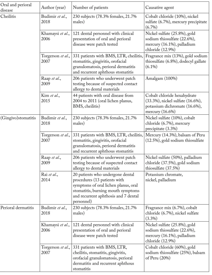
Table 1. Causative allergens for each oral and perioral disease
Oral and perioral
disease Author (year) Number of patients Causative agent
Cheilitis Budimir et al.,
2018 230 subjects (78.3% females, 21.7% males) Cobalt chloride (10%), nickel sulfate (6.7%), mercury precipitate (6.7%)
Khamaysi et al.,
2006 121 dental personnel with clinical presentation of oral and perioral disease were patch tested
Nickel sulfate (25.8%), gold sodium thiosulfate (22.6%), mercury (16.1%), palladium chloride (12.9%)
Torgerson et al.,
2007 331 patients with BMS, LTR, cheilitis, stomatitis, gingivitis, orofacial granulomatosis, perioral dermatitis and recurrent aphthous stomatitis
Fragrance mix (13%), gold sodium thiosulfate (6.8%), dodecyl gallate (6.1%)
Raap et al.,
2009 206 patients who underwent patch testing because of suspected contact allergy to dental materials
Amalgam (100%) Kim et al.,
2015 44 patients with oral disease from 2004 to 2011 (oral lichen planus, BMS, cheilitis)
Cobalt chloride hexahydrate (33.3%), nickel sulfate (16.6%), potassium dichromate (16.6%), mercury (16.6%)
(Gingivo)stomatitis Budimir et al.,
2018 230 subjects (78.3% females, 21.7% males) Nickel sulfate (10%), cobalt chloride (6.7%), mercury precipitate (3.3%)
Torgerson et al.,
2007 331 patients with BMS, LTR, cheilitis, stomatitis, gingivitis, orofacial granulomatosis, perioral dermatitis and recurrent aphthous stomatitis
Mercury (14.3%), balsam of Peru (12.5%), gold sodium thiosulfate Raap et al.,
2009 206 patients who underwent patch testing because of suspected contact allergy to dental materials
Nickel sulfate (50%), palladium chloride (37.5%), gold sodium thiosulfate (37.5%)
Rai et al.,
2014 20 patients who undergone dental procedures (13 patients with symptoms of oral lichen planus, oral stomatitis, burning mouth symptoms and recurrent aphthosis and 7 dental personnel)
Potassium chromate, nickel, palladium
Perioral dermatitis Budimir et al.,
2018 230 subjects (78.3% females, 21.7% males) Fragrance mix (6.7%), cobalt chloride (6.7%), nickel sulfate (3.3%)
Khamaysi et al.,
2006 121 dental personnel with clinical presentation of oral and perioral disease were patch tested
Nickel sulfate (25.8%), gold sodium thiosulfate (22.6%), mercury (16.1%), palladium chloride (12.9%)
Torgerson et al.,
2007 331 patients with BMS, LTR, cheilitis, stomatitis, gingivitis, orofacial granulomatosis, perioral dermatitis and recurrent aphthous stomatitis
Cobalt chloride (60%), gold sodium thiosulfate (25%), balsam of Peru (20%)
Oral and perioral
disease Author (year) Number of patients Causative agent
Burning mouth
syndrome Budimir 2018 et al., 230 subjects (78.3% females, 21.7% males) Cobalt chloride (13.3%), P-phe- nylenediamine colophony (3.3%) Khamaysi et al.,
2006 121 dental personnel with clinical presentation of oral and perioral disease were patch tested
Nickel sulfate (15.9%), mercury (15.8%), palladium chloride (10.5%), gold sodium sulfate (10.5%)
Torgerson et al.,
2007 331 patients with BMS, LTR, cheilitis, stomatitis, gingivitis, orofacial granulomatosis, perioral dermatitis and recurrent aphthous stomatitis
Potassium dicyanoaurate (16.4%), nickel sulfate hexahydrate (12.3%), gold sodium thiosulfate (10.9%)
Raap et al.,
2009 206 patients who underwent patch testing because of suspected contact allergy to dental materials
Gold sodium thiosulfate (66.6%), nickel sulfate (66.6%), palladium chloride (33.3%), cobalt chloride (33.3%)
Rai et al.,
2014 20 patients who underwent dental procedures (13 patients with symptoms of oral lichen planus, oral stomatitis, burning mouth symptoms and recurrent aphthosis and 7 dental personnel)
Methylhydroquinone
Kim et al.,
2015 44 patients with oral disease from 2004 to 2011 (oral lichen planus, BMS, cheilitis)
Cobalt chloride hexahydrate (25%)
Oral lichen planus (lichenoid tissue reaction)
Budimir et al.,
2018 230 subjects (78.3% females, 21.7% males) Cobalt chloride (6.7%), gold (3.3%), thimerosal (3.3%) Khamaysi et al.,
2006 121 dental personnel with clinical presentation of oral and perioral disease were patch tested
Gold sodium thiosulfate (11.8%), nickel sulfate (5.8%), mercury (5.8%)
Torgerson et al.,
2007 331 patients with BMS, LTR, cheilitis, stomatitis, gingivitis, orofacial granulomatosis, perioral dermatitis and recurrent aphthous stomatitis
Potassium dicyanoaurate (28%), fragrance mix (17.1%), gold sodium thiosulfate (15.1%) Raap et al.,
2009 206 patients who underwent patch testing because of suspected contact allergy to dental materials
Palladium chloride (44.4%), nickel sulfate (22.2%), gold sodium thiosulfate (22.2%)
Rai et al.,
2014 20 patients who underwent dental procedures (13 patients with symptoms of oral lichen planus, oral stomatitis, burning mouth symptoms and recurrent aphthosis and 7 dental personnel)
Nickel, potassium chromate, copper sulfate, amalgam
Kim et al.,
2015 44 patients with oral disease from 2004 to 2011 (oral lichen planus, BMS, cheilitis)
Gold sodium thiosulfate (33.3%), nickel sulfate (33.3%), potassium dichromate (33.3%), cobalt chloride hexahydrate (8.3%) BMS = burning mouth syndrome; LTR = lichenoid tissue reaction
subjects with certain oral diseases and healthy con-trols22. This was shown to be true for BMS and its as-sociation to nutritive allergens and food additives, for oral lichen planus and inhalants, and for cheilitis and contact allergens such as cobalt-chloride and nickel-sulfate found in dental alloys. These findings contrib-ute to the knowledge of the etiology of these diseases and the justification of using skin tests in these par-ticular oral diseases.
According to Torgerson et al., positive contact al-lergies were established in 44.7% of patients that un-derwent patch testing, as well as possible multiple positive reactions due to cross-reactions, which has also been noted in other studies12,22.The frequency of positive patch tests to dental materials was higher in some other studies, even reaching 70.5%10.Many stud-ies show various frequencstud-ies of positive patch tests for allergens in certain oral and perioral diseases. Particu-larly common allergens established with patch testing are metals found in dental materials24. According to Khamaysi et al., the most common contact allergens established with patch testing in their study were gold sodium thiosulfate (14.0%), nickel sulfate (13.2%), mercury (9.9%), palladium chloride (7.4%), cobalt chloride (5.0%) and 2-hydroxyethyl methacrylate (5.8%)9.In a study by Kim Tae-Wook et al., the most common contact allergic reactions in oral and perioral diseases were established in oral lichen planus (75%), cheilitis (75%), BMS (25%), and other oral diseases (75%)10. [In the study by Khamaysi et al., these were cheilitis (41.9%), perioral dermatitis (41.9%) and li-chenoid reactions (35.3%)]9.In addition to patch tests, immediate hypersensitivity tests such as prick tests and serum tests for determination of specific IgE can also be used sometimes (Fig. 2)7,11.
Review of Oral and Perioral Diseases and Causative Allergens
Cheilitis (inflammation of the lips) includes many
clinical types and is possibly related to many allergens (Table 1). Cheilitis can present alone or be associated with stomatitis or perioral eczema2,14,21,26. According to the latest classification of cheilitis, proposed by Lugović-Mihić et al., it can be divided into three groups, as follows: mainly reversible (simplex, angular/ infective, contact/eczematous, exfoliative, drug relat-ed); mainly irreversible (actinic, granulomatous,
glan-dular, plasma cell); and cheilitis connected to dermato-ses and systemic diseadermato-ses (lupus, lichen planus, pem-phigus/pemphigoid group, angioedema, salivation disorder, etc.)26. Contact/eczematous cheilitis is the result of an irritating or allergic contact effect with various substances, such as medications, toothpaste in-gredients (e.g., sodium lauryl sulfate), cleaning agents for dentures (potassium-persulfate), dental floss (col-ophony), nail polish, cosmetics (e.g., lipstick, lip gloss), food and flavors, musical wind instruments (nickel, wood), etc. (Fig. 3)1,27. Allergies and allergens can sometimes be difficult to establish in some patients. A study conducted in cheilitis patients established irri-tant contact dermatitis in 36% of patients having un-dergone patch testing, allergic contact dermatitis in 25%, atopic eczema in 19%, and unknown causes in 9%27.According to a recent study by Budimir et al., patients with cheilitis showed a statistically signifi-cantly higher frequency of positive patch tests (26.7%) compared to healthy controls, and the common aller-gens were cobalt-chloride (10%), nickel-sulfate (6.7%) and mercury precipitate (6.7%)22. Torgerson et al. ob-served a similar frequency of positive patch tests in their patients (25.9%), whereas Kim et al. report an even higher frequency (75%), particularly when metals used in dental medicine were involved10,12.
Angioedema can be induced by various factors and
allergens, such as drugs, foodstuffs, preservatives, cos-metics, etc. (Fig. 4). It predominantly appears as a hy-persensitivity reaction type I, or sometimes type IV, and such reactions can occur after contact with latex, dental products, etc. during dental treatment (when dentist’s glove comes in contact with the lip, or in con-tact with cinnamaldehyde, menthol or eugenol in toothpaste), etc.18,19. In dental practice, also possible are allergic reactions to formaldehyde (used for disin-fection in root canals), immediate-type allergies to lo-cal anesthetics or delayed-type allergies from longer operative procedures (e.g., additives from the glove rubber or rubber dam)1,7. These reactions should be ex-amined for both immediate and delayed hypersensitiv-ity reactions, usually by skin allergy tests, which are conducted during remission of angioedema and when the patient is not under anti-allergic therapy. Also, food ingredients, e.g., benzoates, antioxidants or spic-es, can be the possible causes of angioedema. Accord-ing to the results of the study by Budimir et al., addi-tive allergens were confirmed in 23.3% of angioedema
patients, and physicians were advised to monitor pa-tient conditions after allergen elimination22.In addi-tion, facial edema sometimes occurs due to metals in the oral cavity (e.g., crowns with palladium), and re-moval of such metals has proved beneficial. In a recent study by Budimir et al., patch test was positive in 6.7% of angioedema patients, and the most frequent contact allergens were cobalt-chloride (3.3%) and nickel-sul-fate (3.3%)22. In the study by Khamaysi et al., the num-ber of patients positive to gold and nickel was high (13.2%), but one half of the patients positive to nickel were also positive to palladium chloride and cobalt chloride, which most probably indicated cross reaction with nickel9. According to Budimir et al., recent results on allergic reactions in the oral and perioral regions show that the risk of angioedema is 3-fold higher in subjects with established allergies and in men. Fur-thermore, the risk increases with age. It was also estab-lished that patients with angioedema exhibited reac-tions to more allergens than other patients22.
Perioral dermatitis is sometimes, although rarely,
associated with allergic reactions and is possibly con-nected to many allergens (Table 1)22. The disease is be-nign and it is usually contact dermatitis caused by sub-stances in toothpaste, gum, lipstick, or medications28. While some studies suggest that metals (e.g., nickel and chrome) in dental appliances can be the cause or aggravating factor for this disease, others did not re-cord adverse reactions in patients allergic to nickel fol-lowing application of dental crowns or bridges29,30. Torgerson et al. report positive patch tests in 80% of patients with perioral dermatitis, but positive patch tests were less frequent in the study by Budimir et al. (16.7%)12,22. Fragrances, cobalt-chloride and nickel-sulfate were the most common contact allergens in both these studies12.It is possible thatperioral lesions are similar toallergic contact dermatitis and irritant contact dermatitis. Allergic contact dermatitis is a manifestation of type IV hypersensitivity reaction to agents that come in contact with the skin, and patients may present with inflammatory papules, vesicles, weeping or crusting, while distribution of lesions is de-pendent upon the specific sites of contact with the ini-tiating agent. Unlike perioral dermatitis, intense pruri-tus is usually present, scaling is often prominent, and lesions fail to improve with antibiotic therapy. On the other hand, in irritant contact dermatitis, clinical find-ings vary based upon the nature of the external trigger
and site of involvement, and usually include papules, vesicles, scales, erythema or edema, and a burning rather than itching sensation, unlike allergic contact dermatitis but similar to perioral dermatitis. In irri- tant contact dermatitis, patient history is of value for identifying this diagnosis (as an example, irritant der-matitis re lated to chronic lip-licking behavior can re-semble perioral dermatitis).
Oral lichenoid reactions (OLR) are often associated
with contact allergies and positive patch tests (Table 1). In patients with oral lichenoid lesions, determina-tion of metal sensitivity is quite important12,31-33. There are many studies that confirm the effect of metal aller-gies in oral lichenoid lesions, especially in dental resto-rations and orthodontic devices. Laine et al. established allergies to metals in 67.7% of OLR patients using the patch test, particularly to mercury (66.1%), gold (9.3%), cobalt (3.3%), and others (tin, silver, palladium and chrome)31. Studies often point out allergic reac-tions to mercury, although their percentages vary33,34. Irritant contact reactions to mercury are possible in cases when patch test is negative; thus, removal of an adjacent amalgam can initiate improvement due to the fact that amalgam releases mercury7,32,33. Dunsche et al. report that 27.7% of 134 patients with oral lichenoid lesions showed positive patch test results to inorganic mercury or amalgam. Amalgam removal led to im-provement in 97.1% of patients33. However, two stud-ies (performed by Budimir et al. and Kim et al.) ob-served no positive reactions to mercury, which may be connected to the use of amalgam as restorative mate-rial, as shown previously by Choi et al.10,22,35. Scalf etal. report that 49% of 51 patients with lichenoid lesions in different regions and tissues (oral, genital, cutane-ous) had positive patch test with at least one mercurial allergen36. In the same study, positive patch test reac-tions were detected to chromate, gold and thimerosal exposure, and interestingly, 100% of patients declared improvement after metal replacement. Torgerson et al. established positive patch tests in 55.9% of OLR pa-tients, whereas Budimir et al. found positive patch test (10%) less frequently, and the commonest contact al-lergens were cobalt-chloride (6.7%), gold sodium thio-sulfate (3.3%) and thimerosal (3.3%)12,22. In OLR, a connection with other metals (particularly gold, chrome and cobalt) is also sometimes observed22. Gold can cause various oral difficulties (including OLR, fa-cial dermatitis and oral burning symptoms); in such
cases, removal of gold can result in improvement37. This leads to a conclusion that contact allergy to differ-ent metals is more common among people having li-chenoid tissue changes. Taking all the above into con-sideration, performing patch tests before dental proce-dures or implanting orthodontic materials is quite important. In addition, for instance, after consumption of sour or spicy food or drinks, people with OLR can react to other substances in various ways, ranging from pricking sensation to severe pain. Because these pa-tients often complain of oral sensitivity and an un-pleasant burning sensation in the mouth, the immedi-ate hypersensitivity test is also useful7,38. By using prick tests in different oral diseases, recently we established that allergic reactions were most common in lichen (53.3%), and also more frequently occurred in atopic patients, thus appropriate diagnostics should be car-ried out to establish possible OAS22.
Gingivostomatitis is a disease of oral cavity that can
also be associated with contact allergies after exposure to dental materials (e.g., metals or plastics in dentures), as it has been shown that stomatitis is associated with partial dentures and some dental metals (e.g., palladi-um, gold or manganese) (Table 1)7,39,40. Gingivostoma-titis as a reaction to acrylates is also possible (e.g., due to allergy to 2-hydroxyethyl methacrylate, HEMA, used in dentures). In this case, a patient can have tin-gling sensation or feel jaw pain, but symptoms gradu-ally disappear after acrylate has been removed41. As for the etiology in cases of gingivostomatitis, reactions to food additives (e.g., benzoic acid) and flavors (e.g., cnamaldehyde) are also possible, which can cause in-flammation of oral mucosa. Avoiding these additives and flavors is the only therapy. According to the results reported by Budimir et al., patch test was positive to at least one contact allergen in 16.7% of patients with gingivostomatitis, while Torgerson et al. established statistically more frequent contact allergies in patients with stomatitis and gingivitis (30.8% and 64% of test-ed patients, respectively)12,22. The most common con-tact allergens in our recent study of patients with gin-givostomatitis were nickel-sulfate, cobalt-chloride and mercury precipitate, although with no statistical sig-nificance in comparison to healthy controls22.
Burning mouth syndromeis a disease of unknown
etiology and is possibly related to some allergens (Ta-ble 1). Some authors differentiate BMS into primary
and secondary type, primary being idiopathic and sec-ondary determined by local, systemic or psychological factors2. There is a controversy of connecting BMS with contact allergy although sometimes, various sub-stances like foodstuffs (instant coffee, peanuts, chest-nuts), additives (benzoic acid, sodium metabisulfite) metals (cadmium, mercury, nickel, cobalt-chloride), plastics (epoxy resins, benzoyl peroxide, bisphenol A), etc. are mentioned as potential causes7,21. It is therefore necessary to rule out all possible etiologic factors, in-cluding allergy. A few studies have indicated that pa-tients with BMS exhibit clinically relevant contact al-lergies to gold and nickel, even small quantities of them in dental materials can cause these allergies25. While Torgerson et al. report positive patch test in 42.1% of BMS subjects, according to Budimir et al. positive test was less frequent (20% of patients)12,22. The most frequent contact allergens were cobalt-chlo-ride, Ursol™ and colophony12. Some studies also indi-cated that in part of BMS patients, immediate-type allergies to nutritive allergens were established in the same patients12. Based on the results of the study con-ducted by Budimir et al., the risk of BMS was higher in atopic patients, although with no statistical signifi-cance22. However, it is possible that an established al-lergy may not be related to oral symptoms, so positive allergy tests should be taken with caution and addi-tional diagnostic workup should be considered. Importance of Examination for Allergy and Patch Test Usefulness
Given the different results of individual studies on the usefulness of performing allergy tests in diagnos-ing oral and perioral diseases with non-specific sensa-tions, it is justified in the cases of unknown etiology to carry out allergy tests in order to establish possible al-lergies (mostly using patch tests and prick tests). Skin patch testing is a simpler procedure and preferred to mucosal testing. Other reasons for choosing skin test before mucosal are the higher specificity and sensitiv-ity of the skin patch test and the requirement of a sig-nificantly higher concentration of allergens for muco-sal testing, which often results in many adverse reac-tions42. Taking into account the fact that burning symptoms in the oral cavity can be caused by allergic cross-reactions between food and inhalants (OAS), it is also justified to examine the possible immediate
al-lergic hypersensitivity, usually by prick testing13. The choice of the allergens to be tested is also important; it varies by studies, countries and number of allergens. Allergy unit prepares testing samples according to pa-tient history and in consultation with dentists.
One should also keep in mind that patch tests have a few limitations and pitfalls when it comes to the sig-nificance for oral diseases2. This is due to different al-lergen concentrations and standard preparations for patch testing, as well as due to different pH of the skin and oral mucosa, which may result in either false-pos-itive/negative reactions or non-specific irritant reac-tions2. When carrying out patch test and establishing reactions, it is important to consider that standard reading may be insufficient and subsequent test (in 10 days or more) should be read in case of false-positive results (e.g., up to one-third of patients allergic to mer-cury)1. When establishing reactions, other potential factors should be considered, as well as the possibility that oral difficulties are in no way connected with the suspected dental materials or that the same substances can cause different reactions in one patient (either al-lergic or irritant). In such cases, it is possible that, re-gardless of a negative patch test, such a substance in-duces an irritant (non-allergic) reaction. Here it is crucial to emphasize that occasionally, some patients may have negative results to patch to metals on stan-dard reading and positive results on delayed reading after 10 days (this often refers to allergy to mercury)14.
Clinical relevance of positive results to haptens in patch test is also difficult to evaluate because of the high number of products and their contents that are in contact with patient oral cavity and mucosa every day, which makes it difficult to prove one particular hapten blameworthy by avoiding method2. However, patch tests with dental screening series are worth consider-ing for oral diseases, especially for oral lichen planus10. In a study by Holmstrup, the author suggests indica-tions for patch testing, including OLRs and mucositis resistant to treatment, objective and evident relation between allergen and lesions, and absence of symmetry in lesions, in order to prevent adverse effects and sen-sitization in these patients43. There aresome other di-agnostic tools that could be helpful in the future for determination of allergies in the oral and perioral area, such as persistent manifestations. Some studies sug-gest that histology would be useful in case of revealing whether the specific T cells for certain allergen are
present at the lesion site, or that the flow cytometry technique could demonstrate increase in the concen-tration of T cells in blood after exposure to allergen, which would improve both diagnostic and treatment aspects of oral and perioral manifestations/diseaes15,44. There also are some other new methods for detecting type IV hypersensitivity to metals, which are promis-ing but not yet widely available, such as memory lym-phocyte immunostimulation assay or MELISA14. Conclusion
In conclusion, we would like to point out that in the cases of non-specific oral difficulties, it is impor-tant to examine patient medical histories (e.g., occur-rence of symptoms after dental therapy or food con-sumption) and in consultation with their dentist, carry out allergy tests to the specific dental allergens that are used or planned to be used in subsequent treatment. One should always keep in mind that the same sub-stances can cause undesirable (either allergic or irri-tant) and different reactions in patients. Therefore, it is important to follow up patients and determine wheth-er elimination of cwheth-ertain substances will contribute to the elimination of ailments.
References
1. Bakula A, Lugović-Mihić L, Šitum M, Turčin J, Šinković A. Contact allergy in the mouth: diversity of clinical presentations and diagnosis of common allergens relevant to dental practice. Acta Clin Croat. 2011;50:553-61.
2. Minciullo PL, Galati P, Isola S, Lombardo G, Gangemi S, Di Leo E, Nettis E, Mallamace A, Vacca A. The role of dental se-ries patch tests in oral mucosal diseases. Dermatitis. 2010; 21:123-4.
3. Rai R, Dinakar D, Kurian SS, Bindoo YA. Investigation of con-tact allergy to dental materials by patch testing. Indian Derma-tol Online J. 2014;5:282-6. doi: 10.4103/2229-5178.137778. 4. Linauskienė K, Malinauskienė L, Blažienė A. Metals are
im-portant contact sensitizers: an experience from Lithuania. Biomed Res Int. 2017;2017:3964045. doi: 10.1155/2017/ 3964045.
5. Rochford C, Milles M. A review of the pathophysiology, diag-nosis, and management of allergic reactions in the dental office. Quintessence Int. 2011;42:149-56.
6. Johansson SG, Bieber T, Dahl R, Friedmann PS, Lanier BQ, Lockey RF, Motala C, Ortega Martell JA, Platts-Mills TA, Ring J, Thien F, Van Cauwenberge P, Williams HC. Revised nomenclature for allergy for global use: Report of the
Nomen-clature Review Committee of the World Allergy Organization, October 2003. J Allergy Clin Immunol. 2004;113:832-6. 7. Gawkrodger DJ. Investigation of reactions to dental materials.
Br J Dermatol. 2005;153:479-85.
8. Ahlgren C, Isaksson M, Möller H, Axéll T, Liedholm R, Bruze M. The necessity of a test reading after 1 week to detect late positive patch test reactions in patients with oral lichen lesions. Clin Oral Investig. 2014;18:1525-31. doi: 10.1007/s00784-013-1122-0.
9. Khamaysi Z, Bergman R, Weltfriend S. Positive patch test re-actions to allergens of the dental series and the relation to the clinical presentations. Contact Dermatitis. 2006;55:216-8. 10. Kim TW, Kim WI, Mun JH, Song M, Kim HS, Kim BS, Kim
MB, Ko HC. Patch testing with dental screening series in oral disease. Ann Dermatol. 2015;27:389-93. doi: 10.5021/ad.2015. 27.4.389.
11. Syed M, Chopra R, Sachdev V. Allergic reactions to dental ma-terials – a systematic review. J Clin Diagn Res. 2015;9:ZE04-9. doi: 10.7860/JCDR/2015/15640.6589.
12. Torgerson RR, Davis MD, Bruce AJ, Farmer SA, Rogers RS 3rd. Contact allergy in oral disease. J Am Acad Dermatol. 2007;57:315-21.
13. Kelava N, Lugović-Mihić L, Duvančić T, Romić R, Šitum M. Oral allergy syndrome – the need of a multidisciplinary ap-proach. Acta Clin Croat. 2014;53:210-9.
14. Lugović-Mihić L, Šitum M, et al., editors. Skin Diseases with Facial and Oral Lesions. Zagreb: Medicinska naklada, 2019; p. 16-27.
15. Di Tola M, Marino M, Amodeo R, Tabacco F, Casale R, Por-taro L, Borghini R, Cristaudo A, Manna F, Rossi A, De Pità O, Cardelli P, Picarelli A. Immunological characterization of the allergic contact mucositis related to the ingestion of nickel-rich foods. Immunobiology. 2014;219:522-30. doi: 10.1016/j.im-bio.2014.03.010.
16. Levi L, Barak S, Katz J. Allergic reactions associated with met-al met-alloys in porcelain-fused-to-metmet-al fixed prosthodontic de-vices – a systematic review. Quintessence Int. 2012;43:871-7. 17. Agrawal A, Bhatt N, Kk S, Singh K, Chaudhary H, Asawa K.
Prevalence of allergy to latex gloves among dental professionals in Udaipur, Rajasthan, India. Oral Health Prev Dent. 2010; 8:345-50.
18. Japundžić I, Lugović-Mihić L. Skin reactions to latex in dental professionals – first Croatian data. Int J Occup Saf Ergon. 2017;14:1-6. doi:10.1080/10803548.2017.1388026.
19. Japundžić I, Vodanović M, Lugović-Mihić L. An analysis of skin prick tests to latex and patch tests to rubber additives and other causative factors among dental professionals and students with contact dermatoses. Int Arch Allergy Immunol. 2018; 177:238-44. doi: 10.1159/000490181.
20. Aalto-Korte K, Henriks-Eckerman ML, Kuuliala O, Jolanki R. Occupational methacrylate and acrylate allergy – cross-reac-tions and possible screening allergens. Contact Dermatitis. 2010;63:301-12. doi: 10.1111/j.1600-0536.2010.01760.x.
21. Eisen D, Eisenberg E. Oral lichen planus and the burning mouth syndrome. Is there a role for patch testing? Am J Con-tact Dermatitis. 2000;11:111-4.
22. Budimir J, Mravak-Stipetić M, Bulat V, Ferček I, Japundžić I, Lugović-Mihić L. Allergic reactions in oral and perioral dis-eases – what do allergy skin test results show? Oral Surg Oral Med Oral Pathol Oral Radiol. 2019;127:40-8. doi: 10.1016/j. oooo.2018.08.001.
23. Chakravarthi S, Padmanabhan S, Chitharanjan AB.Allergy and orthodontics. J Orthodont Sci. 2012;1:83-7. doi: 10.4103/ 2278-0203.105871.
24. Mittermüller P, Hiller KA, Schmalz G, Buchalla W. Five hun-dred patients reporting on adverse effects from dental materi-als: frequencies, complaints, symptoms, allergies. Dent Mater. 2018;34:1756-68. doi: 10.1016/j.dental.2018.09.012.
25. Pigatto PD, Brambilla L, Guzzi G, Spadari F. Burning lips syndrome. Contact Dermatitis. 2007;57:344-6.
26. Lugović-Mihić L, Pilipović K, Crnarić I, Šitum M, Duvančić T. Differential diagnosis of cheilitis – how to classify cheilitis? Acta Clin Croat. 2018;57:342-51. doi: 10.20471/acc.2018. 57.02.16.
27. Freeman S, Stephens R. Cheilitis: analysis of 75 cases referred to a Contact Dermatitis Clinic. Am J Contact Dermatitis. 1999;10:198-200.
28. Lopez CM, Mendez MD. Food allergies. In: StatPearls [Inter-net]. Treasure Island (FL): StatPearls Publishing; 2018. Avail-able at: https://www.ncbi.nlm.nih.gov/books/NBK482187/ 29. de Silva BD, Docherty V. Nickel allergy from orthodontic
ap-pliances. Contact Dermatitis. 2000;42:102-3.
30. Spiechowicz E, Glantz PO, Axell T, Grochowski P. A long-term follow-up of allergy to nickel among fixed prostheses wearers. Eur Prosthodont Restor Dent.1999;7:41-4.
31. Laine J, Kalimo K, Happonen RP. Contact allergy to dental restorative materials in patients with oral lichenoid lesions. Contact Dermatitis. 1997;36:141-6.
32. Thanyavuthi A, Boonchai W, Kasemsarn P. Amalgam contact allergy in oral lichenoid lesions. Dermatitis. 2016;27:215-21. doi: 10.1097/DER.0000000000000204.
33. Dunsche A, Kästel I, Terheyden H, Springer IN, Christophers E, Brasch J. Oral lichenoid reactions associated with amalgam: improvement after amalgam removal. Br J Dermatol. 2003; 148:70-6.
34. Wong L, Freeman S. Oral lichenoid lesions (OLL) and mer-cury in amalgam fillings. Contact Dermatitis. 2003;48:74-9. 35. Choi EJ, Jung TR, Kim CC, Kim YJ. The changes in practice
pattern and patient distribution for the last 5 years (2000-2005) in the Department of Pediatric Dentistry at Seoul Na-tional University Dental Hospital. J Korean Acad Pediatr Dent. 2006;33:673-7.
36. Scalf LA, Fowler JF Jr, Morgan KW, Looney SW. Dental met-al met-allergy in patients with ormet-al, cutaneous and genitmet-al lichenoid reactions. Am J Contact Dermatitis. 2001;12:146-50.
37. Möller H. Contact allergy to gold as a model for clinical-exper-imental research. Contact Dermatitis. 2010;62:193-200. doi: 10.1111/j.1600-0536.2010.01671.x.
38. Thongprasom K, Carrozzo M, Furness S, Lodi G. Interven-tions for treating oral lichen planus. Cochrane Database Syst Rev. 2011;CD001168. doi: 10.1002/14651858.CD001168. pub2.
39. Pardo J, Rodriguez-Serna M, de la Cuadra J, Fortea JM. Al-lergic contact stomatitis due to manganese in a dental prosthe-sis. Contact Dermatitis. 2004;50:41.
40. Yoshimura FC, Cunha Vdo E, Hahnstadt RL, Pires MC. Evalu-ation of dental material series from patients with dental prosthe-ses and suspicion of delayed-type hypersensitivity. An Bras Der-matol. 2016;91:141-8. doi: 10.1590/abd1806-4841.20164116.
41. Martin N, Bell HK, Longman LP, King CM. Orofacial reac-tion to methacrylates in dental materials: a clinical report. J Prosthet Dent. 2003;90:225-7.
42. McParland H, Warnakulasuriya S. Oral lichenoid contact le-sions to mercury and dental amalgam – a review. J Biomed Bio-technol. 2012;2012:589569. doi: 10.1155/2012/589569. 43. Holmstrup P. Oral mucosa and skin reactions related to
amal-gam. Adv Dent Res. 1992;6:120-4.
44. Shigematsu H, Kumagai K, Kobayashi H, Eguchi T, Kitaura K, Suzuki S, Horikawa T, Matsutani T, Ogasawara K, Hamada Y, Suzuki R. Accumulation of metal-specific T cells in inflamed skin in a novel murine model of chromium-induced allergic contact dermatitis. PLoS One. 2014;9:e85983. doi: 10.1371/ journal.pone.0085983.
Sažetak
UČESTALE ALERGIJE I ALERGENI U ETIOLOGIJI ORALNE I PERIORALNE SLUZNICE I KOŽE
L. Lugović-Mihić, I. Ilić, J. Budimir, N. Pondeljak i M. Mravak Stipetić
Alergijske reakcije ponekad sudjeluju u razvoju perioralnih i oralnih bolesti, što ukazuje na potrebu određivanja potenci-jalnih alergena. Ovaj pregledni članak govori o trenutnim saznanjima o potencijalnim alergijskim reakcijama na različite dentalne materijale u bolesnika s oralnim i perioralnim bolestima. Uz alergije na razne dentalne materijale mogu se pojaviti slične nealergijske, neimunske kontaktne reakcije (nadražujuće ili toksične). Među zubnim materijalima zubne legure naj-češći su alergeni, a slijede ih gumeni materijali, polimeri i akrilati. Alergijske reakcije na zubne legure koje sadrže nikal, kobalt i amalgam osobito su česte, jer ih stomatolozi koriste za protetičke i druge restauracije. Postoji širok spektar oralnih i peri-oralnih bolesti koje su vjerojatno povezane s alergijama, poput lihenoidnih reakcija, heilitisa, perioralnog dermatitisa, osjeća-ja pečenosjeća-ja itd. Unatoč određenim ograničenjima epikutani test je presudan u diosjeća-jagnozi i prepoznavanju uzročnih alergena, jer otkriva kontaktne alergije i još je superiorniji u razlikovanju alergijskih i iritativnih kontaktnih reakcija. Važno je uzeti detalj-nu anamenzu bolesnika (npr. pojavu simptoma nakon stomatološke terapije ili konzumiranja hrane) i uz savjetovanje sa stomatologom provesti alergološko testiranje na specifične stomatološke alergene koji se koriste ili se planiraju koristiti u sljedećem liječenju.
Ključne riječi: Alergija; Sindrom pečenja usta; Heilitis; Gingivostomatitis; Bolesti usne šupljine; Oralne lihenoidne reakcije; Epikutani test