www.elsevier.com/locate/jinsphys
The structural mechanism of trypsin-induced intrinsic motility in
Manduca sexta spermatozoa in vitro
Michael Friedla¨nder
a,*, Ananya Jeshtadi
b, Stuart E. Reynolds
baDepartment of Life Sciences, Ben Gurion University of the Negev, Beer Sheva 84105, Israel bDepartment of Biology and Biochemistry, Bath University, Claverton Down, Bath BA2 7AY, UK
Received 31 January 2000; accepted 16 June 2000
Abstract
Lepidopteran males produce eupyrene (nucleate) and apyrene (anucleate) spermatozoa, but in the female only eupyrene spermato-zoa leave the spermatheca and fertilize the eggs. Both kinds of spermatospermato-zoa lack intrinsic motility in the male genital duct. They become motile in the spermatophore, in a process involving proteases from the male duct. In vitro, trypsin induces immotile spermatozoa to become motile. We studied the changes spermatozoa of Manduca sexta undergo during trypsin-induced motility and found that (a) they mimick rather closely those occurring in vivo during normal sperm maturation in genital ducts and (b) they are time- and dose-dependent. As in vivo, they comprise, successively, (a) disappearance of an extracellular matrix that maintains the integrity of eupyrene bundles in the seminal vesicle, (b) dispersion of the eupyrene bundles and intermingling of eupyrene and apyrene spermatozoa and (c) “hatching” of eupyrene spermatozoa from individual enclosing envelopes that are formed in the seminal vesicle. “Hatching” may not directly be related to motility since eupyrene spermatozoa become motile before “hatching” and motile apyrene spermatozoa never “hatch”. Rather “hatching” may be related to the capacitation of eupyrene spermatozoa to either leave the spermatheca or fertilize the eggs, or both, as neither apyrene spermatozoa, nor those eupyrene spermatozoa that fail to “hatch”, leave the spermatheca. 2001 Elsevier Science Ltd. All rights reserved.
Keywords: Lepidopteran sperm motility; Eupyrene–apyrene; Trypsine-induced sperm motility; Seminal vesicles; Spermatheca
1. Introduction
All lepidopteran males produce two kinds of sperma-tozoa, nucleate (eupyrene) and anucleate (apyrene) (Meves, 1903). Both kinds of spermatozoa are trans-ferred to the inseminated female but only the eupyrene ones leave the spermatheca and fertilize the eggs (Holt and North, 1970; Friedla¨nder and Gitay, 1972). Both types of spermatozoa lack intrinsic motility during their migration throughout the male genital duct, only becom-ing motile within the spermatophore (Shepherd, 1974). It has been suggested that the acquisition of motility by either eupyrene or apyrene spermatozoa is regulated by different mechanisms (e.g. Thibout, 1980; Osanai et al., 1987). A number of secretions produced by the male genital duct have been reported to be involved in the induction of sperm motility (Omura, 1938; Shepherd,
* Corresponding author. Fax:+972-7-6472890.
E-mail address: [email protected] (M. Friedla¨nder).
0022-1910/01/$ - see front matter2001 Elsevier Science Ltd. All rights reserved. PII: S 0 0 2 2 - 1 9 1 0 ( 0 0 ) 0 0 1 0 9 - 8
lepidop-teran species (Shepherd, 1974; Osanai and Kasuga, 1990). Then, we studied (c) which, if any, are the struc-tures of the spermatozoa manifestly affected by trypsin, and (d) whether eupyrene and apyrene spermatozoa are affected differently by trypsin. We correlate our findings with the changes that occur in the spermatozoa of intact-untreated individuals, during their passage through the male seminal vesicle and the female spermatheca, their last station before moving towards the vestibulum, where the eggs are fertilized.
2. Material and methods
2.1. Insects
Tobacco hornworms, M. sexta (Lepidoptera: Sphingidae) were reared at 25°C, under a 17 h light:7 h dark photoperiod, on a wheat germ-based artificial diet (Bell and Joachim, 1976). Males were isolated 1 day before adult eclosion.
2.2. Chemicals
All the chemicals and reagents were from Sigma (Poole, UK). Trypsin was from bovine pancreas; chemo-trypsin activity was less than 0.04%.
2.3. DAPI staining
The material was macerated and stained in 100µl of a solution of 100µg/ml−lof 4,6-diamidino-2-phenylindole (DAPI) in 45% acetic acid in a 0.5 ml-microcentrifuge tube. The content of the tube was mixed thoroughly but gently and 1 µl aliquots from the preparation were spread on microscope slides. The slides were studied under an epifluorescent microscope, using an excitation wavelength of 380 nm. The sperm bundles and the dis-persed spermatozoa were classified into either eupyrene, which displayed fluorescent nuclei, or apyrene, which did not.
2.4. Activation of motility
2.4.1. Sperm preparations
Individual seminal vesicles were excised from 0 to 1 day old unmated adult males. The seminal vesicle was carefully teased apart with fine forceps and the content mixed with 50µl of 0.3M HEPES–KOH, pH 7.0 buffer (HEPES buffer) containing 20 mg/ml bovine serum albumin, on a piece of parafilm. The mixture was kept at room temperature, enclosed in a petri dish containing moist filter paper.
2.4.2. Effects of trypsin on spermatozoa
A stock solution of commercial bovine trypsin was made up in the HEPES buffer and a series of trypsin solutions of final concentrations of 0.0001, 0.001, 0.01, 0.1, and 10 mg/ml were prepared. A 10µl aliquot of the relevant trypsin solutions was added to 50 µl of sperm suspension. In control experiments, the trypsin solution was replaced by 10 µl HEPES buffer. Eupyrene sperm bundle dissociation and sperm motility were quantified in aliquots of 5µl of sperm suspension containing appro-priate concentrations of trypsin, transferred to a micro-scope slide and closed with a cover slide. The slides were studied under a phase contrast microscope at con-stant intervals of incubation, as indicated in Section 3. To record the results, five fields on the microscope were randomly selected, each containing at least one eupyrene bundle. The degree of dissociation of eupyrene bundles was given a score between 0 and 4, in which 0 indicates intact bundles and 4 dispersed bundles, with arbitrary intermediate values. The actual amount of motile sperm-atozoa in each field was determined as the number of dissociated eupyrene spermatozoa that had acquired motility.
2.5. Ultrastructural studies
Testes and seminal vesicles of 1-day old unmated adult males and spermatheca of 2–3 days old insemi-nated females were removed and used for both controls and experimentation. For controls, the tissues were fixed immediately after being removed. For experimentation, other seminal vesicles were torn open with fine forceps and incubated either in 0.1, 1 or 5 mg/ml bovine trypsin solutions in HEPES buffer for different periods, as indi-cated in Section 3. The periods of incubation were selec-ted according to our experimental results on the effects of trypsin on spermatozoa motility. For additional con-trols, other disrupted seminal vesicles were incubated in clean HEPES buffer containing no trypsin, for the per-iods indicated in Section 3.
After incubation, the samples were centrifuged at 10,000 g for 30 s, the pellets washed in clean buffer and subsequently, fixed and processed for electron microscopy. All tissues and pellets were successively (a) fixed in 2% glutaraldehyde in 0.2 M cacodylate buffer, pH 7.3, (b) washed in the same clean buffer, (c) post-fixed in 1% OsO4 in the same buffer, (d) dehydrated and (e) embedded in an epoxy resin. Thin sections were contrasted in aqueous uranyl acetate and lead citrate and studied by electron-microscopy.
3. Results
3.1. Induction of sperm motility by trypsin
comprise bundles of eupyrene sperm and fully dis-sociated apyrene sperms. The bundles of eupyrene sperm and the dissociated apyrene sperm were identified as such by their size, presence or absence of nuclei, which react positively for DNA with the DAPI stain, and by electron-microscopy.
Eupyrene spermatozoa remain in bundles and both kinds of spermatozoa stay immotile during a period of 30min incubation in HEPES buffer (Fig. 1). But when
Fig. 1. Spermatozoa from seminal vesicle of 1 day old unmated M. sexta, showing an intact eupyrene bundle and isolated apyrene sperm (arrows) after 30 min culture in clean HEPES buffer. Scale bar=0.1 mm. Fig. 2. Eupyrene bundle from seminal vesicle of 1 day old B. mori, showing partial dissociation after 15 min culture in 1 mg/ml trypsin solution in HEPES buffer. Scale bar=0.2 mm. Fig. 3. Eupyrene bundle from seminal vesicle of 1 day old B. mori, showing almost complete dissociation after 30 min culture in 1 mg/ml trypsin solution in HEPES buffer. Scale bar=0.2 mm.
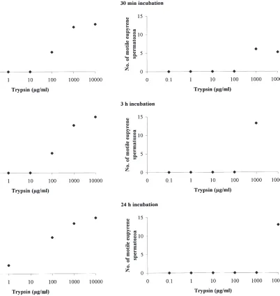
Fig. 4. Effects of concentration of trypsin in HEPES buffer solutions and incubation time in eupyrene bundle dissociation. 0, no dis-sociation; 4, complete disdis-sociation; 2 and 3, arbitrary intermediary values. Points are means of five replicates pooled data.
3.2. Spermatozoa of M. sexta
The architecture of Manduca sexta spermatozoa is similar in principle to that of other lepidopteran species. We report here changes that occur in the spermatozoa and adjunct structures that are overtly related to the acquisition of intrinsic motility of the spermatozoa. For further information and references on other aspects of lepidopteran spermatozoa see Friedla¨nder and Gitay (1972), Riemann (1971) and Friedla¨nder (1997).
3.3. Intratesticular spermatozoa
The spermatozoa are assembled in bundles, which contain exclusively either eupyrene or apyrene cells.
Fig. 5. Effects of concentration of trypsin in HEPES buffer solutions and incubation time on spermatozoa intrinsic motility. Points are means of five replicates pooled data.
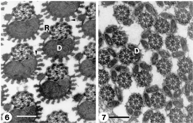
Fig. 6. Transverse section through an intra-testicular eupyrene bundle showing lacinate appendages (arrows) made of stacks of parallel lamellae resembling rays of a rising sun and reticular appendages (R) connected to the cell body by radial thin lamellae. D, mitochondrial derivatives. Scale bar=200 nm. Fig. 7. Transverse section through an intra-testicular apyrene bundle. Spermatozoa lack any noticeable surface ornamentation. D, mitochondrial derivatives. Scale bar=200nm.
3.4. Spermatozoa in the seminal vesicle
The cyst cells, previously enclosing the sperm bundles, are now absent. The eupyrene spermatozoa, however, retain the integrity of cystic configuration and appear as bundles of spermatozoa embedded in a matrix formed by an entanglement of electron-opaque wavy fibres, criss-crossing among them. This matrix material appears more densely packed at the periphery of the bun-dle, close to where the cyst cells were previously located (Figs. 8 and 9). In contrast, no apyrene bundles are present in the seminal vesicle and the apyrene spermato-zoa, lacking any particular pattern of distribution, are dispersed amongst the eupyrene bundles (Fig. 8).
The surface of the eupyrene spermatozoon displays profound modifications. The lacinate appendages are no longer noticeable and two concentric new envelopes now cover the spermatozoon (Fig. 11). In transverse sections, the inner envelope appears as an uneven, asymmetric ring, or a closed crescent. The outer envelope is rela-tively thin and has a longitudinal slit running along the spermatozoon that disrupts its otherwise perfect radial symmetry (Fig. 8, see also Figs. 16 and 17). The slit is located next to the bulkiest portion of the internal envel-ope; the reticular appendage bulges through this slit (Fig. 8, see also Figs. 16 and 17). In tangential sections, the outer envelope displays a regular pattern of equidistant, parallel striations that represent either gyres of a spiral or a series of consecutive closely packed rings (Fig. 9; see also Fig. 11).
The apyrene spermatozoon is covered by only one envelope, which is similar in appearance to the outer envelope of the eupyrene spermatozoon. But in contrast,
the envelope of the apyrene spermatozoon lacks the slit that is found in the eupyrene outer envelope (Fig. 8; see also Fig. 12). A flocculent material is found between the cell membrane and the envelope.
3.5. Spermatozoa in the spermatheca
The electron-opaque matrix, previously found among the eupyrene spermatozoa, is no longer present. The eup-yrene spermatozoa, like the apeup-yrene ones, are no longer associated in bundles but, like the apyrene ones, are now dispersed.
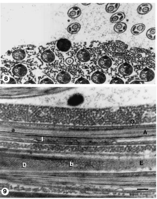
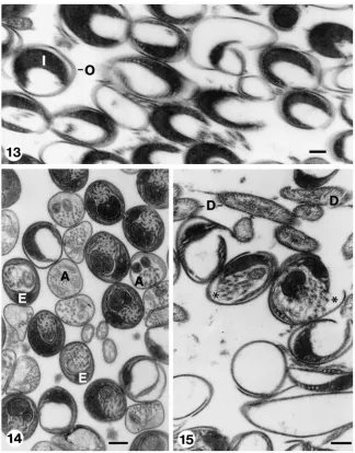
Fig. 8. Transverse section through seminal vesicle. Eupyrene spermatozoa remain in bundles and are embedded in a matrix of entangled electron-opaque wavy fibres. Note that somatic cyst cells are absent from the bundle periphery. Each spermatozoon is surrounded by inner and outer envelopes. Reticular appendages (arrows) protrude through a longitudinal slit of the outer envelope. Apyrene cells (*) are dispersed around the eupyrene bundle (cf. Figs 16 and 17). Scale bar=300 nm. Fig. 9. Longitudinal sections through flagellae of eupyrene spermatozoa in seminal vesicle. The upper flagellum (a) displays axoneme (A) and inner (I) and outer (O) envelopes. A tangentially oblique sectioned flagellum (b) displays mitochondrial derivatives (D) and a regular pattern of equidistant parallel striations of outer envelope (E). Scale bar=200 nm.
envelopes and appear scattered within the spermatheca (Fig. 15).
The apyrene spermatozoa remain in the spermatheca within their envelopes, which are sometimes covered by spikes or spheres (Fig. 12), and finally decay (Fig. 14).
3.6. Trypsin-treated spermatozoa
The incubation times and trypsin concentrations used in the following experiments were chosen so as to com-pare with those used in the earlier part of this study.
The structure of the spermatozoa, kept in clean HEPES buffer solution for 30 min (Fig. 16), is similar to that of the control spermatozoa that were fixed immediately after being dissected (cf. Figs. 8 and 9).
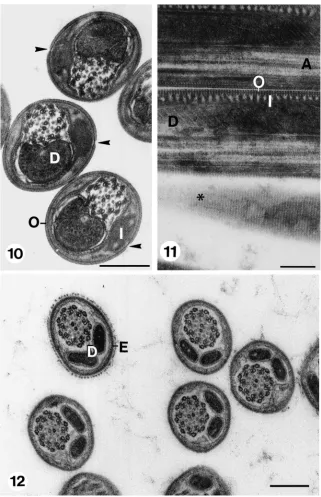
Fig. 10. Transverse sections through eupyrene spermatozoa in spermatheca. Note absence of both extra cellular matrix and reticular appendages. A longitudinal slit (arrows) in the outer envelope marks the location at which the reticular appendages bulged while spermatozoa were in seminal vesicle. D: mitochondrial derivatives, I, inner envelope; O, outer envelope. Scale bar=200 nm. Fig. 11. Longitudinal sections through eupyrene spermatozoa in spermatheca. Note the parallel, equidistant striation in tangential section of spermatozoon (*) and connections between outer (O) and inner (I) envelopes. A, axoneme; D, mitochondrial derivative. Scale bar=200 nm. Fig. 12. Transversal sections through apyrene spermatozoa in spermatheca. The sections are similar in appearance to those of apyrene spermatozoa in seminal vesicle (cf. Fig. 8). Note absence of slit in envelope (E). D, mitochondrial derivatives. Scale bar=200 nm.
treated samples remain similar in appearance to controls untreated with the enzyme.
Samples treated with 1 mg/ml trypsin solution for 1 min, resemble those few bundles, as described above, that react to treatments with 0.1 mg/ml trypsin solution for 20 min (Fig. 18). They display a reduced eupyrene inner envelope and a very reduced matrix surrounding
these spermatozoa. The reticular appendages are separ-ated from the cell body. Additionally, the external envel-ope expands and the volume of the cell increases.
Fig. 13. Spermatheca showing transverse sections of empty envelopes from which eupyrene spermatozoa “hatched”, before leaving spermatheca to fertilize the eggs. I, inner envelope; O, outer envelope. Scale bar=200 nm. Fig. 14. Section of spermatheca showing various stages of decay of both eupyrene (E) and apyrene (A) spermatozoa within their envelopes. Scale bar=200 nm. Fig. 15. Section through spermatheca showing eupyrene spermatozoa at their late stages of decay. Note open envelopes still containing spermatozoa (*), empty envelopes and isolate mitochondrial deriva-tives (D). Scale bar=200 nm.
Treatments with 5 mg/ml trypsin solution for 1 min pro-duce results similar to those just described above. How-ever, samples treated with 5 mg/ml trypsin solution for 15 min, in addition to the alterations the eupyrene sperm-atozoa endured, as indicated before, show: (a) Mixed eupyrene and apyrene spermatozoa (Fig. 20); (b) outer eupyrene envelopes opened up at the slit where the bulg-ing reticular appendages was previously attached (Figs. 20 and 21); (c) “naked” eupyrene spermatozoa; and (d) empty outer eupyrene envelopes (Fig. 21). Apyrene sper-matozoa are structurally unaffected by trypsin treatments at either 1 or 5 mg/ml and remain similar in appearance to controls that were not exposed to trypsin treatments (Figs. 20 and 21; cf. Fig. 8).
4. Discussion
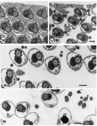
Fig. 16. Transverse section through an eupyrene bundle from seminal vesicle, after incubation in clean HEPES buffer for 30 mins. Sperm structure and extracellular matrix are similar to those of corresponding untreated spermatozoa in seminal vesicle (cf. Fig. 8). Scale bar=200 nm. Fig. 17. Transverse section through eupyrene bundle from seminal vesicle, after incubation in 0.1 mg/ml trypsin solution in HEPES buffer for 1 min, showing dispersion of the extra-cellular matrix. I, inner envelope; O, outer envelope. Scale bar=200 nm. Fig. 18. Transverse section through eupyrene bundle from seminal vesicle, after incubation in 0.1 mg/ml trypsin solution in HEPES buffer for 20 min, showing lack of extra-cellular matrix, decay of inner envelope (I) and detachment of reticular appendages (R). Scale bar=200 nm. Fig. 19. Transverse sections through eupyrene bundle from seminal vesicle, after incubation in 1 mg/ml trypsin solution in HEPES buffer for 30 min showing disappearance of the inner envelope and increase in size of the detached reticular appendages (R). Scale bar=200 nm.
then break up and the cystic configuration of the eupyr-ene cells disappears. The reticular appendage discon-nects from the cell surface and, eventually, the eupyrene spermatozoa “hatch” from their inner and outer envel-opes. In contrast, the structure of the apyrene spermato-zoa apparently does not change; they keep the basic con-formation that they displayed in the seminal vesicles, as they do during their normal evolution.
Moreover, the various distinct events leading to the acquisition of intrinsic sperm motility, appear to be time-and dose-dependent. The lower the trypsin concentration and the shorter the period of incubation, the fewer the
Figs. 20 and 21. Transverse sections through spermatozoa from seminal vesicle after incubation in 5 mg/ml trypsin solution in HEPES buffer for 15 min. Fig. 20 shows beginning of opening of slit of outer envelope of eupyrene spermatozoa (arrows). Apyrene spermatozoa (A) remain unchanged (cf. Fig. 8). Fig. 21 shows outer envelope of eupyrene spermatozoa widely open (arrows). Note empty envelope of eupyrene spermatozoa (*). Apyrene spermatozoa (A) remain unchanged (cf. Fig. 8). Scale bar=200 nm.
low concentration of trypsin for an extended period of incubation, produced an effect similar to that of the use of a high concentration of trypsin for a short period.
In our experiments, we used a single exogenous pro-teolytic enzyme, bovine trypsin, to induce in vitro a cas-cade of sequential structural changes that is involved in inducing sperm motility. It is possible that in vivo, more than one endogenous protease is involved in the cascade, as has been reported for Bombyx mori (Osanai et al., 1987). In vivo, these enzymes may act sequentially or together. If the action is sequential, the difference in their timing of activity may be reflected in our findings of different effects caused by the different times of incu-bation and concentration of trypsin.
Our results show that the acquisition of intrinsic motility by the eupyrene spermatozoa is correlated with the disappearance of the electron-opaque extra-cellular material, which is found among the spermatozoa in the bundles taken from the seminal vesicle. We suggest that this material is responsible for the maintenance of the integrity of the sperm bundle, in the absence of the envelope of cyst cells that enclose the intra-testicular bundles. However, the “hatching” of the eupyrene sper-matozoon from its envelopes is probably not directly related to motility, as the spermatozoa can move even before “hatching”. Rather, “hatching” may be related to capacitation of the eupyrene spermatozoa, either enabling them to leave the spermatheca or to enter the egg, or even both. This is in accordance with our data showing that neither apyrene spermatozoa, which never “hatch”, nor those eupyrene spermatozoa that fail to “hatch”, leave the spermatheca. And, eventually, both decay within the spermatheca.
Acknowledgements
We thank Rina Jeger for her help in the preparation of the materials. This work was supported by a grant from the European Commission (No. CI1*-CT94-0094).
References
Bell, R.A., Joachim, F.G., 1976. Techniques for rearing laboratory col-onies of tobacco hornworms and pink ballworms. Annals of the Entomological Society of America 69, 365–373.
Friedla¨nder, M., 1997. Control of the eupyrene–apyrene spermatogen-esis dimorphism in Lepidoptera. Journal of Insect Physiology 43, 1092–1097.
Friedla¨nder, M., Gitay, E., 1972. The fate of the normal-anucleated spermatozoa in inseminated females of the silkworm Bombyx mori. Journal of Morphology 138, 121–129.
Holt, G.G., North, D.T., 1970. Effects of gamma irradiation on the mechanism of sperm transfer in Trichoplusia ni. Journal of Insect Physiology 16, 2211–2222.
Meves, F., 1903. Ueber oligopyrene und apyrene Spermien und u¨ber ihre Enstehlung, nach Beobachtungen an Paludina und Pygaera. Archives den mikroskopische Anatomie 61, 1–82.
Omura, S., 1938. Studies on the reproductive system of male Bombyx
mori. II. Post-testicular organs and post-testicular behavior of the
spermatozoa. Journal of Faculty of Agriculture of Hokkaido Imperial University Sapporo 40, 129–170.
Osanai, M., Baccetti, B., 1993. Two-step acquisition of motility by insect spermatozoa. Experientia 49, 593–595.
Osanai, M., Kasuga, H., 1990. Role of endopeptidase in motility induc-tion in apyrene silkworm spermatozoa; micropore formainduc-tion in the flagellar membrane. Experientia 46, 261–264.
Osanai, M., Kasuga, H., Aigaki, T., 1987. Physiological role of apyr-ene spermatozoa of Bombyx mori. Experientia 43, 593–596. Phillips, D.M., 1971. Morphogenesis of the lacinate appendages of
lep-idopteran spermatozoa. Journal of Ultrastructure Research 34, 567–585.
Riemann, J.G., 1971. Metamorphosis of sperm of the cabbage looper
Trichoplusia ni, during passage from the testes to the female
sperm-atheca. In: Baccetti, B. (Ed.), Comparative Spermatology. Aca-demic Press, New York, pp. 321–331.
Shepherd, J.G., 1974. Sperm activation in saturniid moths: some aspects of the mechanism of activation. Journal of Insect Physi-ology 20, 2321–2328.
Shepherd, J.G., 1975. A polypeptide sperm activator from male satur-niid moths. Journal of Insect Physiology 21, 9–22.
Takamura, Y., Kanda, T., Horie, Y., 1999. Artificial insemination using trypsin-treated sperm in the silkworm Bombyx mori. Journal of Insect Physiology 45, 471–477.
Thibout, E., 1980. Evolution and role of apyrene sperm cells in lepi-dopterans: their activation and denaturation in the leek moth,
Acro-lepiopsis assectella (Hypomeutoı¨dea). In: Clark, W.H., Adams,