The maize streak virus coat protein transcription unit exhibits
tissue-specific expression in transgenic rice
G. Mazithulela
1, D. Sudhakar
2, T. Heckel
3, L. Mehlo, P. Christou, J.W. Davies,
M.I. Boulton *
John Innes Centre,Norwich Research Park,Colney,Norwich NR4 7UH, UK
Received 20 October 1999; received in revised form 29 October 1999; accepted 3 December 1999
Abstract
Maize streak geminivirus (MSV) is a single-stranded DNA virus that infects cereals and other grasses. A promoter region incorporating the MSV large intergenic region and movement protein gene sequence was ligated to the gus (b-glucuronidase) reporter gene which replaced the virus coat protein (CP) gene. The CP promoter activity was analysed in transgenic rice plants (Oryza sati6aL.) and was compared with that obtained in plants transformed with thegusgene downstream of the cauliflower mosaic virus (CaMV) 35S promoter. The MSV CP promoter activity varied in the five plant lines tested, but was always less than that of the CaMV promoter. Histochemistry showed that the MSV CP promoter was active in cells of regenerating callus but in regenerated plants it provided an expression pattern restricted to the vascular tissues of the root, stem, leaf and floral organs. Expression was highest in phloem-associated tissues of the vegetative organs and was absent from the tip and elongation region of seedling roots. Thus, the MSV CP promoter shows a degree of developmental regulation and can be used to confer tissue-specific expression in transgenic rice plants. © 2000 Elsevier Science Ireland Ltd. All rights reserved.
Keywords:GUS; Tissue-specific expression; Maize streak virus; MSV; Coat protein promoter; Transgenic rice
www.elsevier.com/locate/plantsci
1. Introduction
The 35S promoter of cauliflower mosaic virus (CaMV) has been widely used to provide expres-sion of foreign genes in transgenic plants. How-ever, there is a need for a wider range of promoters with different characteristics, for exam-ple, to obtain different levels of transgene expres-sion, expression at different developmental stages, or in specific organs and cell types. Furthermore, although the CaMV promoter is suitable for ex-pression of foreign genes in transgenic rice, it
provides only low expression in other economi-cally important cereals and grasses of the family Poaceae. Recent successes with the genetic trans-formation of maize [1 – 4], rice [5,6], wheat [7], oat (reviewed in [8]), and barley [9] have required the identification of appropriate promoters for the genetic modification of these crops.
Isolation of promoters from plants is ongoing but plant DNA virus promoters are now accessible and, as was the case for CaMV, may contain cis
regulatory elements able to confer specific patterns of transgene expression in plants [10]. The pro-moters of other pararetroviruses such as cassava vein mosaic virus (CsVMV, [11]), Commelina yel-low mottle virus (CoYMV, [12]), sugarcane bacilli-form virus (ScBV, [13]) and rice tungro bacillibacilli-form virus (RTBV, [14,15]) have been investigated for their ability to promote gene expression in trans-genic cereals. All but CsVMV naturally infect monocots, but only ScBV and RTBV infect mem-* Corresponding author. Tel.: +44-1603-452571; fax: +
44-1603-456844.
E-mail address:[email protected] (M.I. Boulton)
1Present address: Dupont Agricultural Enterprise, Stine-Haskell
Research Centre, P.O. Box 30, Newark, DE 19711, USA.
2Present address: Centre for Plant Molecular Biology, Tamil Nadu
Agricultural University, Coimbatore 641003, India.
3Present address: RDP, Ens-Lyon, 46 Allee d’Italie, F-69364 Lyon
Cedex, France.
bers of the Poaceae. The pattern of transgene expression, identified by b-glucuronidase (GUS) staining, was not identical for the promoters of these viruses, with the CsVMV and ScBV pro-moters being active in all organs tested and providing near constitutive expression, whereas the RTBV and CoYMV promoters were specific to the vascular tissue. Only the RTBV promoter was tested in its natural host species. Another plant DNA virus family, the Geminiviridae, contains a genus (Mastre6irus, [16]) whose species infect members of the Poaceae. These viruses include maize streak virus (MSV), wheat dwarf virus (WDV) and Chloris striate mosaic virus (CSMV). Of these, MSV has the widest host range, infecting most of the economically important Poaceae [17]. However, the activity of the Mastre6irus pro-moters in transgenic cereals has not yet been reported.
MSV possesses a single-stranded circular DNA genome but transcription of the double-stranded viral DNA is bidirectional, initiating mainly in the large intergenic region (LIR), and terminating in the small intergenic region (SIR) which contains polyadenylation signals [18 – 20]. The LIR contains the rightward promoter element (rpe1) [21] from which the virion sense genes encoding the move-ment protein (MP) and coat protein (CP) are expressed [20].
To study the strength and tissue specificity of the MSV CP promoter region in transgenic rice, a
cp-gus reporter gene replacement construct was made. The construct contained the MSV LIR and SIR, the mp gene sequence and parts of the repli-cation associated genes and therefore constituted an MSV CP ‘extended promoter’. This MSV pro-moter conferred tissue specific expression and showed some developmental regulation in trans-genic rice plants. The properties of this promoter are compared with those of the CaMV 35S pro-moter, and other virus promoters, in the context of their use for tissue-specific expression in trans-genic rice.
2. Methods
2.1. Constructs for gene transfer
Two plasmids were used for transformation of rice immature embryos. Plasmid WRG4517 [22] contained the hpt gene (conferring hygromycin resistance) and was used to select transgenic callus. Plasmid MSVCPp-gus contained the gus reporter gene, encoding GUS, in place of the MSV CP coding region. This construct was designed to allow GUS to be produced from a MSV expres-sion unit that contained all identified MSV
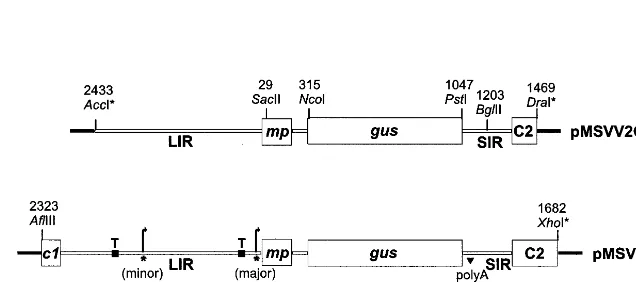
tran-Fig. 1. Construction of pMSVCPp-gus. Plasmid MSVCPp-gus used for bombardment of rice cultures was constructed from pMSVV2 [23] and a clone containing a tandem repeat of the Maize streak geminivirus (MSV)-Ns genome. The position of restriction enzyme sites used for the construction of pMSVV2 and pMSVCPp-gus are shown above the plasmids, the MSV co-ordinates are those reported in Ref. [18]. Restriction sites destroyed during the cloning procedure are suffixed by *. The position of the MSV andgusgenes are shown by boxes, below these the MSV coat protein (CP) extended promoter region, and the proteins likely to be translated in transgenic plants, are shown. Virion-sense transcript initiation sites mapped to the MSV-Ns genome [20] are indicated by * and curved arrows show the direction of transcription. TATA boxes are marked ‘T’. Thec1 gene product is also known as RepA, and the C2 open reading frame (ORF) product forms the C-terminus of the C1:C2 (Rep) protein.
scription regulatory sequences, thereby comprising an ‘extended’ CP promoter (Fig. 1). Up-stream of the gus gene there is the 5% region of the MSV c1 (replication-associated protein A, repA) gene, the MSV LIR and the MSVmpgene (in which the MSV virion sense intron is situated, [20]). The SIR and the MSV C2 open reading frame (ORF) are down-stream ofgus. Plasmid MSVCPp-guswas produced from a previously constructed plasmid pMSVV2-GUS ([23] and Fig. 1). Plasmid MSVV2pMSVV2-GUS, based on pUC19, contained MSV (strain Ns, [24]) se-quences between the AccI and DraI sites of MSV (coordinates 2433 – 1469, according to [18]), except that thecpsequence was replaced by theNcoI –PstI
gus gene fragment (a gift from V. Citovsky). This replacement was facilitated by site-directed mutage-nesis at thecptranslation initiation and termination sites to produce NcoI and PstI sites, respectively. As pMSVV2GUS did not contain the entire LIR, a 2 kbp AflIII –XhoI MSV fragment (comprising MSV sequences between co-ordinates 2323 and 1682) was introduced intoAflIII andSalI-digested pUC19 to generate construct pMSVPUC19. The
SacII –BglII fragment from pMSVV2GUS, which contained thegusreplacement, was then introduced in place of the homologous segment of pMSVPUC19 to generate pMSVCPp-gus.
2.2. Particle bombardment and regeneration of
transformed plants
Immature rice embryos (Oryza sati6a L., geno-type ITA212) were transformed by particle bom-bardment and cultured as described [5] and modified [25]. Plasmids pMSVCPp-gus and pWRG4517 were co-precipitated onto 0.95mm gold particles at a ratio of 3:1 by weight. Transformed callus was selected and regenerated on CCM3 medium containing 35 mg l−1hygromycin.
Regen-erated rice plants were transferred to a loam-based compost in a controlled-environment growth cham-ber (80% humidity, 27°C) before transfer to a glasshouse maintained at 25°C (95°C) and ap-proximately 80% humidity. Daylight was supple-mented to a 16-h photoperiod. Plants regenerated from tissue culture were designated as the T0
generation. Plants regenerated from the same im-mature embryo-derived callus (each callus was des-ignated a ‘clone’) were given the same line number. T1plants were derived by self-pollination of the T0
plants.
2.3. Southern hybridisation analysis of transgenic
plants
Nuclear DNA was isolated from the leaves of rice plants using the Nucleon-Phytopure™ DNA Extraction Kit (Amersham). Purified DNA was estimated spectrophotometrically and also by comparison with ethidium bromide-stained control DNA samples, following electrophoresis in a TBE agarose gel [26]. Genomic DNA (5mg) was digested with SacII, which cuts at a single site in pMSVCPp-gus. Southern transfer of the separated DNA fragments to Hybond N+ membranes (Amersham) and hybridisation to a high specific activity a-[32P]dCTP-labelled NcoI –PstI gus
fragment from pMSVV2GUS (Fig. 1), were carried out according to the method of Ref. [27]. Hybridisation was visualised using phosphor-imaging.
2.4. Aseptic germination of seeds
Rice seeds (T1 generation or non-transformed)
were dehusked by rubbing laterally with sandpaper (grit number 50, English Abrasives). After surface-sterilisation, the seeds were placed on sterile, damp filter paper in Petri plates. Incubation was in the dark at 37°C for 16 – 24 h and then germination was completed at 25 – 27°C for up to 8 days. Seeds were used for GUS assays or were transferred to compost if mature plants were required.
2.5. Analysis of GUS acti6ity in rice plants
2.5.1. Chemiluminescent analysis
Quantitative detection of GUS in the young (approx 5 cm long) leaves of T0plants was carried
stan-Table 1
b-glucuronidase (GUS) activity in leaves and fertility of trans-formed (T0) rice plants
Mean RLUa
Line c Seed production/plant
(and fertility)b
aRelative light units (RLU) are presented as the mean for
each line (n=number of siblings tested) and are expressed per 10mg protein min−1. Data were analysed using an analysis of variance test (95% confidence; Microsoft Excel 1997©).
bFertility is shown as the number of plants producing seed:
number of sterile plants. cND, not determined.
dThis line was transformed with pWRG4517.
cording to the manufacturer’s instructions. Sam-ples were infiltrated for 5 min under vacuum and incubated for a further 4 h with 50% Historesin™, 50% absolute ethanol, before transfer to 100% resin for 24 h. The resin was polymerised overnight at 4°C. Samples were sectioned with glass knives using a Leica RM2055 microtome. Sections of 2 mm (root) or 7 mm (leaf) tissue were taken, viewed under a Nikon microscope with bright field illumination, and photographed with a Nikon camera using Kodak Ektachrome 160 T film. Identification of cell type and tissue was according to Refs. [32,33].
3. Results
3.1. Regeneration of rice transformed with
pMSVCPp-gus
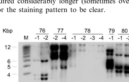
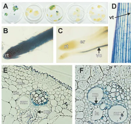
Sixteen clones of ITA212 hygromycin-resistant callus were obtained following co-bombardment of 200 immature embryos with pWRG4517 (carry-ing the hygromycin resistance gene) and pMSVCPp-gus. To determine whether the MSV CP promoter was active in callus tissue, clones were subjected to GUS histochemical assay 6 weeks after the start of selection. After staining for 16 h, all but five of the clones showed some blue colouration although the intensity of staining varied considerably and in most clones there were unstained regions (examples of the staining are shown in Fig. 3A). Seventeen plants (derived from five clones) were regenerated (Table 1); sibling plants were identified by their line number fol-lowed by an individual number (e.g. 76-1 – 76-5). Plants were not obtained from the two clones exhibiting the highest levels of GUS expression. Southern blot hybridisation analysis of selected regenerants from each clone confirmed that all plants were transformed. All lines showed complex patterns of transgene integration and the pattern for each line was unique. However, the patterns for siblings were identical (Fig. 2), showing that each line resulted from a single transformation event. GUS activity, measured by chemilumines-cent assay of leaf tissue, varied between plant lines, but similar levels were observed in sibling plants. GUS activity from plants transformed with the pMSVCPp-gus construct was markedly below that in plants transformed with the CaMV 35S dards using the Titertek Multiscan™ plate reader
and software.
2.5.2. Histochemical staining and microscopical analysis of tissues
Histochemical staining of rice tissue for GUS activity was done essentially as previously de-scribed [28,29] in stain containing 0.05 mM potas-sium ferricyanide and 0.05 mM potassium ferrocyanide. In order to wet the tissues and aid absorption of the stain, a drop of Nonidet-P40 (Sigma) was added to each 10 ml of stain. The stain was vacuum-infiltrated for five minutes to aid penetration into all tissues and samples were incubated for 12 – 16 h at 37°C. When necessary, stained tissues were cleared of pigment by incuba-tion in chlorallactophenol for 24 – 48 h as de-scribed [30]. For chemiluminescent and histo-chemical staining, rice plants (Koshihikari, [31] or ITA 212 plant 15-1, Duc Le Tan, John Innes Centre, unpublished data) transformed with a CaMV 35S-gus construct served as positive con-trols for assay techniques.
ac-promoter (Table 1). Subsequent histochemical as-say showed that this was caused, at least in part, by the restriction of activity to the vascular tissues compared with ubiquitous expression from the CaMV promoter. Line 76 had high GUS activity relative to the very low expression seen in lines 77 – 80.
Plants had a normal phenotype and seeds were obtained from all lines, although three of the five line 76 plants and one each from lines 77 and 78 were sterile. Lines 76 and 80 produced fewer seeds than lines 77 – 79. Thirty-five seeds each from plants 76-2 (high GUS) and 77-2 (low GUS) were germinated aseptically and stained to determine whether GUS activity was reproducible and heri-table. All of the T1seedlings of line 76-2 and 33 of
line 77-2 showed GUS staining (most easily as-sessed in the roots). These data are indicative of integration of the transgene into more than one locus. The level of GUS expression remained higher for the line 76 derived seedlings than for the remaining lines suggesting that expression from the MSV CP promoter was stable through at least one generation. Unlike plants transformed with CaMV 35S-guswhich showed staining within 2 – 4 h, tissues transformed with pMSVCPp-gus re-quired considerably longer (sometimes overnight) for the staining pattern to be clear.
3.2. The MSV ‘extended’ CP promoter confers a
6ascular expression pattern in transgenic rice
The gross GUS expression pattern conferred by the MSV extended CP promoter in mature leaf tissue and primary roots was determined for one plant from each T0 line when plants were 2
months old. All showed GUS staining associated with the vascular tissue, although the intensity varied, with plant 76-1 showing the most intense staining (Fig. 3C,D). No endogenous GUS activity was seen in leaves or roots of plants transformed with pWRG4517 alone, or in tissue culture-derived non-transformed plants (not shown). Since the leaves and roots of all lines transformed with pMSVCPp-gusrevealed a vascular-specific expres-sion pattern, only plants 76-1 and 77-2 were sub-jected to further histochemical analysis. The pattern of expression controlled by the MSV CP promoter in plant 76-1 is summarised in Fig. 3. A similar expression pattern was seen for plant 77-2, but the lower expression level made detection of staining difficult, especially in cells with little cyto-plasm. In thin sections of leaf tissue, GUS activity was strongest in phloem and phloem-associated cells (phloem parenchyma and companion cells, Fig. 3E) although stain could also be seen in xylem parenchyma cells. Apparent staining of the epidermal cells was also seen in non-transformed controls and therefore was not attributed to ex-pression from the CP promoter. No exex-pression was detected in outer bundle sheath, mesophyll or sclerenchyma cells.
The expression pattern in the vascular bundles of the stem was similar to that found in the leaf, except that the xylem parenchyma was more strongly stained (Fig. 3F). The MSV CP promoter was also expressed in flowers; staining was limited to the vascular tissue of the palea and lemma, which are considered to comprise tissues similar to the leaf sheath [33]. Approximately 50% of pollen grains showed GUS activity (not shown).
In primary roots, GUS expression was limited to the vascular cylinder of the mature region; no activity was detected in the meristematic (root tip) or elongation regions despite the presence of vas-cular tissue in the elongation region (Fig. 3C). However, these regions were stained strongly in plants transformed with CaMV 35S-gus (Fig. 3B) even after a short staining period, showing that stain was able to penetrate these regions. The
Fig. 3. Histochemical localisation ofb-glucuronidase (GUS) activity in rice callus and plants transformed with pMSVCPp-gus(A, C – F) or CaMV 35S-gus(B). Tissues were stained for 16 h. (A) Transformed callus; (B, C) primary roots; (D) part of a mature leaf blade; (E) transverse section through the central conducting element of a leaf midrib (adaxial region); (F) transverse section through a vascular bundle of the stem. rt, root tip; er, elongation region; vc, vascular cylinder; vt, vascular tissue; mc, mesophyll cells; mx, metaxylem; p, phloem; la, xylem lacuna; xp, xylem parenchyma.
vascular-specific expression of the MSV CP pro-moter was maintained in lateral roots of older seedlings (not shown).
4. Discussion
Although following co-bombardment of rice im-mature embryos with pMSVCPp-gus and pWRG4517 16 hygromycin-resistant callus clones were obtained, plants could be regenerated from only five of the 11 clones that showed GUS
activ-ity, and plants could not be obtained from the two lines which exhibited the highest activity. Further-more, the two fertile T0 plants of line 76, which
as well as the entire mp gene, are present within pMSVCPp-gus, but only the first 17 amino acids of the C1 protein may be produced and C2 ORF should not be transcribed. It may be that expres-sion of the MSV mp gene, situated immediately upstream of the cp gene in MSV, could affect regeneration efficiency if expressed at ‘high’ level. For each line, the level of GUS is likely to reflect that of the MP as both genes are expressed from the same transcription unit. However, it is clear that the variability in GUS expression levels be-tween different transformed rice lines is influenced by factors other than the promoter sequences in the MSV LIR. Such variation in expression is generally related to the chromosomal location of the transgene, the extent to which it is methylated and the transgene copy number [34]. It has been shown that the MSV MP is toxic when over expressed in transgenic N. benthamiana (Pitak-sutheepong, Davies and Boulton, unpublished). If failure to regenerate plants with high GUS activity is determined by the toxicity of the MP, then disruption of the mp may result in transgenic plants with higher levels of expression from the MSV CP promoter.
The complex transgene integration pattern present in the pMSVCPp-gus transformed rice is similar to that reported in other studies [35] and also was seen in the CaMV35S-gus transformed rice used as controls in the current study [31]. It is ascribed to the presence of multiple copies of both intact and fragmented plasmid sequences. The complexity does not prevent comparison of the strength or expression pattern of the MSV CP promoter, as all lines produced the same tissue-specific expression pattern. Furthermore, many other studies of promoter expression in cereals have been carried out using plants with multiple transgene insertions or in plants for which the integration patterns were not characterised [13,36,37].
Unlike the CaMV promoter that directs expres-sion in most cell types, the MSV CP promoter directed vascular-specific expression in all plant tissues examined. However, expression was also seen in undifferentiated callus tissue (Fig. 3A). A number of viral promoters have been reported to direct vascular-specific expression in monocots (e.g. RTBV [14,15], CoYMV, [12] and banana bunchy top virus (BBTV, [38]), although in the case of RTBV and BBTV expression was also seen
in the actively dividing cells of the root tip. Strong activity of the BBTV DNA-6 promoter in undif-ferentiated, actively dividing cells (root and callus) was suggested to be a result of a motif with homology to the hexamer motif of plant histone promoters which confers S-phase specific expres-sion. A motif with homology to the octamer motif of the maize histone 4 gene (H4C14) was identified in MSV and other mastreviruses [39]. Although this motif is situated at MSV co-ordinates 181 – 197, within the mpgene (and the CP promoter), it clearly does not direct S-phase expression in rice roots. However, the differential activity of the MSV CP promoter in developmentally distinct tissues of the same cell lineage in the root, suggests that the MSV CP promoter exhibits a degree of developmental regulation in transgenic rice.
Further work will be required to determine whether expression from the MSV CP promoter can be enhanced, and to identify signals conferring tissue and developmental specificity. Nevertheless, this study shows that the MSV CP promoter pro-vides unique gene expression patterns of use for the genetic manipulation of a wide range of eco-nomically important cereals and grasses.
Acknowledgements
We would like to thank Brian Wells for assis-tance with histological procedures. TheNcoI –PstI
gusgene fragment was a gift from Dr V. Citovsky, State University of New York, Stony Brook. G. Mazithulela and L. Mehlo are recipients of Rocke-feller Foundation studentships and we acknowl-edge further financial support from the UK/SA Science and Technology Research Fund. D. Sud-harkar was sponsored by the Rockefeller Founda-tion Career Fellowship programme. The John Innes Centre is grant-aided by the Biotechnology and Biological Sciences Research Council. MSV was held and manipulated under the conditions of MAFF licences PHL 11/2300 and PHL11A/ 2627(6/1998). We thank Dr R. Twyman for cor-rections to the manuscript.
References
[1] M.E. Fromm, F. Morrish, C. Armstrong, R. Williams, J. Thomas, T.M. Klein, Inheritance and expression of chimeric genes in the progeny of transgenic maize plants, Bio/Technology 8 (1990) 833 – 839.
[2] W.J. Gordon-Kamm, T.M. Spencer, M.L. Mangano, T.R. Adams, R.J. Daines, W.G. Start, J.V. O’Brien, S.A. Chambers, W.R. Adams Jr, N.G. Willetts, T.B. Rice, C.J. Mackey, R.W. Krueger, A.P. Kausch, P.G. Lemaux, Transformation of maize cells and regeneration of fertile transgenic plants, Plant Cell 2 (1990) 603 – 618.
[3] D.A. Walters, C.S. Vetsch, D.E. Potts, R.C. Lundquist, Transformation and inheritance of a hygromycin phos-photransferase gene in maize plants, Plant Mol. Biol. 18 (1992) 189 – 200.
[4] H. Zhong, B. Sun, D. Warkentin, S. Zhang, R. Wu, T. Wu, M.B. Sticklen, The competence of maize shoot meristems for integrative transformation and inherited expression of transgenes, Plant Physiol. 110 (1996) 1097 – 1107.
[5] P. Christou, T.L. Ford, M. Kofron, Production of trans-genic rice (Oryza sati6a L.) plants from agronomically
important Indica and Japonica varieties via electric dis-charge particle acceleration of exogenous DNA into
immature zygotic embryos, Bio/Technology 9 (1991) 957 – 962.
[6] Y. Ishida, H. Saito, S. Ohta, Y. Hiei, T. Komari, T. Kumashiro, High efficiency transformation of maize (Zea mays L.) mediated by Agrobacterium tumefaciens, Nat. Biotechnol. 14 (1996) 745 – 750.
[7] V. Vasil, A.M. Castillo, M.E. Fromm, I.K. Vasil, Herbi-cide-resistant fertile transgenic wheat plants obtained by microprojectile bombardment of regenerable embryo-genic callus, Bio/Technology 10 (1992) 667 – 674. [8] D.A. Somers, W. Rines, K.A. Torbert, W.P. Pawlowski,
S.K.C. Milach, Genetic transformation in A6ena sati6a
L. (oat), in: Y.P.S. Bajaj (Ed.), Biotechnology in Agricul-ture and Forestry: Plant Protoplasts and Genetic Engi-neering VII, vol. 38, Springer-Verlag, Berlin, 1996, pp. 178 – 190.
[9] A. Ritala, K. Aspegren, U. Kurten, M. Salmenkallio-Marttila, L. Mannonen, R. Hannus, V. Kauppinen, T.H. Teeri, T.-M. Enari, Fertile transgenic barley by particle bombardment of immature embryos, Plant Mol. Biol. 24 (1994) 317 – 325.
[10] P.N. Benfey, L. Ren, N.-H. Chua, The CaMV 35S enhancer contains at least two domains which can confer different developmental and tissue-specific expression patterns, EMBO J. 8 (1989) 2195 – 2202.
[11] B. Verdaguer, A. de Kochko, R.N. Beachy, C. Fauquet, Isolation and expression in transgenic tobacco and rice plants, of the cassava vein mosaic virus (CVMV) pro-moter, Plant Mol. Biol. 31 (1996) 1129 – 1139.
[12] K.A. Torbert, M. Gopalraj, S.L. Medberry, N.E. Ol-szewski, D.A. Somers, Expression of the Commelina
yellow mottle virus promoter in transgenic oat, Plant Cell Rep. 17 (1998) 284 – 287.
[13] I. Tzafrir, K.A. Torbert, B.E.L. Lockhart, D.A. Somers, N.E. Olszewski, The sugarcane bacilliform badnavirus promoter is active in both monocots and dicots, Plant Mol. Biol. 38 (1998) 347 – 356.
[14] M. Bhattacharyya-Pakrasi, J. Peng, J.S. Elmer, G. Laco, P. Shen, M.B. Kaniewska, H. Kononowicz, F. Wen, T.K. Hodges, R.N. Beachy, Specificity of a promoter from the rice tungro bacilliform virus for expression in phloem tissues, Plant J. 4 (1993) 71 – 79.
[15] Y. Yin, R.N. Beachy, The regulatory regions of the rice tungro bacilliform virus promoter and interacting nu-clear factors in rice (Oryza sati6a L.), Plant J. 7 (1995) 969 – 980.
[16] M.A. Mayo, C.R. Pringle, Virus taxonomy — 1997, J. Gen. Virol. 79 (1997) 649 – 657.
[17] J. Stanley, M.I. Boulton, J.W. Davies, The Geminiviri-dae, In: Embryonic Encyclopedia of Life Sciences, Na-ture Publishing Group, London, www.els.net.
[18] P.M. Mullineaux, J. Donson, B.A.M. Morris-Krsinich, M.I. Boulton, J.W. Davies, The nucleotide sequence of maize streak virus DNA, EMBO J. 3 (1984) 3063 – 3068. [19] B.A.M. Morris-Krsinich, P.M. Mullineaux, J. Donson, M.I. Boulton, P.G. Markham, M.N. Short, J.W. Davies, Bidirectional transcription of maize streak virus DNA and identification of the coat protein gene, Nucleic Acids Res. 13 (1985) 7237 – 7256.
virion- and complementary-sense gene expression, Plant J. 12 (1997) 1285 – 1297.
[21] C. Fenoll, J. Schwarz, D.M. Black, M. Schneider, S.H. Howell, The intergenic region of maize streak virus con-tains a GC-rich element that activates rightward tran-scription and binds maize nuclear factors, Plant Mol. Biol. 15 (1990) 865 – 877.
[22] J. Cooley, T. Ford, P. Christou, Molecular and genetic characterization of elite transgenic rice plants produced by electric-discharge particle acceleration, Theor. Appl. Genet. 90 (1995) 97 – 104.
[23] T. Heckel, Pathogenicity determinants and gene expres-sion of maize streak virus, PhD thesis, University of East Anglia, UK, 1996.
[24] M.I. Boulton, D.I. King, J. Donson, J.W. Davies, Point substitutions in a promoter-like region and the V1 gene affect the host range and symptoms of maize streak virus, Virology 183 (1991) 114 – 121.
[25] P. Vain, B. Worland, M.C. Clarke, G. Richard, M. Beavis, H. Liu, A. Kohli, M. Leech, J. Snape, P. Chris-tou, H. Atkinson, Expression of an engineered cysteine proteinase inhibitor (Oryzacystatin-IDD86) for nematode resistance in transgenic rice plants, Theor. Appl. Genet. 96 (1998) 266 – 271.
[26] J. Sambrook, E.F. Fritsch, T. Maniatis, Molecular Cloning: A Laboratory Manual, Cold Spring Harbor Laboratory Press, New York, 1989.
[27] H. Funatsuki, H. Kuroda, M. Kihara, P.A. Lazzeri, E. Muller, H. Lorz, I. Kishinami, Fertile transgenic barley generated by direct DNA transfer to protoplasts, Theor. Appl. Genet. 91 (1995) 707 – 712.
[28] R.A. Jefferson, Assaying chimeric genes in plants: the GUS fusion system, Plant Mol. Biol. Rep. 4 (1987) 387 – 405.
[29] R.A. Jefferson, T.A. Kavanagh, M.W. Bevan, GUS fu-sions: b-glucuronidase as a sensitive and versatile gene fusion marker in higher plants, EMBO J. 6 (1987) 3901 – 3907.
[30] T. Beeckman, G. Engler, An easy technique for the clearing of histochemically stained plant tissue, Plant Mol. Biol. Rep. 12 (1994) 37 – 42.
[31] A. Kohli, M. Leech, P. Vain, D.A. Laurie, P. Christou, Transgene organization in rice engineered through direct DNA transfer supports a two-phase integration mecha-nism mediated by the establishment of integration hotspots, Proc. Natl. Acad. Sci. USA 95 (1998) 7203 – 7208.
[32] K. Esau, Anatomy of Seed Plants, second ed., Wiley, New York, 1977.
[33] T. Matsuo, K. Hoshikawa, Science of the Rice Plant, Morphology, vol. 1, Food and Agriculture Policy Re-search Centre, Tokyo, 1993.
[34] K. Weising, J. Schell, G. Kahl, Foreign genes in plants: transfer, structure, expression, and applications, Ann. Rev. Genet. 22 (1988) 421 – 477.
[35] M. Takano, H. Egawa, J.-E. Ikeda, K. Wakasa, The structures of integration sites in transgenic rice, Plant J. 11 (1997) 353 – 361.
[36] R. Terada, K. Shimamoto, Expression of CaMV35S-GUS gene in transgenic rice plants, Mol. Gen. Genet. 220 (1990) 389 – 392.
[37] M.-J. Cornejo, D. Luth, K.M. Blankenship, O.D. Ander-son, A.E. Blechl, Activity of a maize ubiquitin promoter in transgenic rice, Plant Mol. Biol. 23 (1993) 567 – 581. [38] B. Dugdale, P.R. Beetham, D.K. Becker, R.M. Harding,
J.L. Dale, Promoter activity associated with the inter-genic regions of banana bunchy top virus DNA-1 to -6 in transgenic tobacco and banana cells, J. Gen. Virol. 79 (1998) 2301 – 2311.
[39] S.Y. Morozov, A. Merits, B.K. Chernov, Computer search of transcription control sequences in small plant virus DNA reveals a sequence highly homologous to the enhancer element of histone promoters, DNA Sequence 4 (1994) 395 – 397.
[40] A.P. Lucy, M.I. Boulton, J.W. Davies, A.J. Maule, Tissue specificity ofZea maysinfection by maize streak virus, Mol. Plant-Microbe Interact. 9 (1996) 22 – 31. [41] M.I. Boulton, D.I. King, P.G. Markham, M.S. Pinner,
J.W. Davies, Host range and symptoms are determined by specific domains of the maize streak virus genome, Virology 181 (1991) 312 – 318.
[42] G. Mazithulela, The activity of MSV virion sense pro-moters and their use in the transformation of cereals, PhD thesis, University of East Anglia, UK (1998). [43] S.L. Medberry, B.E.L. Lockhart, N.E. Olszewski, The
Commelina yellow mottle virus promoter is a strong promoter in vascular and reproductive tissues, Plant Cell 4 (1992) 185 – 192.
[44] G. Sunter, D. Bisaro, Regulation of a geminivirus coat protein promoter by AL2 protein (TrAP): evidence for activation and depression mechanisms, Virology 232 (1997) 269 – 280.
[45] J.M.I. Hofer, E.L. Dekker, H.V. Reynolds, C.J. Wool-ston, B.S. Cox, P.M. Mullineaux, Coordinate regulation of replication and virion-sense gene expression in wheat dwarf virus, Plant Cell 4 (1992) 213 – 223.
[46] X.C. Zhan, K.A. Richardson, A. Haley, B.A.M. Morris, The activity of the coat protein promoter of Chloris striate mosaic virus is enhanced by its own and C1 – C2 gene products, Virology 193 (1993) 498 – 502.
[47] S. Collin, M. Fernandez-Lobato, P.S. Gooding, P.M. Mullineaux, C. Fenoll, The two non-structural proteins from wheat dwarf virus involved in viral gene expression and replication are retinoblastoma-binding proteins, Vi-rology 219 (1996) 324 – 329.