SURVEY ON THE OCCURRENCE OF VIRUSES INFECTING CUCURBITS IN YOGYAKARTA AND CENTRAL JAVA
SURVEI VIRUS YANG MENYERANG LABU-LABUAN DI YOGYAKARTA DAN JAWA TENGAH
Budi Setiadi Daryono*
Laboratory of Genetics, Faculty of Biology, Gadjah Mada University, Yogyakarta 55281, Indonesia Keiko T. Natsuaki
Laboratory of Tropical Plant Protection, Graduate School of Agriculture, Tokyo University of Agriculture, Japan *Corresponding author. E-mail: [email protected]
ABSTRACT
Cucurbits are grown throughout the Java Island as dry season crops. Plants having mosaic, mottling, chlorosis and leaf distortion symptoms were frequently found in most of the cucurbit fields during the survey which conducted in Central Java including Sleman, Kulon Progo, and Klaten during July–September 2000 and 2001. Using double antibody sandwich enzyme-linked immunosorbent assay (DAS-ELISA); Cucumber mosaic virus (CMV), Cucumber green mottle mosaic virus (CGMMV) and Kyuri green mottle mosaic virus (KGMMV) were found infecting cucurbits. CMV was widespread, infecting 48.9% of the samples tested followed by CGMMV (12.8%) and KGMMV (6.4%), while others samples (31.9%) were not tested, double infections were common with 8.5 % of the samples being infected with two viruses (CGMMV and KGMMV) and 34% with three viruses (CMV, CGMMV, and KGMMV). Severe mosaic and mottle symptoms were associated most often with single infection of CGMMV and KGMMV respectively. In addition, these are the first detections of CGMMV and KGMMV infecting cucurbit plants in Indonesia.
Key words: CGMMV, CMV, Cucurbits, DAS-ELISA, KGMMV
INTISARI
Tanaman labu-labuan umumnya tumbuh sepanjang musim kemarau di Pulau Jawa. Tanaman labu-labuan dengan gejala mosaik, klorosis,mottlingdan bentuk daun serta buah yang berubah banyak dijumpai selama survei yang dilakukan di Kulon Progo, Sleman dan Klaten pada bulan Juli sampai September tahun 2000 dan 2001. Deteksi menggunakan metodedouble antibody sandwich enzyme-linked immunosorbentassay (DAS-ELISA) telah berhasil mengetahui keberadaan dan infeksi Cucumber mosaic virus (CMV), Cucumber green mottle mosaic virus (CGMMV) dan Kyuri green mottle mosaic virus (KGMMV) pada tanaman labu-labuan di tiga kabupaten tersebut. CMV menginfeksi tanaman labu-labuan tinggi yaitu 48,9% dari jumlah sampel tanaman yang dikoleksi, kemudian CGMMV (12,8%) dan KGMMV (6,4%), sedangkan sebanyak 14 sampel tanaman (31,9%) tidak dideteksi. Infeksi ganda banyak ditemukan dan 8,5 % sampel tanaman terinfeksi oleh dua jenis virus (CGMMV dan KGMMV) sedangkan 34% sampel tanaman terinfeksi oleh tiga jenis virus (CMV, CGMMV, dan KGMMV). Gejala mosaik danmottlingsering terjadi pada tanaman labu-labuan yang terinfeksi ganda oleh CGMMV dan KGMMV. Hasil penelitian merupakan deteksi pertama CGMMV dan KGGMV pada tanaman labu-labuan di Indonesia.
Kata kunci: CGMMV, CMV, Cucurbits, DAS-ELISA, KGMMV
INTRODUCTION
Cucurbit plant species, including angled loofah (Luffa acutangula L.), bitter gourd (Momordica charantia L.), chayote (Sechium edule Swartz), cucumber (Cucumis sativusL.), melon (C. meloL.), pumpkin (Cucurbita maxima L.), watermelon (Citrullus lanatus L.), and the wax gourd (Benincasa hispidaL.) are cultivated throughout the year in Indonesia. Java is one of the main cucurbit growing areas of Indonesia and more than 80% of national production produced by five provinces in
squash, and other cucurbits such as angled loofah, chayote, bitter gourd, and wax gourd in small number.
Recently, viruses have become a problem in cucurbits production in Indonesia. The most common symptoms in infected plants are leaf mosaic and distortions, reduction in fruit size, and abnormal fruit color and shape. In efforts to identify the major virus diseases and their incidence in cucurbits, in 1992, Somowiyarjo et al. (1993) collected 83 samples of cucurbit plants excluding melon plants, showing foliar mosaic or related symptoms from fields in Yogyakarta. Refer to this report,Cucumber mosaic virus (CMV) and three potyviruses,Zucchini yellow mosaic virus(ZYMV), Watermelon mosaic virus (WMV) and the watermelon strain of Papaya ringspot virus (PRSV-W) were the cause to severe losses in cucurbits. Despite this information, there is no data about the incident and distribution of these viruses in melon-growing fields in Indonesia. Therefore, this study was conducted to identify the major virus diseases and their incident in melon production center area in Java.
MATERIALS AND METHODS
Surveys and Sample Collection
Samples were collected during July–August 2000 and August–September in 2001 from 3 melon-growing regions in Java including Sleman, Kulon Progo, and Klaten. Fields were selected randomly from various locations in three villages in Sleman, one village in Kulon Progo, and two villages in Klaten. Surveys were performed in nine fields containing melon and 15 fields containing watermelon, cucumber, pumpkin, bitter gourd, chayote, angled loofah, and wax gourd plants.
In each region, melon plants were arbitrarily inspected and selected for virus infection. In addition, other cucurbit plants showing virus symptom were also selected from around/border area of each field to detect virus that may have been missed by the random collections (Sammonset al., 1989). Surveys were preferably conducted during a mid developmental stage of the melon plants. Plants were randomly evaluated base on symptoms thought to be caused by virus infection such as chlorosis, vein clearing, mottle and/or leaf malformation, and fruits that were smaller, malformed, and showed abnormal discoloration on their surface. Young leaves from some symptomatic plants were placed on plastic bags and sent to Japan from Indonesia under Plant Quarantine permission for further serological identification.
Serological Testing
The double antibody sandwich (DAS)-ELISA method was performed according to Clark & Adams (1977). Melon, cucumber, watermelon, pumpkin, bitter gourd, and angled loofah were ground in mortar with extraction buffer (PBST: 0.13 M NaCl, 0.014 M KH2PO4, 0.08 M Na2HPO4, 0.002 M KCL, pH 7.4) containing 0.05% Tween 20 added to wells which were pre-coated with CMV,Cucumber green mottle mosaic virus (CGMMV) and Kyuri green mottle mosaic virus (KGMMV) specific polyclonal antisera respectively and diluted in carbonate buffer (pH: 9.6). Plates (Nunc-ImmunoTM
Plate) were incubated at 4oC overnight and washed five times with PBST-Tween 20 buffer. Then, plates were coated with alkaline phosphatase conjugated antibody diluted in extraction buffer and incubated for 2 hours at room temperature. After washing, p-nitrophenyl phosphate in diethanolamine substrate buffer (1 mg/ml, pH: 9.8) was added to each well and incubated at room temperature for 1 hour. Absorbance values were read at 405 using a microplate reader (Corona MTP-120, Microplate Reader, Katsuta, Ibaraki, Japan). The dilution was 1:200 for the antisera to CMV, CGMMV, and KGMMV, and for the conjugate antibody (Agdia Inc. USA).
Virus free melon and other cucurbit species grown in a growth chamber were used in DAS-ELISA as negative control. Whereas, an Indonesian banana isolate of CMV, called CMV-B2 (Suastika et al., 1995), a Japanese isolate of CGMMV, called CGMMV-K (stocked in the Lab. of Tropical Plant Protection, TUA), and the cucumber isolate of KGMMV (KGMMV-C) infected melon plants were used as virus positive controls to compare absorbance values among plates. The average absorbance values of three wells for each sample was used to evaluate virus infection. Samples were considered to be positive when the absorbance at 405 nm (A405) values exceeded the mean of the negative control by at least a factor of three (more than 3 times of negative control).
RESULTS
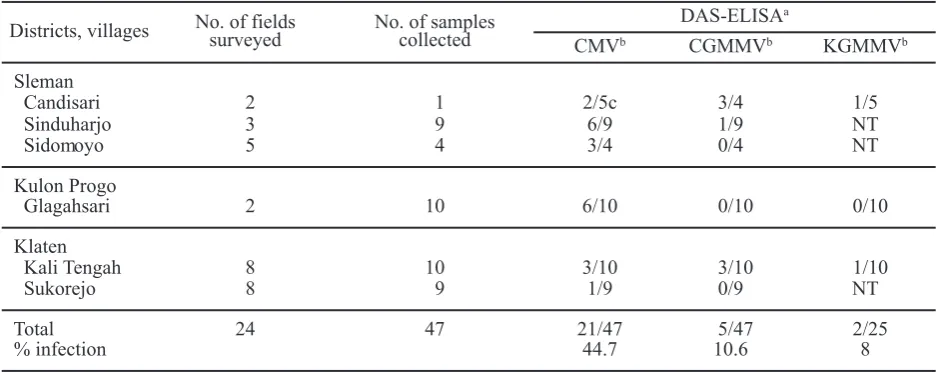
A total of 24 fields were surveyed and 47 plant samples were randomly collected from three melon growing areas in Java-Indonesia during 2000 and 2001. Among 47 plant samples, 28 samples were collected from Sleman and Kulon Progo in Yogyakarta province, while 19 samples were collected from Klaten in Central Java province.
nineteen samples from Klaten. The results of this survey are summarized in Table 1. The results of serological test showed that melon and other cucurbit plants were infected by CMV, CGMMV, and KGMMV in Klaten and Yogyakarta. Tests were conducted using several negative controls of melon and cucumber plants. Therefore, the range of absorbance values of negative controls varied from 0.020 to 0.147 at 405 nm, whereas positive samples gave absorbance value of 0.124 to 2.394 (Table 2). As shown in Table 1, CMV and CGMMV were detected in 21 and 5 of 47 fields, while KGMMV was detected only in 2 of 25 fields. All of fields surveyed had plants infected with at least one of these three viruses. The percentages of single infections were 44.7% (CMV), 10.6% (CGMMV) and 8% (KGMMV) in the samples.
The incident of these viruses in the sample of melon and other cucurbit plants is shown in Table 3.
The result shows that inCucumis meloandCucumis sativus, CMV was the most prevalent virus and was detected in 11 of 21, and 7 of 10 plants respectively. Moreover, CMV was detected in all, 3 of Momordica charantiaplants. InCitrullus vulgaris, and Cucurbita maxima, however, CMV was detected only in 1 of 5 and 1 of 3 plants respectively. Furthermore, CMV was not detected in 5 samples of Luffa acutangula. On the other hand, CGMMV and KGMMV were not detected in Cucumis sativus, Citrullus vulgaris, Cucurbit maxima, and Momordica charantia respectively and only detected in Cucumis melo, and Luffa acutangula plants. InCucumis melo, CGMMV was detected in 4 of 21, while KGMMV was detected only in 1 of 21 plants. In Luffa acutangula, CGMMV and KGMMV were detected in 2 of 5 plants respectively. CMV was the most common virus in all samples collected during the survey and 48.9%
Table 1. Numbers of fields surveyed and samples collected and tested in each district/village during July–August 2000 and August–September 2001
Districts, villages No. of fieldssurveyed No. of samplescollected DAS-ELISA
a
CMVb CGMMVb KGMMVb
Sleman
Candisari 2 1 2/5c 3/4 1/5
Sinduharjo 3 9 6/9 1/9 NT
Sidomoyo 5 4 3/4 0/4 NT
Kulon Progo
Glagahsari 2 10 6/10 0/10 0/10
Klaten
Kali Tengah 8 10 3/10 3/10 1/10
Sukorejo 8 9 1/9 0/9 NT
Total 24 47 21/47 5/47 2/25
% infection 44.7 10.6 8
a DAS-ELISA reading (absorbance at 405 nm) of sample extracts.
bCucumber mosaic virus(CMV),Cucumber green mottle mosaic virus(CGMMV), andKyuri green mottle mosaic
virus(KGMMV).
c Number of infected plant/number of plants sampled; NT: not tested.
Table 2. Range of absorbance values of negative control, positive and negative samples
Plant, virus Absorbance values (n=3)
Negative controls Positive samples Negative samples
Cucumber
CMV 0.046–0.048 0.124–1.026 0.012–0.960
CGMMV 0.020–0.032 0.219–0.463 0.005–0.058
KGMMV 0.067–0.117 0.422–2.219 0.045–0.193
Melon
CMV 0.063–0.159 0.466–2.118 0.021–0.292
CGMMV 0.032–0.105 0.413–1.563 0.088–0.138
Table 3. The incidence of viruses on melon and other cucurbit plants grown in Klaten and Yogyakarta
Species No. of samplestested CMVa CGMMVa KGMMVa
Cucumis melo 21 11 4 1
Cucumis sativus 10 7 0 0
Citrullus vulgaris 5 1 0 NT
Cucurbit maxima 3 1 0 NT
Momordica charantia 3 3 0 0
Luffa acutangula 5 0 2 2
Total 47 23 6 3
Percent infected - 48.9 12.8 6.4
aDAS-ELISA reading (absorbance at 405 nm) of sample extracts.
Figure 1. Cucurbit plants showing mosaic symptoms during survey in 2000–2001. A. Watermelon; B. Pumpkin; C. Melon; D. Bitter gourd; E. Angled loofah
A
B
C
of the samples were infected by CMV. With CGMMV and KGMMV, 12.8% and 6.4% of the samples were infected, respectively.
Disease symptoms showing in leaves of cucurbit plant were mosaic, chlorotic mottle, vein clearing or vein banding, as well as leaf distortion and malformation. Severe mosaic and mottle symptoms were associated most often with single infection of CGMMV and KGMMV respectively. The cucurbit plants showing mosaic symptoms during the survey is presented in Figure 1.
DISCUSSION
During July–August 2000 and August– September 2001, CMV was the most widespread virus in three melon-growing areas in the Yogyakarta and Klaten-Central Java in the number of field and its incident in each location. Similarly, Somowiyarjoet al.(1993) found that CMV was the most prevalent virus among four viruses CMV (37.3%), WMV (30%), ZYMV (12.1%), and PRSV-W (3.6%) in Yogyakarta and 5 fields in Central Java. In addition, they reported unidentified virus in 16.9% of the samples. In 1983, CMV was also reported caused the most severe diseases in squash (Cucurbita pepo) in New Jersey, USA (Davis & Mizuki, 1987). Further, Luis-Arteagaet al. (1998) reported that CMV and WMV-2 were the most frequently found viruses in the main areas for field grown melon production in Spain, both by the number of location and by their incidence in each location. In contrast, Grafton-Cardwellet al. (1996) found that WMV-2 was the most prevalent virus in all three melon-growing regions in the Central Valley of California, both in the number of sites with infected plants and in the proportion of symptomatic plants within each site.
CMV was detected for the first time in Indonesia in 1972 (Suseno & Lumanau, 1972). The disease was widely distributed throughout Indonesia and it infected not only cucurbits but also tobacco (Hartana, 1987). Furthermore, the virus was detected on ornamental and vegetable crops in Yogyakarta-Indonesia (Sulandari, 1990). Since that time, CMV has been one of the most serious viruses attacking agricultural plants and cause in loss of agricultural production in Indonesia (Semangun, 1989).
During the survey, CGMMV and KGMMV were detected only in angled loofah and melon samples. Infected angled loofah and melon showed growth retardation, mottle mosaic symptoms on the leaves
and on the surface of fruit. Moreover, CGMMV infected 12.8% of collected samples and more prevalent than KGMMV (6.4%). Both CGMMV and KGMMV were found in two of five angled loofah samples and according to ELISA result, CMV was not found in all angled loofah samples. On the other hand, CGMMV was detected only in four of 21 melon samples, while KGMMV was found in one of 21 melon samples. Recently, KGMMV have been reported occurred on melon and angled loofah in Indonesia (Daryono et al., 2005 and 2006). Both CGMMV and KGMMV belong to the genus Tobamovirus. These viruses have been reported as a serious disease agent of cucurbit crops and causing significant economical losses in several countries (Tan et al., 2000). CGMMV was found in Europe, India, Japan, Iran, Israel, Korea, and Greece, while KGMMV only reported in Japan and Korea (Ainsworth, 1935; Antignuset al., 2001; Inoueet al., 1967; Kitaniet al., 1970; Komuro et al., 1968; Tanet al., 2000; Ugaki et al., 1991; and Vasudewa et al., 1949). Furthermore, a melon isolate of CGMMV (CGMMV-SH) was reported as soil borne diseases infected greenhouse-grown melons in Japan and caused mosaic and necrotic lesion on the fruit of melon (Ugaki et al., 1991). On the other hand, KGMMV was found in zucchini fields in South Korea (Leeet al., 2000).
Viruses other than CMV, CGMMV, and KGMMV likely infect melon and other cucurbits in Yogyakarta and Klaten. Some samples that showed virus like symptoms only react with antisera used in serological test may react with the other cucurbit viruses such as WMV-2, ZYMV and PRSP which were not available in the present study. In 1992, WMV-2 and ZYMV were highly detected in cucurbits in Yogyakarta (Somowiyarjoet al., 1993). This result indicated that in addition to CMV and potyviruses, CGMMV and KGMMV becomes a potentially important virus in melon growing areas in Indonesia.
Inoue, T., N. Inoue, M. Asatani, & K. Mitsuhata. 1967. Studies on Cucumber Green Mottle Mosaic Virus in Japan (in Japanese).Nogaku Kenkyu51: 175–186.
Kitani, K., A. Kiso, & Y. Shigematsu. 1970. Studies on A New Virus Disease of Cucumber (Cucumber sativusL. var. F1Kurume-Oitai-H type) Discovered in Yodo (in Japanese).Proceeding of the Associa-tion for Plant ProtecAssocia-tion of Shikoku5: 59–66.
Komuro Y., H. Tochihara, R. Fukatsu, Y. Nagai, & S. Yoneyama. 1968. Cucumber Green Mottle Mosaic Virus on Watermelon in Chiba and Ibaraki Prefectures (in Japanese). Annals of the Phytopathology Society of Japan34: 377.
Lee, S.H., Y.G. Lee, J.W. Park, J.U. Cheon, K.W. Lee, & Y.C. Choi. 2000. Nucleotide Sequence of Coat Protein Gene ofKyuri Green Mottle Mosaic Virus (KGMMV) Isolated from Zucchini (Cucurbita pepo) in Korea. Plant Pathology Journal16: 118–124.
Luis-Artega, M., J.M. Alvarez, J.L. Alonso-Prados, J.J. Bernal, F. Garcia-Arenal, A. Lavina, A. Batle, & E. Moriones. 1998. Occurrence, Distribution, and Relative Incidence of Mosaic Viruses Infecting Field-grown Melon in Sapin. Plant Disease 82: 979–982.
Palukaitis, P., M.J. Roossinck, R.G. Dietzgen, & R.I.B. Francki. 1992. Cucumber Mosaic Virus. Advance Virus Research41: 281–348.
Ryu, K.H. 2004. Analysis of Cucurbit-infecting Tobamoviruses and Plant Virus Sequencing in Korea, p. 31–42. In K. Tomaru (ed.), PSJ Plant Virus Disease Workshop Report.No. 7.
Sammons, B., O.W. Barnett, R.F. Davis, & M.K. Mizuki. 1989. A Survey of Viruses Infecting Yellow Summer Squash in South Carolina.Plant Disease 73: 401–404.
Semangun, H. 1989. Penyakit-Penyakit Tanaman Hortikultura di Indonesia Gadjah Mada University Press, Yogyakarta. 124 p.
Somowiyarjo, S., N. Sako, & K. Tomaru. 1993. The Use of Dot Immunobinding Assay for Detecting Cucurbit Viruses in Yogyakarta, p. 3–11. In K. Tomaru & K.T. Natsuaki (eds.), Production of Virus-free Tropical Crops. NODAI Center for International Programs. Tokyo University of Agriculture, Tokyo.
Suastika, G., K. Tomaru, J. Kurihara, & K.T. Natsuaki. 1995. Characteristics of Two Isolates of Cucumber Mosaic Virus Obtained from Banana Plants in Indonesia (abstract in Japanese).Annals of the Phytopathology Society of Japan61: 272. In this study CMV was found to be the more
dominantly widespread virus in infected plants than CGMMV or KGMMV. It was suggested that CMV was more common in three melon growing areas because it is transmitted efficiently from weed hosts to melon and other cucurbits by aphids compared to CGMMV or KGMMV which are transmitted by mechanical and infected seeds.
ACKNOWLEDGEMENT
The authors thank Dr. Tri Joko, Laboratory of Plant Biotechnology, Faculty of Agriculture, Gadjah Mada University-Yogyakarta for sampling assistance.
REFFERENCES
Ainsworth, G.C. 1935. Mosaic Disease of Cucumber.Annals of Applied Biology22: 55–67.
Anonim. 2003. Buku Statistik Indonesia. Badan Pusat Statistik Indonesia, Jakarta. 21 p.
Antignus, Y., Y. Wang, M. Pearlsman, O. Lachman, N. Lavi, & A. Gal-On. 2001. Biological and Molecular Characterization of A New Cucurbit-infecting Tobamovirus.Phytopatology91: 565–571.
Clark, M.F. & A.N. Adams. 1977. Characteristics of the Microplate Method of Enzyme-linked Immunosorbent Assay for the Detection of Plant Viruses.Journal of General Virology34: 475–483.
Daryono, B.S., Somowiyarjo, S., & K.T. Natsuaki. 2005. Biological and Molecular Characterization of Melon-infectingKyuri Green Mottle Mosaic Virus in Indonesia. Journal of Phytopathology 153: 588–595.
Daryono, B.S., S. Somowiyarjo, & K.T. Natsuaki. 2006. Biological Characterization and Complete Nucleotide Sequence of Coat Protein of Kyuri Green Mottle Mosaic VirusIsolated from Angled Loofah in Indonesia. Journal of Agricultural Science,Tokyo University of Agriculture51: 45–52.
Davis, R.F. & M.K. Mizuki. 1987. Detection of Cucurbit Viruses in New Jersey.Plant Disease71: 40–44.
Grafton-Cardwell, E.E., T.M. Perring, R.F. Smith, J. Valencia, & C.A. Farrar. 1996. Occurrence of Mosaic Viruses in Melons in the Central Valley of California.Plant Disease80: 1092–1097.
Sulandari, S. 1990.Deteksi Cucumber mosaic virus dengan Metode Serologi pada Tanaman Hias dan Sayuran di DIY. Laporan Penelitian. Fakultas Pertanian UGM, Yogyakarta.
Suseno, H.R. & J.R. Lumanau. 1972.Occurrence of Cucumber Mosaic Virus on Tobacco Plants in Indonesia. South East Asian Regional Symposium on Plant Diseases in the Tropics, Yogyakarta.
Tan, S.H., M. Nishiguchi, M. Murata, & F. Motoyoshi. 2000. The Genome Structure of Kyuri Green Mottle Mosaic Tobamovirus and its Comparison with that of Cucumber Green Mottle Mosaic Tobamovirus. Archives of Virology 145: 1067–1079.
Ugaki, M., M. Tomiyama, T. Kakutani, S. Hidaka, T. Kiguchi, R. Nagata, T. Sato, F. Motoyoshi, & M. Nishiguchi. 1991. The Complete Nucleotide Sequence of Cucumber Green Mottle Mosaic Virus (SH Strain) Genomic RNA. Journal of General Virology72: 1487–1495.